Digital Poster
Alzheimer's & Dementias I
Joint Annual Meeting ISMRM-ESMRMB & ISMRT 31st Annual Meeting • 07-12 May 2022 • London, UK

| Computer # | ||||
|---|---|---|---|---|
2275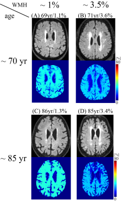 |
11 | Cerebral oxygen extraction fraction (OEF) mapping in cognitively impaired and intact elderly
Junghun Cho1, Gloria C Chiang1, Jonathan Dyke1, Hang Zhang2, Qihao Zhang2, Michael Tokov3, Thanh D Nguyen1, Pascal Spincemaille1, Ilhami Kovanlikaya1, Michael Amoashiy4, Mony de Leon1, and Yi Wang1,2
1Radiology, Weill Cornell Medicine, New York, NY, United States, 2Biomedical Engineering, Cornell University, Ithaca, NY, United States, 3College of Osteopathic Medicine, New York Institute of Technology, Glen Head, NY, United States, 4Neurology, Weill Cornell Medicine, New York, NY, United States
Cerebral metabolic dysfunction is known to underlie cognitive impairment. This study using a novel challenge-free MRI-based oxygen extraction fraction (OEF) mapping method, namely “QQ”, demonstrated that lower OEF in white matter and hippocampus was associated with greater white matter hyperintensities in cognitively impaired, but not cognitively intact, elderly, whereas older age was association with decreased OEF in cortical gray matter in the cognitively intact. This study suggests that QQ-based OEF mapping may be a useful tool readily and widely available for investigating metabolic dysfunction underlying dementia.
|
||
2276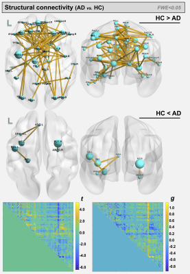 |
12 | Assessment of white matter changes associated with neurodegeneration in Alzheimer’s disease and early Parkinson’s disease
Maurizio Bergamino1, Sana Aslam2, Holly A Shill 2, and Ashley M Stokes1
1Neuroimaging Division, Barrow Neurological Institute, Phoenix, AZ, United States, 2Muhammad Ali Parkinson Center, Barrow Neurological Institute, Phoenix, AZ, United States
The objective of this study was to assess white matter integrity in both Alzheimer’s disease (AD) and early Parkinson’s disease (PD) using diffusion tensor imaging (DTI) metrics and connectivity analysis. Single-shell DTI data were obtained from open-source databases for AD and PD.
|
||
2277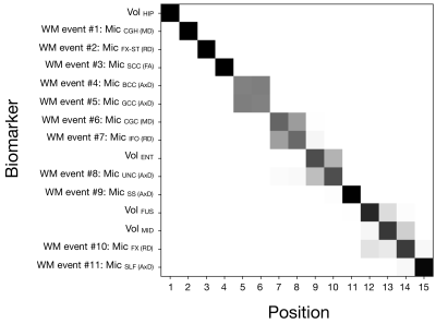 |
13 | Fine-grained characterisation of white matter microstructural abnormality in the pathophysiological cascade of Alzheimer’s disease
Christopher S Parker1, Hui Zhang1, and Neil P Oxtoby1
1Centre for Medical Image Computing, Department of Computer Science, UCL, London, United Kingdom White matter (WM) microstructure abnormalities have been observed in prodromal and manifest Alzheimer’s disease (AD). However, the precise nature and sequence of WM neurodegeneration across the disease course is unclear. Here, we leverage state-of-the-art disease progression modelling to sequence WM microstructure abnormalities in AD, as quantified by diffusion MRI in the ADNI study. We observe WM abnormalities among the earliest AD structural abnormalities and characterise the precise sequence of WM neurodegeneration. This fine-grained approach may be used to identify subjects at early disease stages for participation in therapeutic clinical trials.
|
||
2278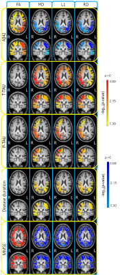 |
14 | Automatic DTI measures in the superficial white matter associated with cognitive decline and CSF amyloid-β42 in neurodegenerative dementia
Valeria Elisa Contarino1, Silvia Siggillino1, Andrea Arighi1, Elisa Scola1, Giorgio Giulio Fumagalli1, Giorgio Conte1,2, Emanuela Rotondo1, Daniela Galimberti1, Anna Margherita Pietroboni1, Tiziana Carandini1, Alexander Leemans3, Anna Maria Bianchi4, and Fabio Maria Triulzi1,2
1Fondazione IRCCS Ca' Granda Ospedale Maggiore Policlinico Milano, Milano, Italy, 2Department of Pathophysiology and Transplantation, Università degli Studi di Milano, Milano, Italy, 3Image Sciences Institute, University Medical Center Utrecht, Utrecht, Netherlands, 4Department of Electronics, Information and Bioengineering, Politecnico di Milano, Milano, Italy
Diffusion tensor imaging (DTI) studies showed superficial white matter (SWM) alterations in Alzheimer’s disease (AD). The study aims to investigate the relationship between DTI in the SWM and CSF biomarkers in neurodegenerative dementia (ND). 93 ND patients were retrospectively recruited and images were processed for automatic segmentation of lobar white matter and Diffusion tensor imaging (DTI). Spearman’s correlation tests were performed between lobar DTI measures in the SWM, CSF biomarkers and clinical data. DTI measures in the SWM strongly correlated with Mini-Mental State Examination (MMSE) and amyloid-β42 suggesting SWM DTI as a candidate in-vivo non-invasive clinical and preclinical biomarker.
|
||
2279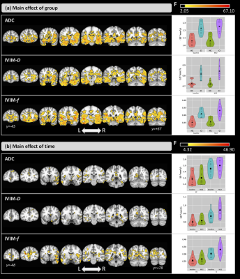 |
15 | Longitudinal Assessment of Intravoxel Incoherent Motion Diffusion-Weighted MRI (IVIM-DWI) Metrics in Alzheimer’s Disease
Maurizio Bergamino1, Lori Steffes2, Anna Burke3, Leslie C Baxter4, Richard J Caselli4, Marwan N Sabbagh5, and Ashley M Stokes1
1Neuroimaging Division, Barrow Neurological Institute, Phoenix, AZ, United States, 21Neuroimaging Division, Barrow Neurological Institute, Phoenix, AZ, United States, 3Division of Neurology, Barrow Neurological Institute, Phoenix, AZ, United States, 4Department of Neurology, Mayo Clinic Arizona, Phoenix, AZ, United States, 5Alzheimer's and Memory Disorders Division, Barrow Neurological Institute, Phoenix, AZ, United States
The objective of this pilot study was to assess the complementary brain microstructural and perfusion changes using the apparent diffusion coefficient (ADC) and the intravoxel incoherent motion-diffusion weighted imaging (IVIM-DWI) parameters in a group of cognitively impaired people over a period of 12 months. Additionally, voxel-based correlations between cognitive assessment scores and DWI imaging parameters were evaluated.
|
||
2280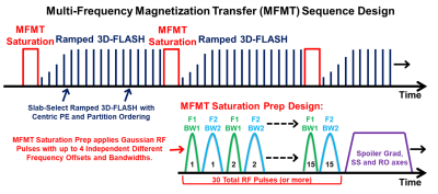 |
16 | Improved 7T High-Resolution Human Hippocampal Imaging with Multi-Frequency Magnetization Transfer (MFMT)
Ronald J Beyers1 and Thomas Denney1
1MRI Research Center, Auburn University, Auburn University, AL, United States
Alzheimer’s disease (AD) and other cognitive debilitating disease drive the need to improve neuro 7T MRI methods. We developed a multi-frequency magnetization transfer (MFMT) method for contrast improvement in 7T 3D MRI of the human hippocampus. MFMT applied to healthy volunteers demonstrated a 2.05 (p < 0.003) factor improvement in hippocampal contrast.
|
||
2281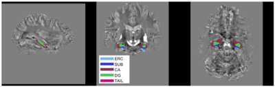 |
17 | REPRODUCIBILITY OF 7T MEASUREMENTS OF THE SUSCEPTIBILITY AND VOLUME OF HIPPOCAMPAL SUBFIELDS Video Permission Withheld
Oluwatobi Folorunsho Adeyemi1,2, Olivier Mougin1, Penny Gowland1, Richard Bowtell1, and Akram Hosseini3
1University of Nottingham, Nottingham, United Kingdom, 2University of Abuja, Abuja, Nigeria, 3Nottingham University Hospital, Nottingham, United Kingdom
This study provides an assessment of the level of reproducibility of measurements of the magnetic susceptibility of the hippocampal sub-fields that can achieved at 7T in a group of healthy volunteers using high-resolution QSM in conjunction with ASHS segmentation. The 2 to 4 ppb range of the values of the average standard deviation of repeated measurements characterises the minimum level of susceptibility change that could be reliably assessed in longitudinal studies, such as those evaluating changes in the iron content of the hippocampal sub-fields in Alzheimer’s disease.
|
||
2282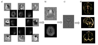 |
18 | Comparison of vein diameter and susceptibility values in individuals with and without APOE ε4 allele
Julia Huck1,2, Nathan Spreng3,4,5, Brittany Intzandt1,2,6,7, PREVENT-AD Research Group8, Sylvia Villeneuve5,9, Mallar Chakravarty9, Pierre-Louis Bazin10,11,12, and Claudine Gauthier1,2,7
1Concordia University, Montreal, QC, Canada, 2PERFORM Centre, Concordia University, Montreal, QC, Canada, 3Laboratory of Brain and Cognition, Department of Neurology and Neurosurgery, McGill University, Montreal, QC, Canada, 4Departments of Psychiatry and Psychology, Neurological Institute, McGill University, Montreal, QC, Canada, 5McConnell Brain Imaging Centre, Montreal Neurological Institute (MNI), McGill University, Montreal, QC, Canada, 6Centre de Recherche de l’Institut Universitaire de Geriatrie de Montreal, Montreal, QC, Canada, 7Centre de Recherche de l’Institut de Cardiologie de Montreal, Montreal, QC, Canada, 8StoP-AD Centre, Douglas Mental Health Institute Research Centre, Montreal, QC, Canada, 9Douglas Mental Health University Institute, McGill University, Montreal, QC, Canada, 10Psychology, University of Amsterdam, Amsterdam, Netherlands, 11Neurophysics, Max Planck Institute for Human Cognitive and Brian Sciences, Leipzig, Germany, 12Neurology, Max Planck Institute for Human Cognitive and Brian Sciences, Leipzig, Germany
Previous studies have shown an increase in oxygen extraction fraction (OEF) in individuals with Alzheimer’s disease (AD). Here, we investigate if vein diameters and susceptibility values measured with quantitative susceptibility maps (QSM), can be used as a biomarker in individuals with the APOE ε4 allele, which confers an increased risk of developing AD for detecting early changes in venous and metabolic properties.
|
||
2283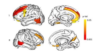 |
19 | Prediction of MCI conversion using sulcal morphometry
Giovanni Sighinolfi1, Micaela Mitolo2,3, Fabrizio Pizzagalli4, Raffaele Lodi1,2, Michelangelo Stanzani-Maserati5, Daniel Remondini6, Sabina Capellari1,5, Rocco Liguori1,5, Caterina Tonon1,2, and Claudia Testa2,6
1Department of Biomedical and NeuroMotor Sciences, University of Bologna, Bologna, Italy, 2Functional and Molecular Neuroimaging Unit, IRCCS Istituto delle Scienze Neurologiche di Bologna, Bologna, Italy, 3Department of Experimental, Diagnostic and Specialty Medicine, University of Bologna, Bologna, Italy, 4Department of Neurosciences "Rita Levi Montalcini", University of Torino, Torino, Italy, 5UOC Clinica Neurologica, IRCCS Istituto delle Scienze Neurologiche di Bologna, Bologna, Italy, 6Department of Physics and Astronomy, University of Bologna, Bologna, Italy We investigated the capabilities of brain sulcal morphometry and cortical thickness (CT) to predict the conversion from MCI stage to AD and explored the correlation with neuropsychological performance. Sulcal surface, mean depth, length and width were analysed. The width showed the best capability to discriminate between HC, MCI and AD (also in comparison to CT), and was able to distinguish at baseline converter MCI from non-converter MCI. Moreover, the width of specific frontal sulci correlated with memory and language performance. Sulcal morphometry emerged as a useful tool, capable of providing potential biomarkers for MCI conversion. |
||
 Back to Meeting Home
 Back to the Program-at-a-Glance
The International Society for Magnetic Resonance in Medicine is accredited by the Accreditation Council for Continuing Medical Education to provide continuing medical education for physicians.
 Back to Meeting Home
 Back to the Program-at-a-Glance
 View the Presentation