Digital Poster
Alzheimer's & Dementias II
Joint Annual Meeting ISMRM-ESMRMB & ISMRT 31st Annual Meeting • 07-12 May 2022 • London, UK

| Computer # | ||||
|---|---|---|---|---|
2385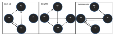 |
14 | Effective Connectivity within the Resting-State Network using Spectral Dynamic Causal Modeling Video Permission Withheld
Fatemeh Mohammadian1, Arash Zare Sadeghi2, Hanieh Mobarak Salari3, Mahsa Talebi1, Hassan Hashemi3, Hamid Reza Saligheh Rad1,4, and Maryam Noroozian5
1Department of Medical physics and Biomedical Engineering, Tehran university of medical sciences, TEHRAN, Iran (Islamic Republic of), 2Medical Physics Department, Iran University of Medical Sciences, TEHRAN, Iran (Islamic Republic of), 3Quantitative MR Imaging and Spectroscopy Group, Research Center for Molecular and Cellular Imaging, Tehran University of Medical Sciences, TEHRAN, Iran (Islamic Republic of), 4Quantitative MR Imaging and Spectroscopy Group, Research Center for Molecular and Cellular Imaging, Tehran University of Medical Sciences, Tehran, Iran (Islamic Republic of), 5Department of Psychiatry, Tehran university of medical sciences, TEHRAN, Iran (Islamic Republic of) Alzheimer's disease (AD) is a network connection dysfunction syndrome. An approximate picture of functional integration and statistical dependence on responses between different regions of the brain can be defined by functional connectivity (FC). Explanation of the statistical dependencies and estimating how the dynamics of neurons affect each other remotely is done by effective connectivity (EC). Studying directional interactions between different regions of the brain plays a key role in our understanding of the functional integration of brain networks. |
||
2386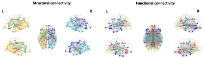 |
15 | Application of Graph Theory in the study of functional and structural connectivity in Mild Cognitive Impairment (MCI) disease
Emilio Cipriano1,2, Laura Biagi1, Paolo Bosco1, Giovanni Cioni1, Alessandro Sale3, Nicoletta Berardi3, Michela Matteoli3, Michela Tosetti1, and the Train the Brain Consortium4
1FiRMLAB, IRCCS Stella Maris, Pisa, Italy, 2Department of Physics, University of Pisa, Pisa, Italy, 3Institute of Neuroscience of the CNR, Pisa, Italy, 4the Train the Brain Consortium, Pisa, Italy
Graph theory approach was used to analyze structural and functional connectivity networks to identify candidate biomarkers of Mild Cognitive Impairment (MCI), a prodromal stage of Alzheimer’s disease (AD), for an early diagnosis and intervention. We proposed a method of brain network analysis to combine structural and functional connectivity networks information. Through graph measures (Segregation, Centrality and Global Efficiency), we evaluated the differences between MCI and healthy control (HC) subjects as well the possible effects of physical/cognitive training, in terms of structural and functional connectivity, in MCI population.
|
||
2387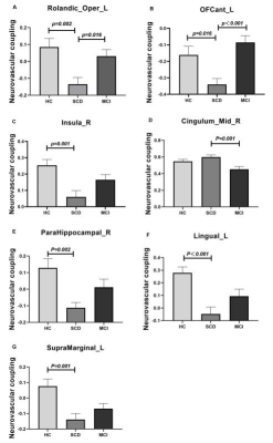 |
16 | Neurovascular dysfunction in patients with subjective cognitive decline: A resting-state fMRI study Video Not Available
Yin Tang1, Ling Zhang1, Hongyuan Ding1, Yi Zhu2, Yaxin Gao3, Long Qian4, Weiqiang Dou4, and Ming Qi1
1Radiology department, The First Affiliated Hospital of Nanjing Medical University, Nanjing, China, 2Rehabilitation Department, The First Affiliated Hospital of Nanjing Medical University, Nanjing, China, 3Rehabilitation Department, The Affiliated Suzhou Hospital of Nanjing Medical University, Suzhou, China, 4MR Research China, GE Healthcare, Beijing, China
The main goal of this study was to investigate the changes of neurovascular coupling (NVC) in patients at early stages of Alzheimer’s disease. Patients with SCD and MCI were enrolled. Significantly changes of neurovascular coupling have been separately found between SCD, MCI patients and HCs in the regions involved the left Rolandic operculum, left anterior orbital gyrus, right insula, right parahippocampal gyrus, left lingual gyrus, median cingulate and paracingulate gyri and left supramarginal gyrus. In addition, these changes of NVC showed strong correlations with multiple clinical scales. Therefore, NVC can be an effective method for early diagnosis of SCD patients.
|
||
2388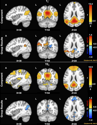 |
17 | Assessment of resting-state networks in Alzheimer’s disease using combined spin- and gradient-echo fMRI
Elizabeth G. Keeling1,2, Maurizio Bergamino1, Nicholas J. Sisco1, Sudarshan Ragunathan1, C. Chad Quarles1, George P. Prigatano1, and Ashley M. Stokes1
1Barrow Neurological Institute, Phoenix, AZ, United States, 2Arizona State University, Tempe, AZ, United States
Functional connectivity, measured with BOLD-fMRI, has been previously used to study changes in aging and Alzheimer’s disease. A combined spin- and gradient-echo (SAGE) acquisition may be able to more sensitively distinguish between disease-related changes in functional connectivity. Therefore, this study aims to implement SAGE in resting-state fMRI to assess differences between macro- and microvasculature-sensitive data, between SAGE maps and single-echo methods, and between healthy aging and cognitively impaired cohorts’ functional networks.
|
||
2389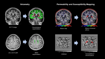 |
18 | Choroid plexus changes in clinically defined Alzheimer disease spectrum: Volumetry, QSM and BBB permeability imaging
Won-Jin Moon1, Jong Duck Choi1, Yeonsil Moon2, Hee Jin Kim3, Younghee Yim4, and Subin Lee5
1Radiology, Konkuk University Medical Center, Seoul, Korea, Republic of, 2Neurology, Konkuk University Medical Center, Seoul, Korea, Republic of, 3Neurology, Hanyang University Hospital, Seoul, Korea, Republic of, 4Radiology, Chung-Ang University Hospital, Seoul, Korea, Republic of, 5Electrical and Computer Engineering, Seoul National University, Seoul, Korea, Republic of
In patients with cognitive impairment spectrum, 3T MRI revealed that choroid plexus volume and permeability was associated with cognitive impairment severity. l In patients with cognitive impairment, choroid plexus (CP) volume followed this order: Alzheimer dementia > late mild cognitive impairment (MCI) > early MCI or subjective cognitive impairment (p<.001) l Large CP volume was associated with lower Ktrans (r=-0.403, p=.006) and Vp (r=-0.543, p=.003) l Increased CP volume was negatively associated with memory function (B=-0.6662, SE=0.2089, p=.002), frontal executive function (B=-0.8973, SE=0.3145, p=.005), and mini-mental status examination score (B=-0.8158, SE=0.3153, p=.01) after adjusting for covariates.
|
||
2390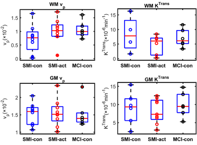 |
19 | DCE-MRI of blood brain barrier leakage in subjective memory and mild cognitive impairment: The Cognitive Ageing, Nutrition & Neurogenesis Trial Video Permission Withheld
Rashed Sobhan1, David R. Willis2, Rachel Gillings1, Michael J. Thrippleton3, Joanna M. Wardlaw3, Anne-Marie Minihane1, Narelle M. Berry1, and Donnie Cameron1,4
1Norwich Medical School, University of East Anglia, Norwich, United Kingdom, 2School of Environmental Sciences, University of East Anglia, Norwich, United Kingdom, 3Centre for Clinical Brain Sciences, University of Edinburgh, Edinburgh, United Kingdom, 4C.J. Gorter Centre for High Field MRI, Department of Radiology, Leiden University Medical Centre, Leiden, Netherlands As part of the CANN trial, we compared blood brain barrier (BBB) integrity in subjective memory impairment (SMI) and mild cognitive impairment (MCI), two early indicators of Alzheimer’s disease. Also, the impact of a combined flavanol and omega-3 fatty acid-based nutritional intervention on BBB leakage was explored for the SMI cohort. Dynamic-contrast-enhanced MRI was performed to obtain fractional plasma volume (vp) and volume transfer constant (KTrans) parameters. The results suggest that BBB leakage is similar in the SMI cohort and the nutritional intervention tends to be associated with improved BBB parameters. |
||
2391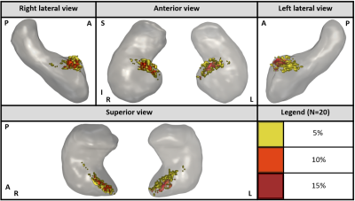 |
20 | Automatic detection of the anterior choroidal artery in TOF-MRA and its trajectory through the hippocampus and its sub-regions
Félix Janelle1,2, Monica Sean2,3, Félix Dumais2, Pascal Tétreault2,3, Christian Bocti1,4,5, and Kevin Whittingstall2
1Faculté de Médecine et des Sciences de la Santé, Université de Sherbrooke, Sherbrooke, QC, Canada, 2Médecine nucléaire et radiobiologie, Université de Sherbrooke, Sherbrooke, QC, Canada, 3Anesthésiologie, Université de Sherbrooke, Sherbrooke, QC, Canada, 4Service de Neurologie, Département de Médecine, CHUS, Sherbrooke, QC, Canada, 5Clinique de la Mémoire et centre de recherche sur le Viellissement, CIUSSS de l'Estrie-CHUS, Sherbrooke, QC, Canada The Anterior Choroidal Artery (AChA) is a small artery in the cerebral vasculature that perfuses the hippocampus. Its role in brain pathologies and cognitive decline is poorly understood. This could be due to the fact that there is no current standard method to analyse this artery in Time-of-Flight Magnetic Resonance Angiography (TOF-MRA). Here, we propose a fully automated approach for accurately localizing and quantifying the AChA in young healthy subjects. We then used this method to analyse the trajectory of the AChA through the hippocampus and its sub-regions. |
||
2392 |
21 | Neuroimaging of a novel mouse model of Alzheimer’s Disease: The hAβKI mouse.
Andre Obenaus1, Amandine Jullienne1, Brenda Patricia Noarbe1, Ashley A Keiser2, Michelle Vy Trinh1, Eniko Kramar2, Joy Beardwood2, Tri Dong2, Marcelo Wood2, and MODEL AD3
1Pediatrics, University of California, Irvine, Irvine, CA, United States, 2Neurobiology and Behavior, University of California, Irvine, Irvine, CA, United States, 3University of California, Irvine, Irvine, CA, United States
Mouse models of Alzheimer’s disease (AD) currently do not recapitulate accurately the human disease. The MODEL-AD consortium has recently developed new mouse models of AD, including the human amyloid β knockin (hAβKI) mouse. Using high resolution diffusion MRI (dMRI) we examined regional changes in the hAβKI male and female mouse across its lifespan (4-18mo). Sex and regional differences were apparent with age, including reduced diffusion metrics in the hippocampus, that mirrored altered learning and memory. Preclinical MR phenotyping allows for cross-species comparisons for biomarker identification.
|
||
2393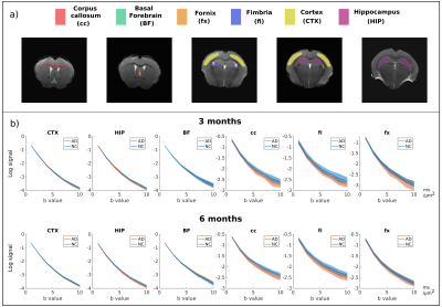 |
22 | Early microstructural aberrations in a mouse model of Alzheimer’s disease detected by Soma and Neurite Density Imaging
Andrada Ianus1, Renata Cruz1, Cristina Chavarrias1, Marco Palombo2,3,4, and Noam Shemesh1
1Champalimaud Research, Champalimaud Centre for the Unknown, Lisbon, Portugal, 2Centre for Medical Image Computing (CMIC), Dept of Computer Science, University College London, London, United Kingdom, 3Cardiff University Brain Research Imaging Centre (CUBRIC), School of Psychology, Cardiff University, Cardiff, United Kingdom, 4School of Computer Science and Informatics, Cardiff University, Cardiff, United Kingdom
Alzheimer’s disease induces early morphological tissue changes which precede the onset of symptoms and can be important targets for neuroimaging. In this work we employ Soma and Neurite Density Imaging (SANDI), a biophysical modelling approach for diffusion MRI, to investigate tissue microstructure alterations in the 3xTg AD mouse model and compare it with the state-of-the-art Diffusion Kurtosis Imaging (DKI).
|
||
2394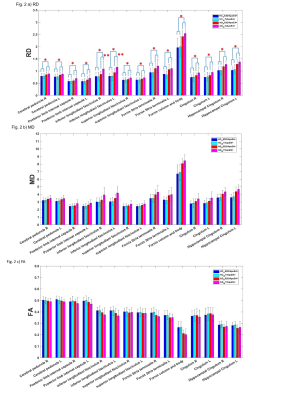 |
23 | DTI to Genetics: The Role of HFE H63D gene mutation in White Matter degeneration in APOE4 carrier of Alzheimer’s disease
Ran Pang1, Jianli Wang2, Prasanna Karunanayaka2, Samgan Kanekar2, Gela Beselia3, Samika Kanekar3, James R. Connor3, and Qing X. Yang4
1Departments of Neurosurgery, Pennsylvania State University College of Medicine, PA, United States; Department of Radiology, Dongfang Hospital, Beijing University of Chinese Medicine, Beijing, China, Hershey, PA, United States, 2Departments of Radiology, Pennsylvania State University Colle, Hershey, PA, United States, 3Departments of Neurosurgery, Pennsylvania State University College of Medicine, PA, United States, Hershey, PA, United States, 4Departments of 1Neurosurgery and 2Radiology, Pennsylvania State University College of Medicine, PA, United States, Hershey, PA, United States
HFE gene mutation involves iron metabolism, which is shown to directly contribute to myelination process. White matter degeneration in bi-genetic mutation (HFEH63D and ApoE4 allele) carriers was investigated using DTI from the AD Neuroimaging Initiative (ADNI) database. Comparing to the HFEWT/ApoE4+ group, mean diffusivity and radial diffusivity of inferior longitudinal fasciculus in the HFEH63D/ApoE4+ group demonstrated less integrity damage and fewer late-myelination loss with aging reflecting a possible mechanism as HFE polymorphism protective role in partly eliminating ApoE4 being the high-risk factor for AD progressing.
|
||
2395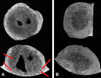 |
24 | Ultra-High Field MRI reveals amyloid plaques-like in a familial Alzheimer’s Disease patient-derived brain organoids
Mathieu David Santin1,2, Benjamin Galet1,3, Clément Daube1,3, and Philippe Ravassard1,3
1Institut du Cerveau – Paris Brain Institute - ICM, Sorbonne Université, INSERM, CNRS, Paris, France, 2Centre de NeuroImagerie de Recherche – CENIR, Paris, France, 3Molecular Pathophysiology of Parkinson’s Disease Group, Paris, France
This study presents preliminary results obtained on APP muted brain organoids. In vitro images were obtained using ultra-high field MRI and Gd passive staining of samples. MR images were compared to histology and immunohistochemistry slices to confirm the presence of amyloid plaques of mutated APP brain organoid compared to isogenic (control) brain organoid.
|
||
 Back to Meeting Home
 Back to the Program-at-a-Glance
The International Society for Magnetic Resonance in Medicine is accredited by the Accreditation Council for Continuing Medical Education to provide continuing medical education for physicians.
 Back to Meeting Home
 Back to the Program-at-a-Glance
 View the Presentation