Digital Poster
Aging & Cognitive Impairment
Joint Annual Meeting ISMRM-ESMRMB & ISMRT 31st Annual Meeting • 07-12 May 2022 • London, UK

| Computer # | ||||
|---|---|---|---|---|
1546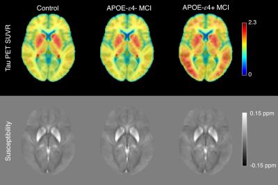 |
1 | Striatal tau deposition in mild cognitive impairment revealed by removal of iron-related off-target binding effects in 18F-AV1451 PET
Jason Langley1, Sumanth Dara2, Ilana Bennett3, and Xiaoping Hu1,2
1Center for Advanced Neuroimaging, University of California Riverside, Riverside, CA, United States, 2Department of Bioengineering, University of California Riverside, Riverside, CA, United States, 3Department of Psychology, University of California Riverside, Riverside, CA, United States
We use tissue susceptibility to control for iron-related off-target binding effects in 18F-AV1451 PET and examine the impact of APOE-ε4 carrier status on striatal tau-PET signal in mild cognitive impairment. We found significant increases in tau-PET SUVR in the putamen (p=0.01) and caudate nucleus (p=0.046) of APOE-ε4 positive participants compared to APOE-ε4 negative participants. Controlling for striatal iron, significant correlations were seen between striatal tau-PET SUVR memory measures in the control (putamen: r=0.435; caudate: r=0.623) and APOE-ε4 positive MCI (putamen: r=0.403; caudate: r=0.648) groups with greater tau burden correlated with greater memory impairment.
|
||
1547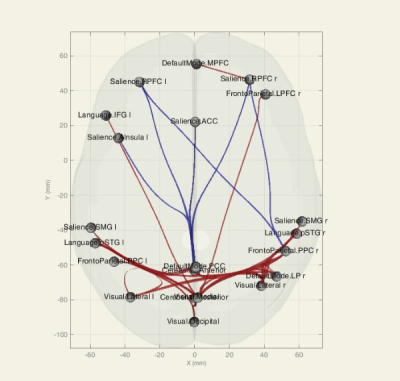 |
2 | Resting-state Functional connectivity to differentiate Mild Cognitive Impairment, Alzheimer's disease, and Normal subjects Video Permission Withheld
Fatemeh Mohammadian1, Maryam Noroozian2, Arash Zare Sadeghi3, Hanieh Mobarak Salari4, Hassan Hashemi5, and Hamid Reza Saligheh Rad1,6
1Department of Medical physics and Biomedical Engineering, Tehran university of medical sciences, TEHRAN, Iran (Islamic Republic of), 2Department of Psychiatry, Tehran university of medical sciences, TEHRAN, Iran (Islamic Republic of), 3Iran University of Medical Sciences, TEHRAN, Iran (Islamic Republic of), 4Quantitative MR Imaging and Spectroscopy Group, Research Center for Molecular and Cellular Imaging, Tehran University of Medical Sciences, TEHRAN, Iran (Islamic Republic of), 5Tehran university of medical sciences, TEHRAN, Iran (Islamic Republic of), 6Quantitative MR Imaging and Spectroscopy Group, Research Center for Molecular and Cellular Imaging, Tehran University of Medical Sciences, Tehran, Iran (Islamic Republic of)
Millions of people around the world suffer from Alzheimer's disease (AD), the most common neurodegenerative disease. Due to the progressive cognitive decline of the disease, early diagnosis can play an important role in the treatment and prevention of disease progression. Because functional and physiological changes occur before structural changes, functional imaging-based biomarkers such as Functional connectivity (FC) can show these changes well.
|
||
1548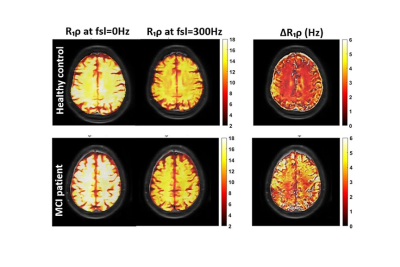 |
3 | T1ρ Dispersion in White Matter Correlates with Quantitative Metrics of Cognitive Impairment
Fatemeh Adelnia1,2, Taylor L Davis2, Lealani Mae Acosta3, Amanda Puckett3, Feng Wang1,2, Zhongliang Zu1,2, Kevin D Harkins1,2, and John C Gore1,2,4
1Vanderbilt University Institute of Imaging Science, NASHVILLE, TN, United States, 2Department of Radiology and Radiological Sciences, Vanderbilt University Medical Center, NASHVILLE, TN, United States, 3Department of Neurology, Vanderbilt University Medical Center, NASHVILLE, TN, United States, 4Department of Biomedical Engineering, Vanderbilt University, Nashville, TN, United States
R1ρ dispersion at weak locking fields (R1ρDiff) has the potential to reveal information on microvascular geometry and density. Our results show ΔR1ρ of white matter extracted from R1ρDiff dispersion is significantly greater in subjects with lower RBANS and MoCA scores. R2 or R1p values measured at a single locking field amplitude have no significant correlation with cognitive impairment scores. This work supports the hypothesis that microvascular impairment in white matter may be one of the causal factors in the progression of cognitive impairment in older adults.
|
||
1549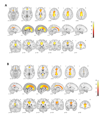 |
4 | Sex-differences in CBF changes as a biomarker of preclinical MCI
Safa Sanami1,2, Brittany N Intzandt2,3,4, Julia Huck1,2, PREVENT-AD Research Group5, and Claudine J Gauthier1,2,4
1Physics, Concordia University, Montreal, QC, Canada, 2PERFORM Centre, Concordia University, Montreal, QC, Canada, 3Centre de Recherche de l’Institut Universitaire de G´eriatrie de Montr´eal, Montreal, QC, Canada, 4Centre de Recherche de l’Institut de Cardiologie de Montr´eal, Montreal, QC, Canada, 5StoP-AD, Douglas Mental Health Institute, Montreal, QC, Canada
Alzheimer’s Disease (AD) is the most common form of dementia, preceded by a mild cognitive impairment (MCI) prodromal stage. There are sex differences in the prevalence and severity of MCI and AD. However, the presence of sex differences in cerebral hemodynamics during the transition from normal cognition to MCI is unknown. This is the first study to assess whether sex differences exist in cerebral blood flow (CBF) in the years prior to MCI diagnosis in patients with a familial history of AD.
|
||
1550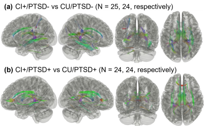 |
5 | Post-traumatic stress disorder may affect the progression of cognitive impairment in World Trade Center responders
Juin W. Zhou1, Minos Kritikos2,3, Chuan Huang4,5, Sean A. P. Clouston2,3, Roberto G. Lucchini6, Samuel E. Gandy7,8, Benjamin J. Luft9,10, and Evelyn J. Bromet5
1Biomedical Engineering, Stony Brook University, Stony Brook, NY, United States, 2Program in Public Health, Renaissance School of Medicine at Stony Brook University, Stony Brook, NY, United States, 3Department of Family, Population, and Preventive Medicine, Renaissance School of Medicine at Stony Brook University, Stony Brook, NY, United States, 4Department of Radiology, Renaissance School of Medicine at Stony Brook University, Stony Brook, NY, United States, 5Department of Psychiatry, Renaissance School of Medicine at Stony Brook University, Stony Brook, NY, United States, 6Department of Environmental Medicine and Public Health, Icahn School of Medicine at Mount Sinai, New York, NY, United States, 7Department of Neurology, Icahn School of Medicine at Mount Sinai, New York, NY, United States, 8Department of Psychiatry, Icahn School of Medicine at Mount Sinai, New York, NY, United States, 9Stony Brook World Trade Center Wellness Program, Renaissance School of Medicine at Stony Brook University, Stony Brook, NY, United States, 10Department of Medicine, Renaissance School of Medicine at Stony Brook University, Stony Brook, NY, United States A larger than expected number of World Trade Center (WTC) responders, now at midlife, are experiencing chronic post-traumatic stress disorder (PTSD) and incidence of mild cognitive impairment (MCI). A previous study reported that WTC responders with MCI have altered white matter connectivity when compared to unimpaired responders. However, the effects of PTSD on fiber integrity in this population was not studied due to a limited sample size. In the present study, we analyzed 97 WTC responders with/without CI and/or PTSD. Our results suggest that PTSD may influence the neuropathology of CI within this population. |
||
1551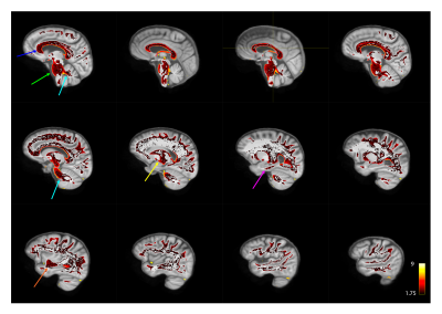 |
6 | Multivariate quantification of brain differences in individuals with family history of Alzheimer's disease and APOE4 genetic risk
Stefanie A Tremblay1,2, Nathan Spreng3,4,5, Amir Pirhadi6, Julia Huck1, Christine Lucas Tardif7,8,9, Mallar Chakravarty10,11, PREVENT-AD Research Group12, Sylvia Villeneuve10,13,14,15, Ilana Ruth Leppert7,8,9, Felix Carbonell16, Yasser Iturria-Medina8,9,17, Christopher J Steele18,19, and Claudine J Gauthier1,2
1Physics, Concordia University, Montreal, QC, Canada, 2Montreal Heart Institute, Montreal, QC, Canada, 3Laboratory of Brain and Cognition, Montreal Neurological Institute, Department of Neurology and Neurosurgery, McGill University, Montreal, QC, Canada, 4McConnell Brain Imaging Centre, Montreal Neurological Institute, McGill University, Montreal, QC, Canada, 5Departments of Psychiatry and Psychology, McGill University, Montreal, QC, Canada, 6Electrical and Computer Engineering, Concordia University, Montreal, QC, Canada, 7Biomedical Engineering, McGill University, Montreal, QC, Canada, 8McConnell Brain Imaging Centre, Montreal Neurological Institute and Hospital, Montreal, QC, Canada, 9Neurology and Neurosurgery, McGill University, Montreal, QC, Canada, 10StoP-AD Centre, Douglas Mental Health Institute Research Centre, Montreal, QC, Canada, 11McGill University, Montreal, QC, Canada, 12StoP-AD Centre, Douglas Mental Health Institute, Montreal, QC, Canada, 13McGill Centre for Integrative Neuroscience, Montreal Neurological Institute, McGill University, Montreal, QC, Canada, 14McConnell Brain Imaging Center, Montreal Neurological Institute, McGill University, Montreal, QC, Canada, 15CRIUGM - Université de Montreal, Montreal, QC, Canada, 16Biospective Inc., Montreal, QC, Canada, 17Ludmer Centre for NeuroInformatics and Mental Health, Montreal, QC, Canada, 18Psychology, Concordia University, Montreal, QC, Canada, 19Neurology, Max Planck Institute for Human Cognitive and Brain Sciences, Leipzig, Germany
Although the APOE4 genotype is known to increase the risk of developing Alzheimer’s disease (AD), the mechanisms through which this occurs are not fully understood. Here, we used a voxel-wise multivariate approach, the Mahalanobis distance (MhD), to quantify the extent to which the white matter microstructure of individuals with the APOE4 genotype differ from those without the E4 allele. The MhD of individuals with the E4 allele deviated significantly from our reference group in several regions, including tracts projecting to the hippocampus, a region known for its involvement in AD. Future work will investigate links with cognition and lifestyle factors.
|
||
1552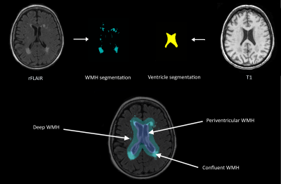 |
7 | The association between white matter hyperintensity shape and long-term dementia outcome in community-dwelling older adults
Jasmin A. Keller1, Sigurður Sigurdsson2, Kelly Klaassen1, Eveline Scholte1, Lydiane Hirschler1, Mark A. van Buchem1, Lenore J. Launer3, Matthias J.P. van Osch1, Vilmundor V. Gudnason2, and Jeroen de Bresser1
1Department of Radiology, Leiden University Medical Center, Leiden, Netherlands, 2Icelandic Heart Association, Kopavogur, Iceland, 3Laboratory of Epidemiology and Population Science, National Institute on Aging, Bethesda, MD, United States
Recently white matter hyperintensity (WMH) shape was introduced as a promising novel marker that may provide a more detailed characterization of WMH than volume alone. We aimed to investigate the association between WMH shape and the occurrence of dementia later in life in community dwelling older adults. WMH shape markers and WMH volumes were determined for periventricular/confluent, and deep WMH. A more complex shape of periventricular/confluent WMH (higher fractal dimension), as well as total and periventricular/confluent WMH volume, were associated with a greater risk of dementia. These results may indicate a prognostic value of WMH shape markers.
|
||
1553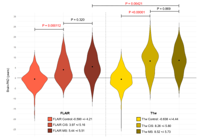 |
8 | Adapting and applying the brain age paradigm for clinical imaging in multiple sclerosis (MS)
Jordan Colman1, Olivia Goodkin1,2, Michael Foster2, Nima Mahmoudi3, Mike Wattjes3, Gabriel Gonzalez-Escamilla4, Sergiu Groppa4, Giuseppe Pontillo2,5, Einar August Høgestøl6,7, Lars T Westlye7, Silvia Messina8, Jacqueline Palace8, Rosa Cortese9, Nicola De Stefano9, Alex Rovira10, Jaume Sastre-Garriga10, Stefan Ropele11, Mara Rocca12, Massimo Filippi13, Ahmed Toosy2,
Olga Ciccarelli2, Tarek Yousry14,15, Ferran Prados1,2,16, Frederik Barkhof1,2,17,18, and James H Cole1,18
1Centre for Medical Image Computing, University College London, London, United Kingdom, 2Queen Square Multiple Sclerosis Centre, Department of Neuroinflammation, UCL Queen Square Institute of Neurology, London, United Kingdom, 3Medizinische Hochschule Hannover, Hannover, Germany, 4Movement Disorders, Neurostimulation and Neuroimaging, University Medicine Mainz, Mainz, Germany, 5Departments of Advanced Biomedical Sciences and Electrical Engineering and Information Technology, University of Naples "Federico II", Naples, Italy, 6Department of Neurology, Oslo University Hospital, Oslo, Norway, 7Department of Psychology, University of Oslo, Oslo, Norway, 8Nuffield Department of Clinical Neurosciences, Medical Sciences Division, University of Oxford, Oxford, United Kingdom, 9Department of Medicine, Surgery and Neuroscience, University of Siena, Siena, Italy, 10Hospital Universitari Vall d’Hebron, Barcelona, Spain, 11Neuroimaging Research Unit, Department of Neurology, Medical University of Graz, Graz, Austria, 12Neuroimaging Research Unit, Division of Neuroscience, IRCCS San Raffaele Scientific Institute, Vita-Salute San Raffaele University, Milan, Italy, 13Neurology Unit, Neurorehabilitation Unit, Neurophysiology Service, and Neuroimaging Research Unit, Division of Neuroscience, IRCCS San Raffaele Scientific Institute, Vita-Salute San Raffaele University, Milan, Italy, 14Lysholm Department of Neuroradiology, UCLH National Hospital for Neurology and Neurosurgery, London, United Kingdom, 15Neuroradiological Academic Unit, 15. Neuroradiological Academic Unit, London, United Kingdom, 16E-Health Center, Universitat Oberta de Catalunya, Barcelona, Spain, 17Department of Radiology and Nuclear Medicine, VU Medical Centre, Amsterdam, Netherlands, 18Dementia Research Centre, UCL Queen Square Institute of Neurology, London, United Kingdom
Brain-predicted age difference (brain-PAD; brain-predicted age – chronological age) is a potential biomarker for neurodegenerative diseases, including multiple sclerosis (MS). Previous models generally rely on T1-weighted (T1w) MRI brain scans. Here, we developed a deep-learning brain-age prediction model on FLAIR MRI. Our Inception-ResNet-V2 model was more accurate than a current state-of-the-art architecture and the FLAIR based model is comparable to a T1w MRI model. We used saliency maps, showing that areas such as the thalamus and ventricles are salient for brain-age prediction. We applied the FLAIR model to patients with MS, finding significantly higher brain-PAD compared to healthy controls.
|
||
 Back to Meeting Home
 Back to the Program-at-a-Glance
The International Society for Magnetic Resonance in Medicine is accredited by the Accreditation Council for Continuing Medical Education to provide continuing medical education for physicians.
 Back to Meeting Home
 Back to the Program-at-a-Glance
 View the Presentation