Digital Poster
Metabolism in Degenerative Diseases & Multiple Sclerosis
Joint Annual Meeting ISMRM-ESMRMB & ISMRT 31st Annual Meeting • 07-12 May 2022 • London, UK

Digital Poster
Metabolism in Degenerative Diseases & Multiple Sclerosis
| Computer # | ||||
|---|---|---|---|---|
1632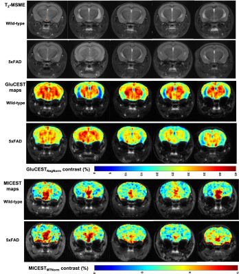 |
1 | In vivo CEST imaging of Glutamate and myo-Inositol for early-stage changes in Alzheimer’s 5xFAD mouse model
Ravi Prakash Reddy Nanga1, Halvor Juul1, Narayan Data Soni1, Blake Benyard1, Anshuman Swain2, Ryan Armbruster1, Paul Jacobs1, and Ravinder Reddy1
1Radiology, Center for Advance Metabolic Imaging in Precision Medicine, Perelman School of Medicine at The University of Pennsylvania, Philadelphia, PA, United States, 2Center for Advance Metabolic Imaging in Precision Medicine, Perelman School of Medicine at The University of Pennsylvania, Philadelphia, PA, United States
In order to probe the metabolism of glutamate and myo-inositol for better understanding of the Alzheimer’s diseases (AD) we have employed the chemical exchange saturation transfer of glutamate (GluCEST) and myoinositol (MICEST) in a fast progressing, 5xFAD mouse model. There were significant differences observed in the GluCEST and MICEST maps of 5XFAD mice when compared to the wild-type (WT) mice for regions of interest drawn on hippocampus and thalamus, which are further supported by the spectroscopy results.
|
||
1633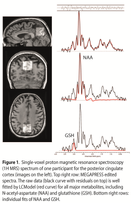 |
2 | Age-related decline of glutathione: a magnetic resonance spectroscopy study in 180 elderly participants
Lars Michels1, Valerie Treyer2,3, Anton Gietl4, and Ruth O’Gorman Tuura5
1Department of Neuroradiology, University Hospital Zurich, Zurich, Switzerland, 2Department of Nuclear Medicine, University Hospital Zurich, Zurich, Switzerland, 3Institute of Regenerative Medicine Zurich, University of Zurich, Zurich, Switzerland, 4Institute of Regenerative Medicine Zurich., University of Zurich, Zurich, Switzerland, 5Center for MR Research, University Children's Hospital, Zurich, Switzerland
Glutathione (GSH) is a brain marker for oxidative stress, which has previously been associated with brain amyloidosis and memory decline. However, to date no study has examined the link between GSH and beta-amyloid in a large cohort of elderly participants. Using simultaneous PET/MRS, we assessed amyloid deposition with amyloid-PET and GSH with MEGAPRESS, in a cohort of 134 healthy elderly and 46 mild cognitively impaired participants. GSH declined with age and showed sex differences, but no significant association between GSH and amyloid status was observed
|
||
| 1634 | 3 | Associations of Aging- and AD-associated 7T MRS Neurochemical Profiles with Cognition and Polygenic Risk for AD Video Permission Withheld
Michael Wolf1, Carlos Cruchaga2, Jillilan Crocker1, Priyanka Gorijala2, Laura Hemmy3, James Hodges1, Samira Mafimoghaddam2, Silvia Mangia1, Malgorzata Marjanska1, Riley McCarten3, Thomas Nichols4, Jeromy Thotland1, Rachel Zilinskas1, and Melissa Terpstra5
1University of Minnesota, Minneapolis, MN, United States, 2Washington University School of Medicine, St. Louis, MO, United States, 3Minneapolis VA Health Care System, Minneapolis, MN, United States, 4University of Oxford, Oxford, United Kingdom, 5University of Missouri, Columbia, MO, United States
Ultra-high field (7T) ultra-short echo time (8 ms STEAM) MRS can consistently provide a 14-neurochemical profile and distinguish aging and AD status. In this project, we generated composite scores from published age- and AD- specific neurochemical profiles and applied them to a typically aging cohort for whom polygenic risk for AD and extensive cognitive performance data were also available. Principal component analysis of the neurochemical profile fully distinguished aging, and AD classification was 58% correct. The largest correlation coefficients (r) were found between MRS and cognition. Correlation between MRS and polygenic risk and between cognition and polygenic risk was smaller.
|
||
1635 |
4 | A Magnetic Resonance Spectroscopy Method in Characterization of Blood Metabolomics for Alzheimer’s Disease
Leo Cheng1, Isabella Muti1, JianXiang Weng1, Anya Zhong1, Pia Kivisäkk2, Bradley Hyman2, and Steven Arnold2
1Massachusetts General Hospital, Charlestown, MA, United States, 2Massachusetts General Hospital, Boston, MA, United States
Currently, a definitive AD diagnosis can only be achieved at autopsy, through pathology examinations of brain tissue. No non- or less-invasive examination can yet diagnose and characterize AD during the patient’s life, in turn limiting insights into potential strategies for countering AD modes of progression. Here, using HRMAS MRS, we studied human blood plasma samples obtained from AD and non-AD subjects to reveal potential AD-associated metabolomic changes measurable in blood, which may assist with AD diagnosis and, more importantly, contribute to the development of precision treatments.
|
||
1636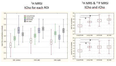 |
5 | MRSI DETECTABLE CHOLINE METABOLISM AS MARKER FOR GENDER DIFFERENCES IN AGING
Ulrich Pilatus1, Gunter P. Eckert2, Nasir Ludin1, Silke Matura1, Johannes Pantel1, Carmen Silaidos2, Lena Wachter2, and Elke Hattingen1
1Goethe-University Frankfurt, Frankfurt, Germany, 2Justus-Liebig-University of Giessen, Giessen, Germany
Combining quantitative (mmol/l) 1H MRSI and 31P MRSI data from the brain of healthy volunteers revealed a choline component (rCho=tCho-(PCho+GPC)) which is visible with 1H MRSI but missing in 31P MRSI. This component, which may account for a fraction of mobile phospholipids, is reduced in young female (mean age 26) subjects. Lower levels would indicate a higher integrity of the membrane phospholipids in the brain of young women. A possible reason is the higher estrogen levels in this group.
|
||
1637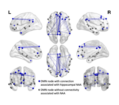 |
6 | The relationship between hippocampal NAA and functional connectivity within the Default Mode Network in Mild Cognitive Impairment
Marilena M DeMayo1,2, Jinglei Lv1,2, Shantel Duffy3,4, Sharon Naismith3,4,5, and Fernando Calamante1,2,6
1School of Biomedical Engineering, University of Sydney, Sydney, Australia, 2Brain and Mind Centre, University of Sydney, Sydney, Australia, 3School of Psychology, University of Sydney, Sydney, Australia, 4Healthy Brain Ageing Program, Brain and Mind Centre, University of Sydney, Sydney, Australia, 5Centre of Research Excellence to Optimise Sleep in Brain Ageing and Neurodegeneration (CogSleep CRE), Sydney, Australia, 6Sydney Imaging, University of Sydney, Sydney, Australia
We investigated the association between two MR markers of Mild Cognitive Impairment (MCI): N-Acetylaspartate (NAA) measured by MR Spectroscopy within the hippocampus; and functional connectivity within the Default Mode Network (DMN). We showed differential relationships between functional coupling/connectivity and NAA in MCI and controls, with an association between these measures in MCI not evident in controls. For those with MCI, we detected 20 connections within the DMN associated with NAA levels, with stronger connectivity associated with lower NAA levels. This suggests change in NAA within the hippocampus is associated with functional change within the DMN for those with MCI.
|
||
1638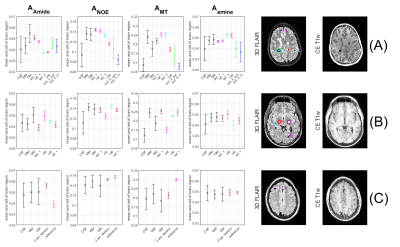 |
7 | 7T multi-pool CEST MRI in multiple sclerosis patients
Moritz Simon Fabian1, Stefan Hock1, Angelika Barbara Mennecke1, Manuel Schmidt1, Arnd Dörfler1, and Moritz Zaiß1,2
1Neuroradiology, University Hospital Erlangen, Erlangen, Germany, 2High-field Magnetic Resonance Center, Max Planck Institute for Biological Cybernetics, Tübingen, Germany
Differentiation of active multiple sclerosis lesions from non-active ones is done in the clinical environment by using contrast enhanced T1 images. CEST MRI has shown to yield correlations with Gadolinium contrast enhancement in tumors and other pathological tissue. In this work, we are trying to gain more insight in the metabolic information regarding the brain of patients suffering from multiple sclerosis.
|
||
 Back to Meeting Home
 Back to the Program-at-a-Glance
The International Society for Magnetic Resonance in Medicine is accredited by the Accreditation Council for Continuing Medical Education to provide continuing medical education for physicians.
 Back to Meeting Home
 Back to the Program-at-a-Glance
 View the Presentation