Digital Poster
Resting-State fMRI in Humans in Disease
Joint Annual Meeting ISMRM-ESMRMB & ISMRT 31st Annual Meeting • 07-12 May 2022 • London, UK

Digital Poster
Resting-State fMRI in Humans in Disease
| Computer # | ||||
|---|---|---|---|---|
2560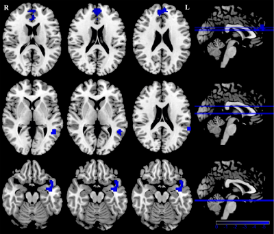 |
1 | Altered Brain Activity and Functional Connectivity between Thalamus and Cortex in Vestibular Migraine: A Resting-State fMRI Study Video Not Available
Xia Zhe1, Yang Huang1, Min Tang1, Kai Ai2, Xiaoyan Lei1, and Xiaoling Zhang1
1Department of MRI, Shaanxi Provincial People’s Hospital, Xi’an, Shaanxi, China, 2Philips Healthcare, Xi’an, China
To investigate the abnormal whole brain functional connectivity patterns in patients with VM by a fully data-driven resting-state functional connectivity methods without any hypothesis. Our findings indicated that whole brain amplitude of low-frequency fluctuation (ALFF) and connectivity abnormalities of these regions may be associated with functional impairments in pain information processing and modulation, transmitting and multisensory integration in patients with VM.
|
||
2561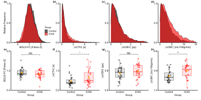 |
2 | Impact of capillary transit time heterogeneity on resting-state BOLD-FC in patients with unilateral asymptomatic carotid artery stenosis
Sebastian Constantin Schneider1,2, Mario Eduardo Archila-Meléndez1,2, Jens Göttler1,2, Stephan Kaczmarz1,2,3, Jan Kufer1,2, Benedikt Zott1,2, Josef Priller4, Michael Kallmayer5, Claus Zimmer1, Christian Sorg1,2, and Christine Preibisch1,2,6
1School of Medicine, Klinikum rechts der Isar, Department of Diagnostic and Interventional Neuroradiology, Technical University of Munich, Munich, Germany, 2School of Medicine, Klinikum rechts der Isar, TUM Neuroimaging Center, Technical University of Munich, Munich, Germany, 3Philips GmbH Market DACH, Hamburg, Germany, 4School of Medicine, Klinikum rechts der Isar, Department of Psychiatry, Technical University of Munich, Munich, Germany, 5School of Medicine, Klinikum rechts der Isar, Department of Vascular and Endovascular Surgery, Technical University of Munich, Munich, Germany, 6School of Medicine, Klinikum rechts der Isar, Department of Diagnostic and Interventional Neuroradiology, School of Medicine, Klinikum rechts der Isar, Department of Neurology, Munich, Germany
Blood oxygenation level dependent functional connectivity (BOLD-FC) is commonly used as a proxy for neuronal connectivity. Therefore, aberrant BOLD-FC in brain disorders is typically interpreted as aberrant neuronal connectivity. However, beyond changes in neuronal connectivity, impairments in neurovascular coupling (NVC) may also impact on BOLD-FC. This study investigates how impaired local NVC, under conditions of preserved neural functioning, influences BOLD-FC in a sample of unilateral asymptomatic internal carotid artery stenosis patients and healthy age-matched controls. We show that timing aspects of local NVC, namely increased capillary transit time heterogeneity, reduces BOLD-FC, without changes in neuronal functioning. |
||
2562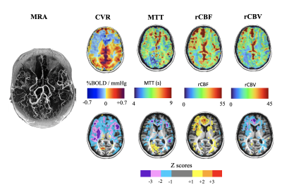 |
3 | Non-invasive measures of resting perfusion and hemodynamic compromise in patients with large vessel steno-occlusive disease
Ece Su Sayin1, James Duffin1,2, Julien Poublanc3, David John Mikulis3, Joseph Arnold Fisher1,2, and Olivia Sobczyk2
1Department of Physiology, University of Toronto, Toronto, ON, Canada, 2Department of Anaesthesia and Pain Management, University Health Network, Toronto, ON, Canada, 3Joint Department of Medical Imaging and the Functional Neuroimaging Lab, University Health Network, Toronto, ON, Canada
Dynamic susceptibility contrast perfusion measures using hypoxia-induced deoxyhemoglobin (dOHb) and BOLD-MRI-CO2 CVR reveals that patients with steno-occlusive disease can have the degree of hymodynamic compromise classified into two severities by the differences in hemodynamic measures at rest and in response to stress.
|
||
2563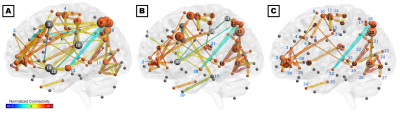 |
4 | The vestibular neuromatrix in patients with post-concussive vestibular dysfunction and healthy controls
Jeremy Lee Smith1, Anna Trofimova1, Vishwadeep Ahluwalia2,3, Jose J. Casado Garrido4, Julia Hurtado5, Rachael Frank5, April Hodge5, Russell K. Gore4,5, and Jason W. Allen1,4,6
1Radiology and Imaging Sciences, Emory University School of Medicine, Atlanta, GA, United States, 2Georgia State University, Atlanta, GA, United States, 3Center for Advanced Brain Imaging, Georgia Institute of Technology, Atlanta, GA, United States, 4Wallace H. Coulter Department of Biomedical Engineering, Georgia Institute of Technology and Emory University, Atlanta, GA, United States, 5Shepherd Center, Atlanta, GA, United States, 6Neurology, Emory University School of Medicine, Atlanta, GA, United States
Convergent clinical and neuroimaging evidence suggests that cognitive-affective and vestibular symptoms are interrelated: affective disorders not only co-occur with vestibular dysfunction but may also influence vestibular processing. However, the topology of the vestibular/cognitive/affective network (‘vestibular neuromatrix’) is not well-defined. The present study leveraged graph theory metrics to assess the functional and structural connectivity among 82 regions of interest in healthy controls and in patients with subacute post-concussive vestibular dysfunction. Patients exhibited deficiencies in connectivity among vestibular, pre- and orbitofrontal, and visual regions, as well as in the integration of visual and vestibular information, visuospatial attention, and monitoring of internal state.
|
||
2564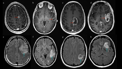 |
5 | Graph theory demonstrates functional reorganization dynamics related to tumor grade and location in glioma
Luca Pasquini1,2, Mehrnaz Jenabi3, Kyung Peck4, and Andrei Holodny1
1Radiology, Memorial Sloan Kettering Cancer Center, New York City, NY, United States, 2NESMOS, La Sapienza University, Rome, Italy, 3Radiology, Memorial Sloan Kettering Cancer Center, New York CIty, NY, United States, 4Medical Physics, Memorial Sloan Kettering Cancer Center, New York City, NY, United States
Brain tumors lead to modifications of brain networks known as functional reorganization or plasticity, which may be compensatory in nature and lead to valuable clinical applications. However, the determinants of plasticity are unclear. We evaluated functional networks of 30 low-grade (LGG) and 30 high-grade (HGG) left-hemispheric gliomas versus 20 healthy controls (HC) through rs-fMRI and graph-theory. We hypothesized that tumor grade and location impact brain connectivity. Our results show that both LGG and HGG develop ipsilateral and contralateral functional network changes. Additionally, tumor location is crucial: frontal and temporal tumors show bilateral modifications; parietal and insular tumors only local effects.
|
||
2565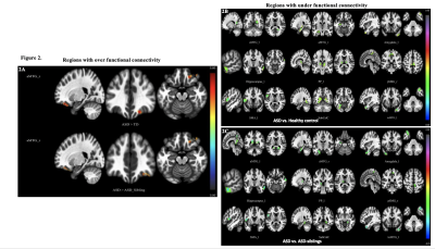 |
6 | Emerging functional connectivity differences in children with autism spectrum disorders and younger siblings affected with Autism
Manoj Kumar1, Sachin Patalasingh1, Chandrakanta Hiremath1, Jitender Saini1, K. John VijaySagar2, BK Yamini3, and Rose Dawn Bhrath1
1Neuroimaging and Interventional Radiology, National Institute of Mental Health and Neurosciences, Bengaluru, India, 2Child and Adolescent Psychiatry, National Institute of Mental Health and Neurosciences, Bangalore, India, 3Speech Pathology and Audiology, National Institute of Mental Health and Neurosciences, Bangalore, India
Resting-state fMRI studies performed during natural sleep indicate that functional-connectivity in ASD-children begins to differentially mature from earliest periods of life and continues to have distinct patterns throughout early development. In current study, we have performed rs-fMRI in younger (12 to 36-months-of-age) cohorts of ASD, ASD-siblings, and typically developing children to look for functional-connectivity patterns. We observed significantly reduced functional-connectivity in language, social-cognition, and motor regions than controls and ASD-siblings. Interestingly, some of these regions are also considerably different when compared between ASD and ASD-siblings. Altered connectivity may use for targeted interventional therapy for better clinical outcomes in ASD-children.
|
||
2566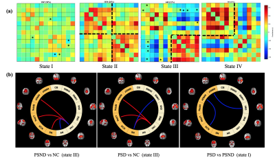 |
7 | Altered dynamic functional network connectivity in post-stroke dementia with subcortical lesion
Qianwen Wu1,2, Huaying Cai3, Linhui Ni3, Guocan Han4, Zhiyong Zhao1, and Dan Wu1
1Key Laboratory for Biomedical Engineering of Ministry of Education, Department of Biomedical Engineering, College of Biomedical Engineering & Instrument Science, Zhejiang University, Hangzhou, China, 2Department of Engineering, University of Oxford, Oxford, United Kingdom, 3Department of Neurology, Neuroscience Center,, Sir Run Run Shaw Hospital, Zhejiang University, Hangzhou, China, 4Department of Radiology, Neuroscience Center,, Sir Run Run Shaw Hospital, Zhejiang University, Hangzhou, China
This study aimed to explore the changes of functional network connectivity (FNC) in post-stroke dementia (PSD) using a dynamic FNC (dFNC) method. We collected resting-state fMRI data from16 PSD patients, 20 post-stroke non-dementia (PSND) patients and 19 normal controls, and identified 13 functional networks. We found PSD-specific decreases in the dFNC between default mode network and executive control network as well as visual network, which were not observed in static FNC analysis. Moreover, PSD showed a significant decrease in time-variance of global efficiency in whole brain compared with PSND. These changes were associated with the clinical assessments of patients.
|
||
2567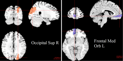 |
8 | Classification of Gulf War Illness Patients vs Control Veterans Using fMRI Dynamic Functional Connectivity
Unal Sakoglu1, Amaresh Mishra1, Kaundinya S Gopinath2, Bruce A Crosson2, and Robert Haley3
1Computer Engineering, University of Houston-Clear Lake, Houston, TX, United States, 2Emory University, Atlanta, GA, United States, 3University of Texas Southwestern Medical Canter at Dallas, Dallas, TX, United States
Around 200,000 veterans suffer from Gulf War Illness (GWI). GWI is characterized by multiple deficits in cognitive, emotion, somatosensory and pain domains. In this study we studied 23 GWI patients and 30 age-matched control veterans with resting-state fMRI in order to classify patients versus controls using dynamic functional connectivity among brain networks. Results show that different brain networks have discriminating power, pointing to widespread impairments in functional connectivity of visual, semantic, multi-sensory, and sensory-motor processing networks in GWI, consistent with multi-symptom nature of GWI.
|
||
2568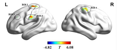 |
9 | Dynamic alterations of functional connectivity density in amyotrophic lateral sclerosis: a resting-state fMRI study Video Not Available
Jia Yan Shi1 and Hua Jun Chen2
1Fujian Medical University Union Hospital, Fuzhou, China, 2Radiology, Fujian Medical University Union Hospital, Fuzhou, China
In this study, we made the first investigation on abnormal pattern of dynamic functional connection density (dFCD) in ALS. We gathered resting state functional magnetic resonance imaging (fMRI) data from 50 patients with ALS and 55 healthy controls (HC). dFCD was analyzed by sliding window correlation method; and the standard deviation of dFCD across the sliding window was calculated voxel-wisely to quantify dFCD variability. Our study has found abnormal dFCD variability in ALS. The indices of dFCD variability showed moderate discrimination power between two groups, suggesting its potential serving as the diagnostic biomarker for ALS.
|
||
2569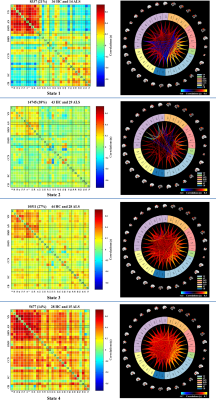 |
10 | Dynamic Changes in Functional Network Connectivity Involving Amyotrophic Lateral Sclerosis and Its Correlation With Disease Severity. Video Not Available
LiMin Cai1 and HuaJun Chen1
1Fujian Medical University Union Hospital, Fuzhou, China
We explored dynamic functional network connectivity (dFNC) in ALS and its correlation with disease severity. FNC states were determined by k-mean clustering, and state-specific FNC and dynamic indices (fraction time/mean dwell time/transition number) were calculated. ALS patients showed increased FNC between DMN-SMN in state 1 and between CCN-SMN in state 4. Patients remained in state 2 (showing the weakest FNC) for a significantly longer time and remained in state 1 (showing a relatively strong FNC) for a shorter time. A significant correlation was observed between ALSFRS-R and mean dwell time in state 2 and transition number.
|
||
2570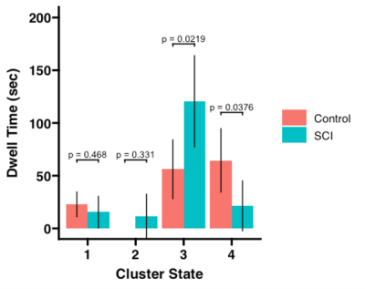 |
11 | Dynamic Functional Connectivity in Pediatric Spinal Cord Injury Patients Versus Healthy Controls Video Not Available
Joseph Schaefer1, Jingya Miao2, Caio Matias2, Feroze Mohamed2, Laura Krisa2, James Harrop2, and Mahdi Alizadeh2
1Neurological Surgery, Thomas Jefferson University, Philadelphia, PA, United States, 2Thomas Jefferson University, Philadelphia, PA, United States
The purpose of this study is to investigate the distributed network properties of subjects with pediatric spinal cord injury using static and dynamic functional connectivity analysis. 36 subjects of which 18 had history of SCI between the ages of 0-18 years underwent 3-Tesla resting state functional MRI (rs-fMRI). Static and dynamic functional connectivity was calculated. Results showed patients with a history of pediatric spinal cord injury did not show widespread changes in static functional connectivity but did have significant changes in dynamic functional connectivity, including spending more time in a relatively hypoconnected brain state and less brain state transitions.
|
||
2571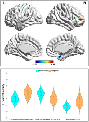 |
12 | Abnormal stability of dynamic functional architecture in amyotrophic lateral sclerosis: a preliminary resting-state fMRI study Video Not Available
Jia Yan Shi1 and Hua Jun Chen2
1Radiology, Fujian Medical University Union Hospital, Fuzhou, China, 2Fujian Medical University Union Hospital, Fuzhou, China
Static and dynamic analyses for identifying functional connectivity(FC)have demonstrated brain dysfunctions in amyotrophic lateral sclerosis(ALS).We are to make the first attempt to perform studies about the stability of dynamic FC analysis based on resting-state fMRI data, in order to provide the new insight into the mechanism in ALS and investigate its correlation with disease severity. Our observation revealed that the abnormal pattern of FC stability involved sensorimotor regions(i.e.left precentral and postcentral gyrus)and non-sensorimotor areas(i.e.right temporal pole and middle/inferior frontal gyrus)in ALS patients, suggesting the potential of functional stability serving as the biomarker for monitoring disease progression in ALS.
|
||
 Back to Meeting Home
 Back to the Program-at-a-Glance
The International Society for Magnetic Resonance in Medicine is accredited by the Accreditation Council for Continuing Medical Education to provide continuing medical education for physicians.
 Back to Meeting Home
 Back to the Program-at-a-Glance
 View the Presentation