Digital Poster
Resting-State Functional Connectivity Methods
Joint Annual Meeting ISMRM-ESMRMB & ISMRT 31st Annual Meeting • 07-12 May 2022 • London, UK

Digital Poster
Resting-State Functional Connectivity Methods
| Computer # | ||||
|---|---|---|---|---|
0994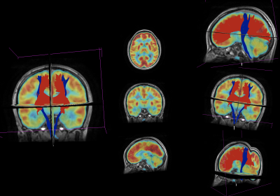 |
1 | Simultaneous FDG-PET and MRI-based connectivity analysis for the detection of epileptogenic zones in non-lesional epilepsy patients
Clara Lisazo1,2, Natalia Noemí Massaccesi Bove1,2, Daniela Zanchi3, María Paula Del Pópolo1,2, Daniel Fino1,2,4, Federico Julián González Nicolini1,4,5, Sergio Mosconi3, Pedro Pablo Ariza1,2, Trinidad Gonzalez Padín1,2, Raúl Otoya6, Roberto Isoardi3,5, Thomas Martin Doring7, and Sebastián Moguilner5,8
1MRI Department, Fundación Escuela de Medicina Nuclear, Mendoza, Argentina, 2MRI Department, Fundación Argentina para el Desarrollo en Salud, Mendoza, Argentina, 3Nuclear Medicine Department, Fundación Escuela de Medicina Nuclear, Mendoza, Argentina, 4Instituto Balseiro, Universidad Nacional de Cuyo, San Carlos de Bariloche, Argentina, 5GQNYCS, Comisión Nacional de Energía Atómica, Ciudad Autónoma de Buenos Aires, Argentina, 6Neuromed, Mendoza, Argentina, 7GE Healthcare, Rio de Janeiro, Brazil, 8Department of Neurology, Massachusetts General Hospital and Harvard Medical School, Boston, MA, United States
Detecting epileptogenic zones (EZ) in subjects with non-lesional epilepsy can represent a challenge, and it involves the analysis of data from various diagnostic procedures. In this study, seed-based structural and functional connectivity matrices were obtained from DWI and resting-state fMRI for each patient and compared with a control group. Then, we assessed whether significantly different regions (p<0.001) coincided with foci of abnormal metabolism on PET images, and if these ROIs corresponded with the subject’s EZ. This reduced the false positive rate and improved specificity of the connectivity analysis, optimizing EZ localization in patients with non-lesional epilepsy.
|
||
0995 |
2 | Comparing sliding window correlation and instantaneous phase coherence in fMRI dynamic functional connectivity analysis
Cláudia Fonseca1, Inês Esteves1, Marta Xavier1, Ana Fouto1, Amparo Ruiz-Tagle1, Nuno A. Silva2, Rita G. Nunes1, Raquel Gil-Gouveia3, Joana Cabral4, and Patrícia Figueiredo1
1Institute for Systems and Robotics, Instituto Superior Técnico, University of Lisbon, Lisbon, Portugal, 2Learning Health, Hospital da Luz, Lisbon, Portugal, 3Neurology Department, Hospital da Luz, Lisbon, Portugal, 4Life and Health Sciences Research Institute, University of Minho, Braga, Portugal
Sliding window Pearson correlation (SW) is the most commonly used approach for estimating dynamic functional connectivity (dFC). However, instantaneous phase coherence (PC) has gained popularity as it yields frame-by-frame dFC estimates. This work aimed to compare both metrics by analysing the mean lifetime, probability of occurrence and spatial similarity of dFC states with the canonical resting-state networks (RSNs). We found that the state lifetimes increase in SW compared to PC and with window length, worsening the detection of RSNs for smaller datasets. These findings indicate that the temporal blurring induced by SW compromises the ability to detect faster network dynamics.
|
||
0996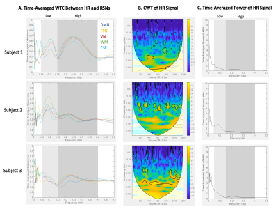 |
3 | Characterizing spectral and temporal effects of heart rate variability on resting-state BOLD signals using wavelet transform coherence
Quimby Lee1, Jingyuan E. Chen2,3, Gregory Wheeler4, and Audrey P. Fan1,4
1Department of Neurology, University of California, Davis, Davis, CA, United States, 2Department of Radiology, Harvard Medical School, Boston, MA, United States, 3Massachusetts General Hospital, Athinoula A. Martinos Center for Biomedical Imaging, Boston, MA, United States, 4Department of Biomedical Engineering, University of California, Davis, Davis, CA, United States
Heart rate variability (HRV), which is reflective of autonomic regulation, induces vascular effects in low (0.05-0.15Hz) and high (0.15-0.4Hz) frequency bands that can influence the blood oxygen level dependent (BOLD) signal of resting-state functional magnetic resonance imaging (rsfMRI). In this study, we utilized a wavelet transform coherence analysis to identify spectral and temporal differences in phased-locked behavior between HRV and resting-state network (RSN) time courses. Subjects differed in the frequency band with greatest time-averaged coherence and percentage of time with significant coherence of HRV and RSN signals.
|
||
0997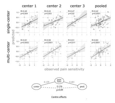 |
4 | Predictive modelling and center effects: towards a robust functional connectivity-based neuromarker of pain sensitivity
Tamas Spisak1, Balint Kincses2,3, Raviteja Kotikalapudi1, Matthias Zunhammer2, Frederik Schlitt2, Tobias Schmidt-Wilcke4,5, Zsigmond Tamas Kincses3, and Ulrike Bingel2
1Laboratory of Predictive Neuroimaging, Institute for Diagnostic and Interventional Radiology and Neuroradiology, University Hospital Essen, Essen, Germany, 2Klinik für Neurologie, University Hospital Essen, Essen, Germany, 3Department of Neurology, University of Szeged, Szeged, Hungary, 4Institut für Klinische Neurowissenschaften und Medizinische Psychologie, Heinrich Heine Universität, Düsseldorf, Germany, 5Neurozentrum, Bezirksklinikum Mainkofen, Deggendorf, Germany Center effects significantly limit the generalizability of brain imaging-based biomarker candidates. Although our previously published resting state functional connectivity-based predictive signature for pain sensitivity (the RPN-signature) showed remarkable out-of-center generalizability, it remained unclear which connectivity features are the most generalizable across study centers. Here, we re-trained the RPN-signature on multi-center data and found that it outperforms the single-center model in all three centers (explained variance: 26-38% vs. 16%-19%). Our results highlight that neurobiological interpretation of feature importance in predictive modelling is constrained both by center-specific artifacts and by certain characteristics (e.g. regularization) of the applied machine learning algorithm. |
||
0998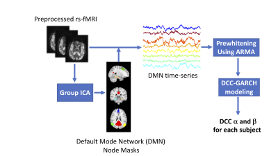 |
5 | A novel method for characterizing dynamic resting state functional connectivity in Alzheimer's Disease
Kun Yue1, Jason M Webster2, Thomas J Grabowski2, Ali Shojaei1, and Hesamoddin Jahanian2
1Department of Biostatistics, University of Washington, Seattle, WA, United States, 2Department of Radiology, University of Washington, Seattle, WA, United States
With advances in experimental therapeutics for Alzheimer's Disease (AD) the need for an accurate, non-invasive and widely available AD biomarker is more pressing than ever. Resting-state functional connectivity in default mode network is a candidate biomarker that is gaining traction in the field. However, the traditional stationary measurement of the default mode network connectivity cannot capture complicated dynamic patterns of functional connectivity that exist in the brain. Here, we have proposed a novel, reliable technique, based on Dynamic Condtional Correlation model, to quantify the dynamic functional connectivity in the brain and evaluated its sensitivity to cerebrospinal fluid biomarkers in AD.
|
||
0999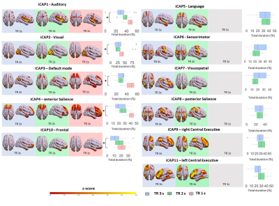 |
6 | Temporal resolution impacts in-vivo human brain dynamic functional MRI connectivity
Francesca Saviola1, Stefano Tambalo1, Dimitri Van De Ville2,3, and Jorge Jovicich1
1University of Trento, Center for Mind/Brain Sciences, Rovereto, Italy, 2Institute of Bioengineering, Center for Neuroprosthetics, Ecole Polytechnique Fédérale de Lausanne (EPFL), Lausanne, Switzerland, 3Department of Radiology and Medical Informatics, University of Geneva (UNIGE), Geneva, Switzerland
The advent of fast fMRI acquisition techniques has enabled whole-brain acquisitions with sub-second temporal resolution enriching the information available throughout the time course. However, there is no consensus about how the application of frame-wise analysis, performed to better understand functional brain fluctuations, could benefit from high temporal resolution fMRI. In this work, we demonstrate the potential need to: (i) gain a finer understanding of signal and noise signatures peculiar to fast acquisition and (ii) extend the models to better estimate dynamic functional connectivity in rapidly sampled fMRI time series.
|
||
1000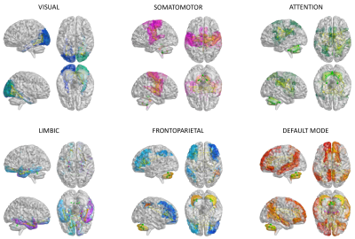 |
7 | First evidence of a link between topology and neurophysiological properties of brain sub-networks.
Anita Monteverdi1,2, Fulvia Palesi2, Claudia AM Gandini Wheeler-Kingshott1,2,3, and Egidio D'Angelo1,2
1Brain Connectivity Center, IRCCS Mondino Foundation, Pavia, Italy, 2Brain and Behavioural Sciences, University of Pavia, Pavia, Italy, 3NMR Research Unit, Queen Square Multiple Sclerosis Centre, Department of Neuroinflammation, UCL Queen Square Institute of Neurology, UCL, London, United Kingdom
A comprehensive assessment of multiple resting-state networks is still lacking and the characterization of their excitatory/inhibitory (E/I) balance is an open field of research. In this exploratory work we performed a first characterization of the E/I balance in resting-state networks (visual, somatomotor, attention, limbic, frontoparietal, DMN) both in healthy and neurodegenerative conditions (Alzheimer’s Disease, Frontotemporal Dementia, Amyotrophic Lateral Sclerosis) taking advantage of a Virtual Brain based approach. Our results provided a new strategy to simultaneously characterize multiple networks at single-subject level, deepening their E/I balance and opening new perspectives for biomarkers research.
|
||
1001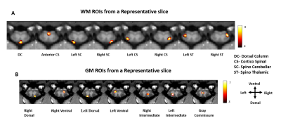 |
8 | Analysis of resting state functional connectivity of white matter tracts in spinal cord of squirrel monkey using Graph Theory
Anirban Sengupta1, Arabinda Mishra1, Feng Wang1, Li Min Chen1, and John C Gore1
1Vanderbilt University Medical Center, Nashville, TN, United States
The goal of this study was to investigate the nature of spontaneous BOLD fluctuations in white-matter (WM) of spinal-cord and to use these to identify the intrinsic functional architecture of WM tracts and their correlations with the gray-matter (GM) hubs. Connectivity measures were obtained using resting state BOLD signals between WM-WM and WM-GM regions, followed by network analysis using graph-theory. We found WM and GM hubs on the dorsal side exhibit greater temporal correlation as exhibited by their stronger node strength in resting state. Also, within segment WM-WM and WM-GM correlations were found to be stronger than those between segments.
|
||
 Back to Meeting Home
 Back to the Program-at-a-Glance
The International Society for Magnetic Resonance in Medicine is accredited by the Accreditation Council for Continuing Medical Education to provide continuing medical education for physicians.
 Back to Meeting Home
 Back to the Program-at-a-Glance
 View the Presentation