Digital Poster
fMRI Studies of Physiology, Metabolism & Evoked Responses
Joint Annual Meeting ISMRM-ESMRMB & ISMRT 31st Annual Meeting • 07-12 May 2022 • London, UK

Digital Poster
fMRI Studies of Physiology, Metabolism & Evoked Responses
| Computer # | ||||
|---|---|---|---|---|
1731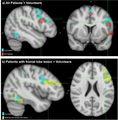 |
1 | How does language fMRI activation in tumour patients compare to healthy volunteers?
Rachael Franklin1,2, Enrico de Vita2, Irène Brumer3, Jozef Jarosz1, and Marco Borri1
1Neuroradiology, King's College Hospital, London, United Kingdom, 2School of Biomedical Engineering & Imaging Sciences, King's College London, London, United Kingdom, 3Computer Assisted Clinical Medicine, Mannheim Institute for Intelligent Systems in Medicine, Medical Faculty Mannheim, Heidelberg University, Heidelberg, Germany
Task-based language fMRI datasets from healthy volunteers (15) and tumour patients (4 with lesions near Wernicke’s area, 6 with lesions near Broca’s area) were compared, to assess the similarity of activation location and extent. Expected areas of the language network were consistently localised in both patients and volunteers. Although patients showed more variation in the extent and strength of activation, our activation metrics for the full patient cohort show similar amount of overlap compared to a size-matched group of volunteers. Furthermore, patient with lesions near Broca’s area had highly consistent activation in specific locations.
|
||
1732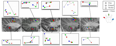 |
2 | A separation between motor and sensory somatotopic maps in the human cerebellum
Emma Brouwer 1, Nikos Priovoulos1, and Wietske van der Zwaag1
1Spinoza Centre for Neuroimaging, Amsterdam, Netherlands
We investigated the organisation of the cerebellar digit map with three distinct tasks (flexing/extending/stroking) using B1-shimmed, high-resolution fMRI at 7T. For all tasks, the positions of the COG of the digit representations formed an orderly progression in at least one of the lobules. The distance between the motor task activations for flexing and extending of the digits were smaller than from either motor task to stroking clusters, indicating a separation between somatosensory and motor clusters in cerebellar lobule V.
|
||
1733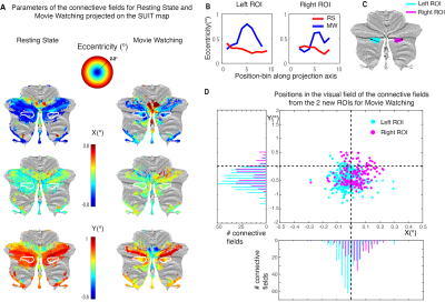 |
3 | Retinotopic connectivity in the cerebellum for different cognitive states Video Permission Withheld
Wietske Zuiderbaan1, Wietske van der Zwaag1, and Tomas Knapen1,2
1Spinoza Centre for Neuroimaging, Amsterdam, Netherlands, 2Behavioral and Movement Sciences, Vrije Universiteit, Amsterdam, Netherlands
We used the retinotopic connective field model to investigate how retinotopic organization in the cerebellum depends on cognitive state. Previous work found three retinotopic clusters in the cerebellum. These regions were found using a standard bar mapping stimulus with fixation task. Here, we show that with a more naturalistic condition, we find an extra cluster to be retinotopically organized. Furthermore, we show that the oculomotor vermis shows a gradient in visual field eccentricity, that was not revealed using the simple stimulus. Our results underline the importance of using a naturalistic condition in studying the retinotopic organization of the cerebellum.
|
||
1734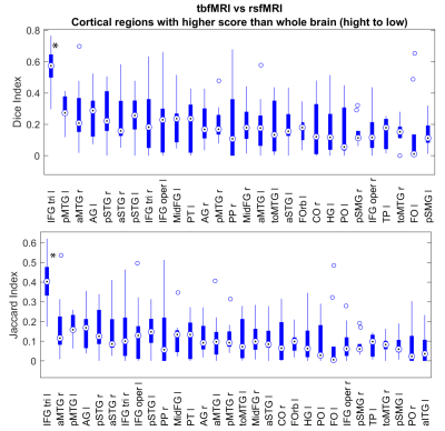 |
4 | Comparison of Language Areas determined by Resting State and Task Based fMRI using healthy subjects HCP data.
Robert Pretorius1, Stefan Sunaert1,2,3, and Ahmed Radwan1
1Department of Imaging and pathology, Translational MRI, Leuven, Belgium, KU Leuven, Leuven, Belgium, 2Leuven Brain Institute, Department of Neurosciences, Leuven, Belgium, KU Leuven, Leuven, Belgium, 3Department of Radiology, University Hospitals Leuven, Leuven, Belgium, University Hospital Leuven, Leuven, Belgium
Adding to the literature of resting state fMRI (rsfMRI) in preoperative planning, we investigated the similarity of a task based fMRI (tbfMRI) language protocol to a pan-language functional connectivity map in 10 healthy subjects. When comparing the whole brain there is relatively low Jaccard and Sorensen-Dice similarity between rsfMRI and tbfMRI language activity, however when looking at separate cortical anatomical regions the language areas are among those having the highest similarity. Additionally, there is a significantly higher similarity in the left IFG pars triangularis. This suggests that rsfMRI could be used reliably to determine Broca’s area in preoperative planning.
|
||
1735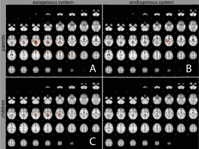 |
5 | Brain-to-brain interaction in parent-child eye contact
Ray Lee1, Joshua Friedman1, Alaa Khadar2, Andrea Karaiskaki1, Nadiya Gura1, Maria O'Brien1, Paul Sajda1, and Nim Tottenham1
1Columbia University, New York, NY, United States, 2NASA, Houston, TX, United States
Parent-child interaction is a significant part of human life, and a large portion of such interaction is carried out by eye contact. However, how the dyadic brain networks enable eye contact is not well understood. Here, we take advantage of fMRI hyperscan and concurrent eye tracking to measure the BOLD responses and pupil sizes during eye contact between parent and child. Our initial analysis begins to reveal some of the brain networks supporting the parent-child bond. A better understanding of such mechanisms may have a significant impact on human social life, and improve parent-child interaction therapy for many psychiatric diseases.
|
||
1736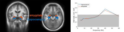 |
6 | Correlation between fMRI signals and oscillatory neuronal responses during audiovisual information processing
Hsin-Ju Lee1,2, Lauri Nummenmaa3,4,5, Hsiang-Yu Yu6,7, Cheng-Chia Lee7,8, Chien-Chen Chou6, Chien Chen6,7, Wen-Jui Kuo7,9, and Fa-Hsuan Lin1,2
1Physical Sciences Platform, Sunnybrook Research Institute, Toronto, ON, Canada, 2Department of Medical Biophysics, University of Toronto, Toronto, ON, Canada, 3Turku PET Centre, University of Turku, Turku, Finland, 4Department of Psychology, University of Turku, Turku, Finland, 5Turku University Hospital, Turku, Finland, 6Department of Neurology, Taipei Veterans General Hospital, Taipei, Taiwan, 7Brain Research Center, National Yang Ming Chiao Tung University, Taipei, Taiwan, 8Department of Neurosurgery, Taipei Veterans General Hospital, Taipei, Taiwan, 9Institute of Neuroscience, National Yang Ming Chiao Tung University, Taipei, Taiwan
We studied how fMRI signal is related to neural oscillations by taking both fMRI and invasive recordings from epilepsy patients. Specifically, we examined the neurovascular coupling during complex naturalistic stimuli processing. A significant negative correlation between gamma-band neural oscillations at hippocampus/amygdala and fMRI signals was found at the amygdala, hippocampus, and inferior occipital lobes while viewing short movie clips. These correlations sustain across gamma, beta, and alpha bands. The left angular gyrus shows a positive correlation between neural oscillations and fMRI dynamics at the theta band.
|
||
1737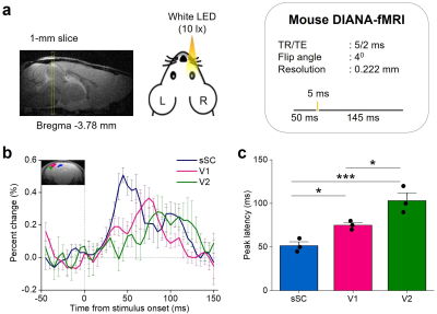 |
7 | In vivo direct imaging of visually evoked neuronal responses at milliseconds temporal resolution
Phan Tan Toi1,2, Jun-Ho Kim1, Jang Woo Park3, and Jang-Yeon Park1,2
1Department of Biomedical Engineering, Sungkyunkwan University, Suwon, Korea, Republic of, 2Department of Intelligent Precision Healthcare Convergence, Sungkyunkwan University, Suwon, Korea, Republic of, 3Korea Radioisotope Center for Pharmaceuticals, Korea Institute of Radiological & Medical Sciences, Seoul, Korea, Republic of
There has been a long-standing demand for high temporospatial resolution in non-invasive neuroimaging. Using our previously proposed approach for direct imaging of neuronal activity (DIANA-fMRI) with a high temporal resolution of milliseconds, we continued to demonstrate DIANA-fMRI performance in mice in vivo at 9.4T using visual stimulation. The DIANA signal change was significantly increased (~0.2-0.4%) in response to flashing light stimulus, and it was found that DIANA responses of sSC, V1, and V2 were sequentially activated in that order. The DIANA response times of sSC, V1, and V2 were consistent with previous electrophysiological studies.
|
||
1738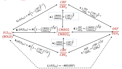 |
8 | Power Laws for Neurovascular Coupling between Cerebral Blood Flow and Oxygen Metabolism
Linqing Li1, Nicholas Blockley2, Sean Marrett1, Andy John Derbyshire1, and Peter Bandettini1
1National Institute of Mental Health, National Institutes of Health, Bethesda, MD, United States, 2University of Nottingham Medical School, School of Life Sciences, Nottingham, United Kingdom
We previously developed (CMRO2/CMRO20)=(CBF/CBF0)(1-α/β)(1-1/β)as special power relation (SPR) for brain activation. In this work, based on different perspective of SPR, we derive that functional activity induces general power relation (GPR) (CMRO2/CMRO20)=(CBF/CBF0)α/β. When α=0.38,β=1.5, both powers (SPR and GPR) are calculated to be 0.25,which is approximately equivalent to inverse of conventional coupling constant of 4. By equalizing two powers, the relationship of α and β parameters can be established. We also show that any coupling pairs between changes of CMRO2,CBF,CBV and OEF can be reduced into power laws. Finally, we propose the power law for quantification CMRO2 changes during CO2 calibration.
|
||
1739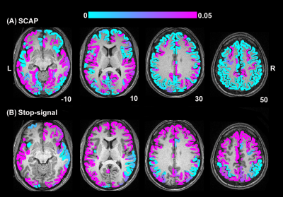 |
9 | Regional variation in the linear relationship between breath-hold cerebrovascular reactivity and BOLD fMRI activation
Rebecca Williams1,2, Jacinta Specht1,2, M. Ethan MacDonald2,3, and G. Bruce Pike1,2
1Department of Radiology, University of Calgary, Calgary, AB, Canada, 2Hotchkiss Brain Institute, University of Calgary, Calgary, AB, Canada, 3Departments of Electrical & Software Engineering, and Biomedical Engineering, Schulich School of Engineering, University of Calgary, Calgary, AB, Canada
Cerebrovascular reactivity (CVR) and task-based BOLD fMRI signals are closely linked. Understanding whether the relationship between CVR and task-based BOLD responses varies across the brain is important for interpreting BOLD, particularly in studies of aging where both CVR and BOLD activation differences are observed. Therefore, this work aimed to investigate the linear relationship between breath-hold (BH) CVR and task-based BOLD across the cerebral cortex to different cognitive tasks. Significant linear relationships were observed in posterior regions independent of task, while anterior regions were task-specific. These findings might contribute to understanding age-related posterior-anterior BOLD activation differences commonly observed in fMRI studies.
|
||
1740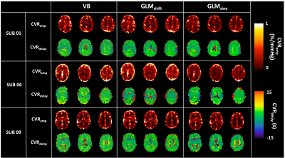 |
10 | Evaluating cerebrovascular reactivity dynamics through a Bayesian inference approach
Joana Pinto1, Martin Craig2,3, Paula Croal2,3, Nicholas P. Blockley2,4, Michael A. Chappell2,3, and Daniel P. Bulte1
1Institute of Biomedical Engineering, Department of Engineering Science, University of Oxford, Oxford, United Kingdom, 2Sir Peter Mansfield Centre, School of Medicine, University of Nottingham, Nottingham, United Kingdom, 3Radiological Sciences, Mental Health & Clinical Neurosciences, School of Medicine, Nottingham, United Kingdom, 4School of Life Sciences, University of Nottingham, Nottingham, United Kingdom
Cerebrovascular reactivity (CVR) has been shown to be an important parameter that is altered in certain pathologies. Analysis strategies using MRI data typically assume similar responses across the brain, neglecting the need and potential of modelling temporal features. In this work, we propose and test a novel method for the evaluation of CVR dynamics based on a variational Bayesian approach. Although this method yields similar CVR results to more standard approaches, it is more time efficient and has the potential to improve modelling by incorporating non-linear and biophysically informed models.
|
||
1741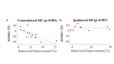 |
11 | Metabolic Changes Immediately Following Sequential Motor Learning: an MRS-fMRI Study
Yasmin Geiger1, Tali Weiss2, Osnat Volovick1, Inbar Aharon1, Rinatia Maaravi-Hesseg3, and Avi Karni3
1Weizmann Institute of Science, Rehovot, Israel, 2Weizmann institute of Science, Rehovot, Israel, 3University of Hifa, Hifa, Israel
Sequential finger tapping (SFT) is a model for motor learning. The initial phase of SFT manifests in changes to behavior, neuronal activation (BOLD) and metabolic concentrations. The metabolic changes and correlations to BOLD and behavioral changes during SFT are still poorly understood. Here we analyze these bilateral changes at the motor cortex using BOLD‑fMRI and 1H-MRS. We show a significant hemispheric‑dependent correlation between changes in GABA+ and behavior, which do not correlate with the BOLD signal. Additionally, GABA+ basal levels differ between hemispheres.
|
||
1742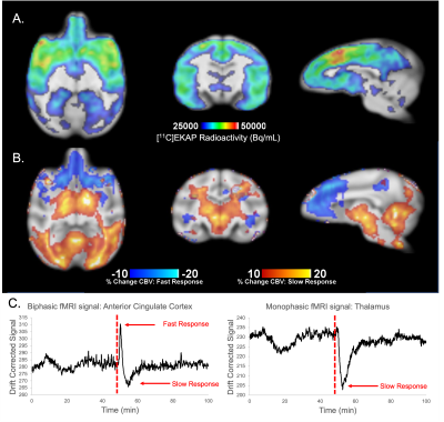 |
12 | Simultaneous PET/fMRI of the Kappa Opioid Receptor System During Psychotropic Drug Challenge
Frederick Andrew Bagdasarian1, Chi-Hyeon Yoo1, Sarah Elizabeth Reid1, Michael Placzek1, and Hsiao-Ying Wey1
1Athinoula A. Martinos Center for Biomedical Imaging, Department of Radiology, Massachusetts General Hospital, Harvard Medical School, Charlestown, MA, United States
This research utilizes simultaneous PET/MR imaging to provide a preliminary analysis of the kappa-opioid receptor (KOR) system in a non-human primate (NHP) brain. We investigated the influence of a psychoactive drug challenge specific to the KOR system in a NHP using the KOR agonist Salvinorin-A. We report regionally specific uptake of a KOR agonist radiotracer ([11C]EKAP) and alterations to BOLD-derived cerebral blood volume (CBV) following the use of Salvinorin-A that are directly related to radiotracer uptake distribution. This work serves a preliminary foundation for KOR imaging and understanding KOR-agonist drug mechanisms through PET/MR.
|
||
1743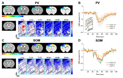 |
13 | Optical and fMRI responses to activation of GABAergic parvalbumin and somatostatin neurons.
Thanh Tan Vo1, Geunho Im2, and Seong-Gi Kim1
1Department of Biomedical Engineering, Sungkyunkwan University, Center for Neuroscience Imaging Research (CNIR), Institute for Basic Science (IBS), Suwon, Korea, Republic of, 2Center for Neuroscience Imaging Research (CNIR), Institute for Basic Science (IBS), Suwon, Korea, Republic of
The GABAergic neurons directly and indirectly regulate hemodynamics. Thus, investigating neurovascular coupling of subtype interneurons is highly important to interpret fMRI data, especially optogenetic fMRI. However, role of subtype interneurons to fMRI is not known. Here, we measured multi-wavelength optical imaging and CBV-weighted fMRI to investigate hemodynamic and metabolic responses of two most common interneuron subtypes, parvalbumin (PV) and somatostatin (SOM). PV induced biphasic hemodynamic responses, initial decrease and later increase, whereas SOM produced only positive hemodynamic responses. In both PV and SOM interneurons, dilation was initiated from the cortical surface.
|
||
 Back to Meeting Home
 Back to the Program-at-a-Glance
The International Society for Magnetic Resonance in Medicine is accredited by the Accreditation Council for Continuing Medical Education to provide continuing medical education for physicians.
 Back to Meeting Home
 Back to the Program-at-a-Glance
 View the Presentation