Digital Poster
High-Resolution fMRI
Joint Annual Meeting ISMRM-ESMRMB & ISMRT 31st Annual Meeting • 07-12 May 2022 • London, UK

| Computer # | ||||
|---|---|---|---|---|
2188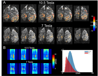 |
1 | Ultra-high resolution human functional imaging using 10.5 Tesla
Luca Vizioli1,2, Steen Moeller1, Andrea Grant1, Nader Tavaf1, Alireza Sadeghi-Tarakameh1, Yigitcan Eryaman1, Jerahmie Radder1, Russell Lagore1, Pierre-François Van de Moortele1, Edward Auerbach1, Gregor Adriany1, Essa Yacoub1, and Kamil Ugurbil1
1CMRR, University of Minnesota, Minneapolis, MN, United States, 2Department of Neurosurgery, University of Minnesota, Minneapolis, MN, United States
With the development of accelerated acquisition protocols, it is possible to achieve functional brain mapping with submillimeter resolution. This permits studying the human brain at the mesoscopic scale. Typical submillimeter images measure 0.8 mm. isotropic voxels, barely enough to study human functional mesoscopic responses. Here we acquire functional images at 10.5 T with the unprecedented spatial resolution of 0.4 mm. isotropic voxels. Using NORDIC to suppress thermal noise, we demonstrate the feasibility of achieving meaningful brain mapping at these ultra-high resolutions, where single voxels contains but a few thousand cells, further bridging the gap between fMRI and optical imaging
|
||
2189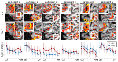 |
2 | Combining Arterial Blood Contrast with BOLD improves fMRI laminar specificity.
Nikos Priovoulos1, Icaro Agenor Ferreira de Oliveira1, Benedikt A Poser2, David Norris3, and Wietske van der Zwaag1
1Spinoza Center for Neuroimaging, Amsterdam, Netherlands, 2MR-Methods group, MBIC, Faculty of Psychology and Neuroscience, Maastricht University, Maastricht, Netherlands, 3Donders Institute for Brain, Cognition and Behaviour, Radboud University Nijmegen, Nijmegen, Netherlands
BOLD fMRI is widely applied in human neuroscience, but is limited in its spatial specificity compared to cerebral blood volume approaches due to its venous bias. Here we added cerebral blood-volume weighting based on magnetization transfer (Arterial Blood Contrast) to a BOLD submillimeter acquisition at 7T. Adding Arterial Blood Contrast helped differentiate the deep and superficial cortical responses in the human primary motor cortex. The results suggest that combining Arterial Blood Contrast with BOLD can improve the fMRI spatial specificity while retaining high sensitivity.
|
||
2190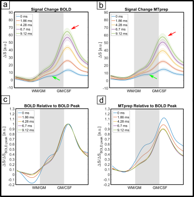 |
3 | Laminar fMRI with MT-prepared multi-echo 3D FLASH
Viktor Pfaffenrot1,2 and Peter J. Koopmans1,2
1Erwin L. Hahn Institute for Magnetic Resonance Imaging, University of Duisburg-Essen, Essen, Germany, 2High Field and Hybrid MR Imaging, University Hospital Essen, University of Duisburg-Essen, Essen, Germany
The GRE-BOLD contrast used in laminar fMRI suffers from suboptimal specificity due to extravascular effects around large veins. CBV-weighting offers improved specificity with signal changes being more confined to microvasculature. CBV-weighting can be achieved through saturation of gray matter signal with magnetization transfer. We use a multi-echo FLASH sequence at 7 Tesla, with and without MT preparation to investigate its feasibility at ultrahigh field. We analyze the TE dependence to determine when the BOLD T2’-effects start to dominate the MT-weighted signal changes. The MTprep profiles exhibit a stronger signal change in gray matter at short TEs and reduced extravascular effects.
|
||
2191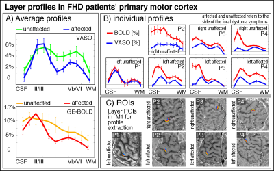 |
4 | Laminar VASO fMRI in hand dystonia patients
Silvina G Horovitz1, Panagiotis Kassavetis1,2, Mark Hallett1, and Renzo Huber3
1HMCS, NINDS - NIH, Bethesda, MD, United States, 2Neurology, University of Utah Health, Salt Lake City, UT, United States, 3Psychology and Neuroscienc, Maastricht University, Maastricht, Netherlands
Mesoscopic fMRI with CBV-sensitive VASO can be a valuable tool for research questions on affected neural processing in patients suffering from neurological diseases. However, its applicability in patient populations remains unclear and is challenged by multiple methodological constraints. Here, we seek to use finger tapping tasks in hand dystonia patients to map affected topographical and laminar fMRI features. Specifically, we described the input-dominated laminar input-output circuits in the primary motor system as well as the ‘scrambled’ finger representations in the somatosensory areas. We built and validated an acquisition and analyses setup for laminar and columnar mapping in patient populations.
|
||
2192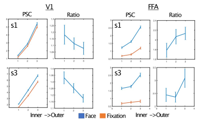 |
5 | Using ultra-high field fMRI to uncover depth-dependent activation in the ventral temporal lobe
Logan T Dowdle1, Geoffrey Ghose2, Clark Chen1, Kâmil Uğurbil3, Essa Yacoub3, and Luca Vizioli1
1Neurosurgery, Center for Magnetic Resonance Research, Minneapolis, MN, United States, 2Neurosciences, Center for Magnetic Resonance Research, Minneapolis, MN, United States, 3Center for Magnetic Resonance Research, Minneapolis, MN, United States We used 7T BOLD functional imaging to acquire 0.7mm voxels and the thermal noise suppression technique NORDIC to evaluate depth-dependent, task related top-down modulations. Four participants viewed face stimuli and performed either a face or fixation task (identical stimuli). We compared depth-dependent task modulations in the primary visual cortex (V1) and fusiform face area (FFA). We found that, relative to the fixation task, face detection led to larger modulations in outer relative to inner layers in the FFA, and the inverse pattern in V1. This is a critical step towards using laminar analyses to examine hierarchical or distributed cognitive processes. |
||
2193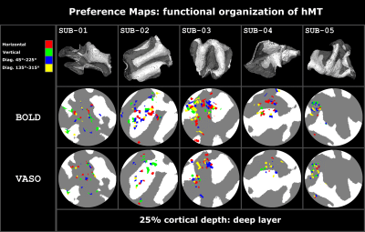 |
6 | Laminar and columnar functional organization of human area MT using VASO at 7T
Alessandra Pizzuti1,2, Renzo Huber1, Omer Faruk Gulban1,2, Amaia Benitez-Andonegui1,3, Judith Peters1, and Rainer Goebel1,2
1Department of Cognitive Neuroscience, Faculty of Psychology and Neuroscience, Maastricht University, Maastricht, Netherlands, 2Brain Innovation, Maastricht, Netherlands, 3National Institutes of Mental Health, MEG Core Facility, Bethesda, MD, United States We investigate the feasibility of using CBV-sensitive VASO fMRI at ultra-high field to study the selective columnar organization to axes of motion stimuli in human area MT. For 5 subjects we found:
|
||
2194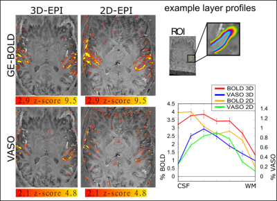 |
7 | CBV-sensitive layer-fMRI in the human auditory cortex at 7T: Challenges and capabilities
Lonike K. Faes1, Omer Faruk Gulban1,2, Benedikt A. Poser1, Federico De Martino1, and Renzo Huber1
1Department of Cognitive Neuroscience, Faculty of Psychology and Neuroscience, Maastricht University, Maastricht, Netherlands, 2Brain Innovation, Maastricht, Netherlands
Laminar-specific fMRI with CBV-sensitive VASO is valuable for neuroscientific questions on hierarchical information processing. While VASO fMRI has already proven its utility in other brain areas, it has not yet been successfully applied in the auditory cortex due to several additional technical challenges in this region. Here, we explore multiple sequences and their effectiveness to mitigate these challenges. Our purpose is to develop an experimental setup that future neuroscientific studies can be built on. Ultimately, we found a stable parameter set for the usage of layer-fMRI VASO in the auditory cortex and validated it in a group of participants.
|
||
2195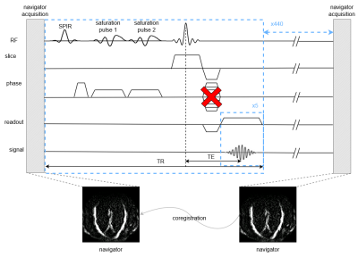 |
8 | Prospective motion correction in multi-echo line-scanning fMRI at 7T: sequence implementation and strategies for functional data analysis
Luisa Raimondo1, Nikos Priovoulos1, Jurjen Heij1, Tomas Knapen1,2, Serge O Dumoulin1, Jeroen JW Siero1,3, and Wietske van der Zwaag1
1Spinoza Centre for Neuroimaging, Amsterdam, Netherlands, 2VU University, Amsterdam, Netherlands, 3Radiology, University Medical Centre, Utrecht, Netherlands
We implemented prospective motion-correction (PMC) for line-scanning fMRI. In line-scanning, online motion correction is needed since 1-dimensional data do not allow motion detection in every dimension and the limited coverage makes this approach sensitive to spin-history artifacts or scanning outside the area of interest, in the presence of motion. We compared three versions of PMC and three ways of managing the T1-driven return to equilibrium (T1-transient) introduced by the navigators, in the functional data analysis. We opted for a water-excitation navigator as the best alternative, and showed that the T1-transient can be successfully overcome in the functional analysis.
|
||
2196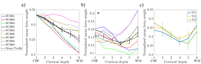 |
9 | Laminar layer 7T fMRI-EEG reveals human alpha oscillations are predominately from superficial and deep layers
Daniel C. Marsh1, Rodika Sokoliuk2, Kevin M. Aquino3,4, Daisie O. Pakenham5, Ross Wilson2, Rosa Sanchez Panchuelo6, Sebastian C. Coleman1, Matthew J Brookes1, Simon Hanslmayr7, Stephen D. Mayhew2, Susan T Francis1, and Karen J Mullinger1,2
1Sir Peter Mansfield Imaging Centre, School of Physics and Astronomy, University of Nottingham, Nottingham, United Kingdom, 2Centre for Human Brain Health, School of Psychology, University of Birmingham, Birmingham, United Kingdom, 3School of Physics, University of Sydney, Sydney, Australia, 4Turner Institute, Monash University, Melbourne, Australia, 5Clinical Neurophysiology, Queens Medical Centre, Nottingham University Hospitals NHS Trust, Nottingham, United Kingdom, 6Queen Elizabeth Hospital, University Hospitals Birmingham NHS Foundation Trust, Birmingham, United Kingdom, 7Centre for Cognitive Neuroimaging,College of Medical, Veterinary and Life Sciences, University of Glasgow, Glasgow, United Kingdom
EEG alpha (8-13Hz) oscillations occur throughout the cortex but the generating mechanisms are poorly understood. Opinion is divided between alpha being driven by bottom-up, top-down or both of these processes. Using simultaneous 7T-fMRI-EEG with an eyes open/closed paradigm, we assess the generator of alpha by performing layer-fMRI analysis of GE-BOLD data to determine the strongest BOLD-alpha negative layer correlations. We show that, after accounting for draining vein effects using spatial deconvolution, alpha-BOLD correlations are strongest in the superficial and deep layers suggesting they are predominately driven by top-down processes.
|
||
2197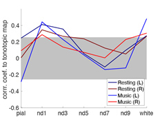 |
10 | Cortical-depth dependent functional connectivity at the human auditory cortex during the resting state and under complex naturalistic stimuli
Hsin-Ju Lee1,2, Hankyeol Lee3, Kamil Uludag3,4, Jyrki Ahveninen5, and Fa-Hsuan Lin1,2
1Physical Sciences Platform, Sunnybrook Research Institute, Toronto, ON, Canada, 2Department of Medical Biophysics, University of Toronto, Toronto, ON, Canada, 3Center for Neuroscience Imaging Research, Institute for Basic Science, Suwon, Korea, Republic of, 4Techna Institute & Koerner Scientist in MR Imaging, Joint Department of Medical Imaging and Krembil Brain Institute, University Health Network, Toronto, ON, Canada, 5A. A. Martinos Center for Biomedical Imaging, Massachusetts General Hospital, Charlestown, MA, United States
We explored the functional connectivity of the human auditory cortex with cortical depth analysis and compared music listening and the resting state using 7T fMRI with 0.8 mm isotropic resolution. Tonotopic maps, principal component analysis, and seed-based correlation revealed network topologies and suggested functions supporting feed-forward and feed-back processing. The feed-forward process is manifested by the reduced correlation to the contralateral auditory cortex during music-listening at the intermediate cortical depths. The feed-back processing is associated with a network like the tonotopic organization, which was stable between the resting and music listening but more correlated with the contralateral auditory cortex.
|
||
2198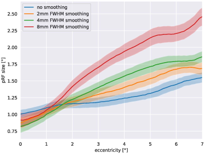 |
11 | Population-receptive field size estimates are biased by acquisition and preprocessing parameters: a 7T fMRI study Video Not Available
Christian Windischberger1, David Linhardt1, Tessa Angerer1, Maximilian Pawloff2, Markus Ritter2, and Ursula Schmidt-Erfurth2
1High Field MR Center, Center for Medical Physics and Biomedical Engineering, Medical University of Vienna, Wien, Austria, 2Department of Ophtalmology, Medical University of Vienna, Wien, Austria
Population receptive field (pRF) mapping is an advanced retinotopic mapping approach. While the size of pRFs has been identified as an important neurophysiological feature, remarkable differences in the pRF sizes across studies has been reported. In this study, we examine the effects of difference in spatial resolution on the pRF size obtained in a group of ten healthy subjects at 7T. Our results clearly show that the amount of spatial dependence between voxels in the visual cortex has a direct effect for the pRF sizes calculated. We can assume similar effects in data sets acquired at different spatial resolutions.
|
||
 |
2199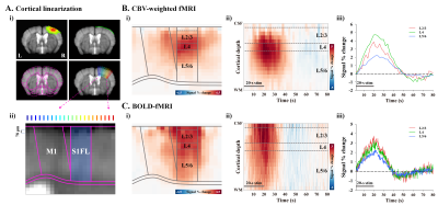 |
12 | Neurophysiological source of laminar fMRI
Won Beom Jung1, Haiyan Jiang1,2, and Seong-Gi Kim1,2
1Center for Neuroscience Imaging Research (CNIR), Suwon, Korea, Republic of, 2Department of Biomedical Engineering, Sungkyunkwan University, Suwon, Korea, Republic of
Layer-dependent fMRI mapping provides insights to understand the directional information flow within brain networks. However, it is unknown whether larimar fMRI responses allows discrimination of activation to cortical input and/or output. To determine neuronal source of hemodynamic fMRI signals, we investigated the somatosensory-evoked laminar fMRI responses and cortical depth-dependent electrophysiological activities in the mouse cortex.
|
|
2200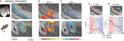 |
13 | Thalamic and cortical input layers in the somatomotor cortex detected by laminar fMRI in non-human primates at 7T Video Permission Withheld
Sangyun Kang1, Min-Jun Han1,2, Seong-Gi Kim1,2, and Eunha Baeg1
1IBS Center for Neuroscience Imaging Research, Sungkyunkwan University, Suwon, Korea, Republic of, 2Department of Biomedical Engineering, Sungkyunkwan University, Suwon, Korea, Republic of
Laminar fMRI has been increasingly used in humans, but its validity needs to be thoroughly confirmed in primate brains. Here, we investigated thalamic and cortical inputs to the somatomotor cortex in the rhesus monkey by adopting tactile stimulation and electrical microstimulation. Compared to 7T BOLD signals, MION-induced CBV-weighted fMRI faithfully captures input layer activities, like the sensory thalamus input to the middle layer and the dense cortical connections in the superficial layers. Taken together, our CBV-weighted fMRI signals revealed different input layers to the somatomotor cortex with high specificity and sensitivity.
|
||
 Back to Meeting Home
 Back to the Program-at-a-Glance
The International Society for Magnetic Resonance in Medicine is accredited by the Accreditation Council for Continuing Medical Education to provide continuing medical education for physicians.
 Back to Meeting Home
 Back to the Program-at-a-Glance
 View the Presentation