Digital Poster
fMRI Methods & Applications in Animals
Joint Annual Meeting ISMRM-ESMRMB & ISMRT 31st Annual Meeting • 07-12 May 2022 • London, UK

Digital Poster
fMRI Methods & Applications in Animals
| Computer # | ||||
|---|---|---|---|---|
1821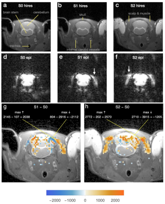 |
1 | Strong non-BOLD Effect of EPI Gradient Switching Sounds on Voxel Intensities Both Inside and Outside Rat Cranium
James W Prichard1 and Samuel Patz2,3
1Yale University, West Tisbury, MA, United States, 2Harvard Medical School, Boston, MA, United States, 3Radiology, Brigham and Women's Hospital, Boston, MA, United States Echoplanar images of rat head with and without blocking of external ear canals revealed large voxel intensity changes outside as well as inside the cranium. These are probably mechanical effects of vibrational energy originating in the tympanic membrane and coupled to other tissue compartments of the head through the auditory ossicles, basilar membrane, and cochlear round window. They are so large that even slight variations in them may distort magnetic resonance imaging measurements of neural activity and water diffusion. Awareness of that possibility may inform measures to remove or compensate for it. |
||
1822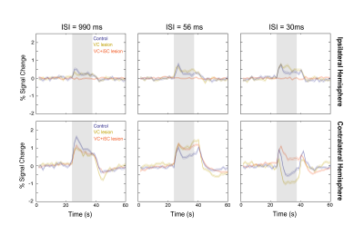 |
2 | Resolving activity suppression in the rat visual pathway using fMRI combined with lesions
Rita Gil1, Mafalda Valente1, Alfonso Renart1, and Noam Shemesh1
1Champalimaud Research, Champalimaud Centre for the Unknown, Lisbon PT, Lisbon, Portugal
Monocular visual stimulation with short inter-stimulus intervals (ISIs) evokes (i) negative BOLD responses (NBRs) in the contralateral superior colliculus (cSC), and (ii) positive BOLD responses (PBRs) in the ipsilateral superior colliculus (iSC). This pattern suggests a potential "push-pull" mechanism between SCs possibly evoked by (mostly inhibitory) tectotectal projections. Here, we mapped activity in the entire visual pathway using fMRI, and modulated cSC inputs through lesions in visual cortex and SC to dissect collicular communication mechanisms. While, cortical context potentiated a putative “push-pull” mechanism, further silencing of iSC resulted in PBRs in cSC, suggesting that iSC exerts suppression on cSC.
|
||
1823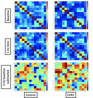 |
3 | Remapping of functional connectivity with optogenetic locus coeruleus activation in rats
Nmachi Anumba1, Michael Kelberman2, Corrie Smith1, Wen Ju Pan1, G Perrin Clavijo1, David Weinshenker2, and Shella Keilholz1
1Biomedical Engineering, Georgia Tech and Emory University, Atlanta, GA, United States, 2Human Genetics, Emory University, Atlanta, GA, United States
High tonic (3-5 Hz) locus coeruleus (LC) activity drives anxiety-like behaviors and the stress response via its dense afferents and receptor distribution profile. Previous studies have utilized DREADD stimulation to understand the global effects of elevating LC activity, but lacked temporal and patterned control of stimulation. Here, we combined optogenetic stimulation of the LC (5 Hz) with functional magnetic resonance imaging to better understand global changes in functional connectivity as a result of high tonic LC activity. We found that LC stimulation at 5 Hz resulted in a shift toward increased connectivity in ChR2 injected animals compared to mCherry controls.
|
||
1824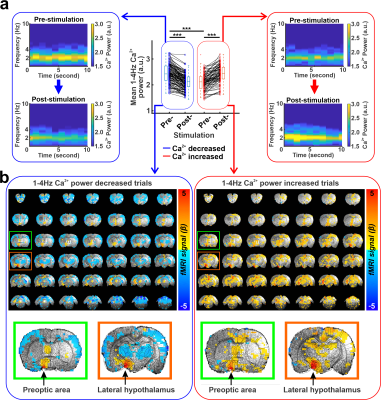 |
4 | Identifying modulation of brain states by optogenetic stimulation of lateral hypothalamus Video Permission Withheld
Kengo Takahashi1, Yi Chen1, Patricia Pais-Roldán2, and Xin Yu3
1High-field Magnetic Resonance, Max Planck Institute for Biological Cybernetics, Tübingen, Germany, 2Institute of Neuroscience and Medicine (INM-4), Forschungszentrum Jülich, Jülich, Germany, 3Martinos Center for Biomedical Imaging, Massachusetts General Hospital and Harvard Medical School, Charlestown, MA, United States
The lateral hypothalamus (LH) plays an important role in regulating the brain state changes during sleep and arousal state fluctuation. Although the anatomical projections of LH have been well-investigated, the brain-wide functional map by LH stimulation under different brain states has not been characterized. Here, we applied fMRI to characterize the whole brain activation maps under different brain states during anesthesia in response to LH optogenetic stimulation. We demonstrate that the underlying brain state determines distinct functional mapping features upon LH activation.
|
||
1825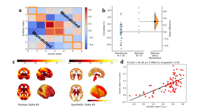 |
5 | Humanized mouse brain biomarkers through transcriptomic conversion
Roël Matthijs Vrooman1, Judith Homberg1, and Joanes Grandjean1
1DCMN, Donders Institute for Brain, Cognition and Behaviour, RadboudUMC, Nijmegen, Netherlands
Since the translation of data between species remains subjective, our goal is to develop a data-driven tool to perform this translation based on the expression patterns of homologous genes. For this, human and mouse fMRI data, which was manually matched based on spatial homology between co-activation patterns, was modelled as a linear addition of gene expression maps. Weighting factors were then used to estimate synthetic brain states based on homologous genes. When comparing the synthetic brain states to their matched biological state, they showed higher correlation than compared to mismatched states, showing the effectiveness of the translational tool.
|
||
1826 |
6 | Impact of day-night cycle on functional brain connectome in mice Video Permission Withheld
Houefa Armelle Lokossou1, Giovanni Rabuffo2, Monique Bernard1, Teodora-Adriana Perles-Barbacaru1, Angèle Viola1, and Christophe Bernard2
1CRMBM UMR 7339, Aix Marseille Université-CNRS, Marseille, France, 2INS, UMR1106, Aix-Marseille University-INSERM-CNRS, Marseille, France
Controlled by the suprachiasmatic nucleus of the hypothalamus, circadian rhythm defines the day-night cycle in most animal species. We evaluated the effect of night and day cycle on the functional connectome by exploring a group of mice under light condition (LC) and another one under night condition (NC) using BOLD rs-fMRI. Our results show a significant increase in functional connectivity in NC group compared to LC group at the pineal gland level. This result is in line with this gland’s function. Furthers studies are needed to explore the effects of the circadian rhythm on both genders at different ages.
|
||
1827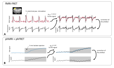 |
7 | Multimodal BOLD and CBV MRI for artifact correction in fluorescence recordings
Henriette Lambers1, Lydia Wachsmuth1, and Cornelius Faber1
1Clinic for Radiology, University Hospital Muenster, Muenster, Germany
Combining fMRI and fluorescence recordings has become an important tool for neuroimaging. However, hemodynamic changes lead to artifacts in fluorescence recordings, since blood absorbs fluorescence light. We investigated the hemodynamic artifact in functional and pharmacological MRI and fluorescence measurements and present an MRI-based correction method. In both stimulation and lactate injection experiments, hemodynamic artifacts in FRET recordings occurred. These artifacts cannot be removed effectively using a standard (purely optical) correction method. An MRI-based correction presented in this work eliminates the artifact while the expected fluorescence signal changes persist.
|
||
1828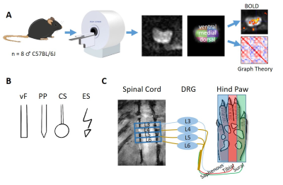 |
8 | fMRI in the Mouse Spinal Cord during peripheral somatosensory Stimulation
Amon Allelein1, Bruno Pradier1, Huifen Chen2, and Cornelius Faber2
1Radiology, UKM, Muenster, Germany, 2UKM, Muenster, Germany
The spinal cord is an important hub for the integration and processing of peripheral somatosensory stimuli within the central nervous system, however, mouse spinal cord functional imaging is not yet established. We successfully performed BOLD fMRI in the mouse spinal cord using 1 electric and 3 different mechanical stimulation modalities in one session. Graph theory-based network analyses showed strong positive correlations within the dorsal spinal cord laminae of L4 and L5. A negative correlation between the dorsal and ventral regions within the same segment suggests the presence of a vascular steal effect.
|
||
1829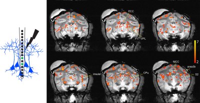 |
9 | Layer-specific microsimulation of S2 cortex revealed distinct functional microcircuits in non-human primate brain
Pai-Feng Yang1,2, Feng Wang1,2, Jamie L. Reed1,2, Zhangyan Yang1,2, John C. Gore1,2, and Li Min Chen1,2
1Institute of Imaging Science, Vanderbilt University, Nashville, TN, United States, 2Radiology and Radiological Sciences, Vanderbilt University Medical Center, Nashville, TN, United States
Intracortical microstimulation is one of the most common techniques used to activate cortical functional networks and probe causal connections. This study investigates whether BOLD signals can detect layer-specific functional circuit responses to electrical brain stimulation. We delivered electrical current to different layers of secondary somatosensory cortex (S2) and detected layer-specific BOLD activations. Effective functional connectivity analysis revealed different networks to different layers of S2 cortex.
|
||
1830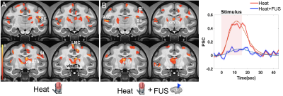 |
10 | Transcranial Focused Ultrasound Suppresses Thalamic Heat Nociceptive Response and Reorganizes Nociceptive Networks in Non-human Primates
Arabinda Mishra1,2, Pai-Feng Yang1,2, Tom Manuel1,3, Allen T Newton1,2, Huiwen Luo1,3, Marshal A Phipps1,2, Michelle Signoa1,3, Jamie L Reed1,2, John C Gore1,2,3, William A Grissom1,3, Charles F Caskey1,2,3, and Li Min Chen1,2
1VUIIS, Vanderbilt University, Nashville, TN, United States, 2Radiology, Vanderbilt University Medical Center, Nashville, TN, United States, 3BME, Vanderbilt University, Nashville, TN, United States
MRI guided transcranial focused ultrasound (MRgFUS) is a neuromodulation tool that can excite or inhibit neural activity with high spatial precision and real-time functional feedback. Our goal is to develop a MRgFUS device for human pain therapy. Here, we studied suppressive effects of FUS on a key hub in the thalamus for transmitting peripheral inputs to cortex for pain perception, and mapped changes in effective functional connectivity of thalamic networks using hierarchical clustering. We show that concurrently delivered FUS of moderate intensity suppressed painful heat responses at the thalamus target and induced reorganization of effective connectivity networks.
|
||
1831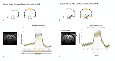 |
11 | Evidence for an intercollicular auditory push/pull mechanism revealed by BOLD fMRI
Frederico Severo1, Mafalda Valente1, and Noam Shemesh1
1Champalimaud Research, Champalimaud Centre for the Unknown, Lisboa, Portugal
The role of the rodent inferior colliculus (IC) in binaural integration is of great interest especially for sound localization and processing of interaural level difference (ILD). Yet, many IC underlying mechanisms remain unclear. Here, we find evidence for a push/pull mechanism in IC, with contralateral (positive BOLD) activity exerting dominance over ipsilateral (negative BOLD) activity, possibly as means of a sound localization/lateralization.
|
||
1832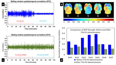 |
12 | Quasiperiodic Patterns in resting state fMRI signal reflect dynamics of cortical slow rhythms
Vahid Khalilzad Sharghi1, Eric A Maltbie1, Wen-Ju Pan1, Shella Keilholz1, and Kaundinya S Gopinath1
1Emory University, Atlanta, GA, United States
In this study, we tested hypothesis advanced by some groups that brain slow rhythms serve as the neurophysiological basis of resting state fMRI (rsfMRI). Putative suppression of cortical rhythms with an established technique, led to significant reduction in the amplitude of rsfMRI quasi-periodic patterns (QPPs), and enhancement in the rsfMRI measures of intrinsic functional connectivity FC in canonical brain function networks in rats. The results indicate cortical slow rhythms serve as the genesis of only the vigilance dependent components (e.g., QPP) of rsfMRI signals. Further attenuation of these non-specific signals enhances delineation of brain function networks.
|
||
 Back to Meeting Home
 Back to the Program-at-a-Glance
The International Society for Magnetic Resonance in Medicine is accredited by the Accreditation Council for Continuing Medical Education to provide continuing medical education for physicians.
 Back to Meeting Home
 Back to the Program-at-a-Glance
 View the Presentation