Digital Poster
Cancer: Metabolites & Molecular Markers
Joint Annual Meeting ISMRM-ESMRMB & ISMRT 31st Annual Meeting • 07-12 May 2022 • London, UK

Digital Poster
Cancer: Metabolites & Molecular Markers
| Computer # | ||||
|---|---|---|---|---|
0831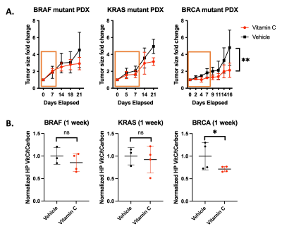 |
56 | Hyperpolarized dehydroascorbate reveals ascorbate-mediated oxidative stress increases flux through the pentose phosphate pathway in cancer
Nathaniel Kim1, Marjan Berishaj1, Elisa de Stanchina2, Manish Shah3, Lewis Cantley4, and Kayvan Keshari1
1Department of Radiology, Memorial Sloan Kettering Cancer Center, New York, NY, United States, 2Antitumor Assessment Core Facility, Molecular Pharmacology Program, Memorial Sloan Kettering Cancer Center, New York, NY, United States, 3Weill Cornell Medicine, New York-Presbyterian Hospital, New York, NY, United States, 4Meyer Cancer Center, Department of Medicine, Weill Cornell Medical College, New York, NY, United States
HP DHA was demonstrated as an imaging agent for probing the biochemical response to ascorbate therapy in patient derived xenograft (PDX) models of cancer. Changes in DHA/ascorbate metabolism were observed in PDX tumors after prolonged treatment, mirroring the observed treatment response to high dose ascorbate. Metabolomic studies showed that high dose ascorbate therapy induced oxidative stress, leading to increased flux through the pentose phosphate pathway (PPP). DNA damage repair mutants exhibit lower baseline flux through the PPP and are more sensitized to ascorbate induced ROS. These results demonstrate a new method for assessing oxidative stress in vivo using HP DHA.
|
||
0832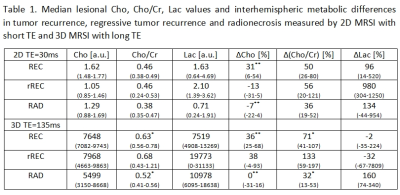 |
57 | Comparison of MR spectroscopy cut-off parameters in differential diagnosis between glioma recurrence and radionecrosis
Dita Pajuelo1, Alberto Malucelli2, Monika Dezortová1, Antonín Škoch1, Robert Bartoš2, Martin Sameš2, and Milan Hájek1
1MR Unit, Department of Diagnostic and Interventional Radiology, Institute for Clinical and Experimental Medicine, Prague, Czech Republic, 2Department of Neurosurgery, JE Purkyne University and Masaryk Hospital, Ústí nad Labem, Czech Republic Differential diagnosis between brain tumor recurrence and radionecrosis is still problematic. MR Spectroscopy Imaging (MRSI) is useful in differentiating between these two histologically and radiologically distinct entities. Six methods with different combination of metabolic parameters were used to compare 37 lesions examined by 2D/3D MRSI. The lesional and contralateral comparison of choline-containing compounds and lactate using long echo time achieved the highest sensitivity and specificity. Choline to creatine ratio showed low specificity. The distinction of regressive recurrence as a separate entity is helpful in the effort to discern the complexity of events leading to new enhancing lesions in neurooncologic patients. |
||
0833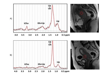 |
58 | The lipid signal in cervical cancer: a 1H MRS pilot study Video Permission Withheld
Rossella Canese1, Miriam Dolciami2, Claudia Testa3, and Lucia Manganaro2
1Core Facilities, Istituto Superiore di Sanita', Rome, Italy, 2Department of Radiological, Oncological and Pathological Sciences, Policlinico Umberto I Sapienza University of Rome, Rome, Italy, 3Department of Physics and Astronomy, University of Bologna, Bologna, Italy, Rome, Italy
Cervical cancer (CC) is the fourth most common malignancy and cancer-related mortality cause in women worldwide. A significant portion of these patients are diagnosed as locally advanced cervical cancer (LACC), thus requiring combined therapies. We investigate the role of the lipid signal derived from 1H magnetic resonance (MR)-spectroscopy in assessing response to neoadjuvant chemotherapy of LACC.
|
||
0834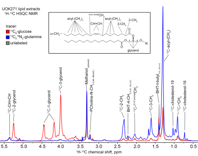 |
59 | Assessment of Lipid Biosynthesis and Turnover in Renal Cell Carcinoma Cells and Tissues Using NMR-Resolved Stable Isotope Experiments Video Not Available
Daniel Robert Crooks1, Ye Yang2, Andrew Lane3, Teresa Fan3, Jeffrey Brender4, Murali C Krishna4, and W. Marston Linehan1
1Urologic Oncology Branch, National Cancer Institute, Bethesda, MD, United States, 2National Cancer Institute, Bethesda, MD, United States, 3Center for Environmental Systems Biochemistry, University of Kentucky, Lexington, KY, United States, 4Radiation Biology Branch, National Cancer Institute, Bethesda, MD, United States
NMR-based analyses of lipids can shed insight into the global complement of cellular lipids, and elucidate the fuel sources and pathways contributing to lipid biosynthesis in cells grown in the presence of 13C-labeled fuels. We utilized 1H-13C HSQC NMR analysis of cellular lipids derived from FH- and mtDNA deficient UOK271 cells and observed robust reductive carboxylation of glutamine that resulted in formation and incorporation of 13C-glutamine-derived acetyl groups into lipid chains.
|
||
0835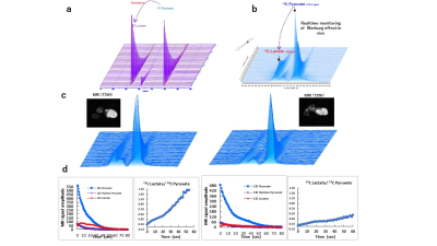 |
60 | Usage of Dissolution DNP Magnetic Resonance Spectroscopy Depicts Efficacy of Chemotherapeutic and Radiotherapeutic Anticancer Interferences Video Permission Withheld
Abdelazim Elsayed Elhelaly1,2, Fuminori Hyodo1, Norikazu Koyasu3, Hiroyuki Tomita4, Masaharu Murata5, Yoshifumi Noda3, Hiroki Kato3, and Masayuki Matsuo3
1Department of Radiology, Frontier Science for Imaging, Gifu University, Gifu, Japan, 2Department of Food Hygiene and Control, Faculty of Veterinary Medicine, Suez Canal University, Ismailia, Egypt, 3Department of Radiology, Gifu University, Gifu, Japan, 4Department of Tumor Pathology, Gifu University, Gifu, Japan, 5Innovation Center for Medical Redox Navigation, Kyushu University, Kyushu, Japan DNP highly improves the sensitivity of magnetic resonance spectroscopy allowing for greater insight into in vivo metabolic activity. We used our dissolution 13C DNP system to monitor the Warburg effect in MIA PaCa human pancreatic carcinoma model mice subjected to radiotherapy or chemotherapy. In vivo production of 13C lactate in tumors showed a significant reduction after radiation therapy. Mice subjected to anticancer drugs also showed a significant reduction in the relative production of 13C lactate at an early stage of the treatment course before any anatomical changes in the structure or size of the tumors were detectable by MRI images. |
||
0836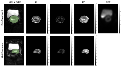 |
61 | IVIM MRI and PET in Locally Advanced Cervical Carcinoma undergoing Concurrent Chemoradiation Therapy
Dante Capaldi1, Jen-Yeu Wang2, Vipul Sheth2, Elizabeth Kidd2, and Dimitre Hristov2
1University of California, San Francisco, San Francisco, CA, United States, 2Stanford University, Stanford, CA, United States
IVIM MRI provides measurements of diffusion and perfusion, which has been shown to be predictive of prognosis, responsive to chemoradiotherapy, and correlated with DCE MRI in cervical cancer. We hypothesized that IVIM in cervical cancer patients would be sensitive to CCRT, and hence our objective was to first investigate changes in IVIM in cervical cancer patients undergoing adjuvant CCRT and evaluate the relationship between these metrics with changes in tumor volume and PET. Preliminary results show that IVIM measurements are related to both PET and changes in overall tumor volume and may reflect changes in the tumor microenvironment during treatment.
|
||
0837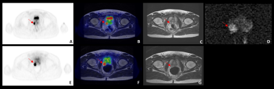 |
62 | 68Ga-PSMA and 68Ga-DOTA-RM2 PET/MRI in staging of high-risk prostate cancer patients: Preliminary analysis on 13 patients
Paola Scifo1, Paola Mapelli1,2, Samuele Ghezzo2, Ana Maria Samanes Gajate1, Erik Preza1, Giorgio Brembilla2,3, Vito Cucchiara2,4, Naghia Ahmed5, Carolina Bezzi1,2, Annarita Savi1, Ilaria Neri1, Ettore Di Gaeta3, Luigi Gianolli1, Massimo Freschi5, Alberto Briganti2,4, Francesco De Cobelli2,3, and Maria Picchio1,2
1Department of Nuclear Medicine, IRCCS San Raffaele Scientific Institute, Milan, Italy, 2Vita-Salute San Raffaele University, Milan, Italy, 3Department of Radiology, IRCCS San Raffaele Scientific Institute, Milan, Italy, 4Department of Urology and Division of Experimental Oncology, URI, Urological Research Institute, IRCCS San Raffaele Scientific Institute, Milan, Italy, 5Department of Pathology IRCCS San Raffaele Scientific Institute, Milan, Italy
The use of PET/MRI scanners is an innovative approach to stage prostate cancer (PCa) and the combined use of both 68Ga-PSMA and 68Ga-DOTA-RM2 radiotracers can improve disease staging by providing different metabolic aspects of the tumour. In the present study, 13 patients with high-risk PCa undergoing 68Ga-PSMA and 68Ga-DOTA-RM2 PET/MRI before radical prostatectomy have been included, with availability of histology as gold standard to validate intraprostatic findings and local extension. Our results suggest that the use of PET/MRI with a dual tracer approach can improve PCa staging, by providing complementary information ameliorating disease characterization.
|
||
0838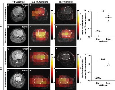 |
63 | Imaging response to radio-chemotherapy in glioblastoma tumor models using deuterium metabolic imaging
Friederike Hesse1, Alan Wright1, Vencel Somai1,2, Flaviu Bulat1,3, and Kevin Brindle1,4
1CRUK CI, University of Cambridge, Cambridge, United Kingdom, 2Department of Radiology, University of Cambridge, Cambridge, United Kingdom, 3Department of Chemistry, University of Cambridge, Cambridge, United Kingdom, 4Department of Biochemistry, University of Cambridge, Cambridge, United Kingdom
Metabolic imaging of brain tumor responses to radio-chemotherapy can give an early indication of treatment outcome. We show here that Deuterium Metabolic Imaging (DMI) with 2H-labeled fumarate is more sensitive than diffusion weighted 1H imaging (DWI) in detecting early evidence of cell death following radio-chemotherapy in orthotopically implanted patient-derived glioblastoma xenografts. 2H spectra were acquired from tumors with a time resolution of 5min, following an injection of 2H-labelled fumarate. Within 48h after the last chemo-radiotherapy treatment (CRT) the rate of tumor malate production from labelled fumarate increased significantly, whereas changes in ADC values were only detectable 7 days post treatment.
|
||
0839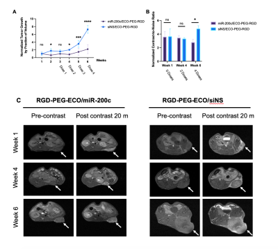 |
64 | MR Molecular Imaging of EDB-Fibronectin for Non-Invasive Monitoring of miR-200c Therapy in Pancreatic Cancer
Victoria Laney1, Ryan Hall 1, Grace Yeung1, Victoria Laney1, Suneel Apte 2, and Zheng-Rong Lu3
1Case Western Reserve University, CLEVELAND, OH, United States, 2Cleveland Clinic Foundation, CLEVELAND, OH, United States, 3Case Western Reserve University, Cleveland, OH, United States
PDAC is a highly aggressive malignant cancer and the 3rd leading cause of cancer related deaths in the US. Currently, treatment options for patients are limited due to late detection of the tumors and lack of effective therapies. The aggressive nature of PDAC is in part due to the desmoplastic tumor microenvironment. MiR-200c regulates epithelial-to-mesenchymal transition and extracellular remodeling for effective treatment of aggressive tumors. In this study, we investigate the effectiveness of MR molecular imaging of extradomain-B fibronectin, an extracellular matrix oncoprotein associated with EMT, for non-invasive monitoring of PDAC tumor response to miR-200c therapy in a mouse model.
|
||
0840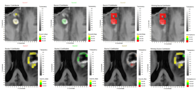 |
65 | MRSI-detected pattern in glioblastoma patients one month after concomitant chemoradiotherapy
Gulnur Semahat Ungan1,2, Albert Pons Escoda3, Daniel Ulinic2, Carles Arús1,2, Alfredo Vellido4, Carles Majós1,3, and Margarida Julià-Sapé1,2
1Centro de Investigación Biomédica en Red, Barcelona, Spain, 2Universitat Autònoma de Barcelona, Barcelona, Spain, 3Institut d'Investigació Biomèdica de Bellvitge (Idibell), Hospital Universitari de Bellvitge, Barcelona, Spain, 4IDEAI-UPC research center, UPC BarcelonaTech, Barcelona, Spain
A blind source separation method (cNMF) was used to extract characteristic metabolic patterns from PRESS MRSI 3T acquired from areas of contrast enhancement in a retrospective set of 31 glioblastoma patients, one month after the end of concomitant chemoradiotherapy with temozolomide. The aim was to evaluate whether these patterns were predictive of true progression or pseudoprogression. They were used as input for supervised classifiers, achieving a maximum of 81% balanced accuracy. A moderate association between extracted patterns and outcome was detected by Cramer’s V. Spatial source distribution with nosologic maps points to MRSI-detected metabolic heterogeneity as cause for classifiers’ performance.
|
||
 Back to Meeting Home
 Back to the Program-at-a-Glance
The International Society for Magnetic Resonance in Medicine is accredited by the Accreditation Council for Continuing Medical Education to provide continuing medical education for physicians.
 Back to Meeting Home
 Back to the Program-at-a-Glance
 View the Presentation