Digital Poster
Advances in Treatment of Body Disorders
Joint Annual Meeting ISMRM-ESMRMB & ISMRT 31st Annual Meeting • 07-12 May 2022 • London, UK

Digital Poster
Advances in Treatment of Body Disorders
| Computer # | ||||
|---|---|---|---|---|
2124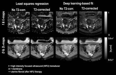 |
26 | Visualizing uterine fibroid perfusion after MR-HIFU ablations using T2-corrected IVIM and deep learning-based fitting – an explorative study.
Derk J. Slotman1,2, Lambertus W. Bartels2, Ingrid M. Nijholt1, Edwin Heijman3,4, Martijn Froeling2, and Martijn F. Boomsma1
1Radiology, Isala Zwolle, Zwolle, Netherlands, 2Image Sciences Institute, Imaging & Oncology Division, University Medical Center Utrecht, Utrecht, Netherlands, 3Faculty of Medicine and University Hospital of Cologne, Institute of Diagnostic and Interventional Radiology, University of Cologne, Cologne, Germany, 4High Tech Campus, Philips Research Eindhoven, Eindhoven, Netherlands Blood volume fraction maps derived from diffusion weighted imaging using intravoxel incoherent motion (IVIM) modeling may be an alternative to contrast-enhanced imaging for visualization of local uterine fibroid perfusion, during magnetic resonance-guided high intensity focused ultrasound (MR-HIFU) ablation procedures. In this study, blood volume fraction maps calculated with a T2-corrected IVIM model were compared to T2-uncorrected parameter maps using two fitting techniques. Based on general inspection, T2-corrected parameter maps from deep learning-based fitting were visually the most appealing. A preliminary evaluation showed a linear association between NPVs delineated on blood volume fraction maps and gadolinium contrast-enhanced T1w scans. |
||
2125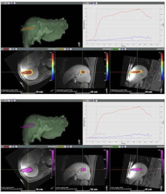 |
27 | Proof of concept of volumetric MR-temperature monitoring during microwave ablation of liver tumor in a patient Video Not Available
Pierre Bour1, Thibaut Faller1, Valery Ozenne2,3,4, Bruno Quesson2,3, Max Seidensticker5, and Olaf Dietrich5
1Certis Therapeutics, Pessac, France, 2INSERM, Centre de Recherche Cardio-Thoracique de Bordeaux, U1045, Bordeaux, France, 3IHU Liryc, Electrophysiology and Heart Modeling Institute, Hopital Xavier Arnozan, Pessac, France, 4UMR5536 CRMSB, Bordeaux, France, 5Department of Radiology, University Hospital, LMU Munich, Munich, Germany
Microwave tumor ablation in the liver is commonly performed in computed tomography scanners with limited imaging capability for real-time monitoring of treatment progression. Performing this procedure under MRI enables to monitor temperature and thermal dose during the ablation. It allows better understanding of the lesion creation and gives better insight of the treatment outcome. In this study, we evaluate a complete MR-guided workflow that include volumetric temperature and dose monitoring during ablation using an ultrafast EPI sequence with respiratory triggering. Results show that spatial extent of the lesion can be monitored and displayed in real-time.
|
||
2126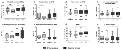 |
28 | Effect of a lifestyle intervention with plant-based diet on liver and muscle lipids in metabolic syndrome-associated osteoarthritis patients
C.S. de Jonge1, C.A. Wagenaar2,3, W. Walrabenstein2,3, M.A. Troelstra1, A.J. Nederveen1, and D. van Schaardenburg2,3
1Amsterdam UMC, University of Amsterdam, Department of Radiology and Nuclear Medicine, Amsterdam Gastroenterology Endocrinology Metabolism, Meibergdreef 9, Amsterdam, Netherlands, 2Amsterdam Rheumatology and Immunology Center, Reade, 1056 AB, Amsterdam, Netherlands, 3Amsterdam UMC, Amsterdam Medical Center, 1105 AZ, Amsterdam, Netherlands
The aim of this study was to evaluate the effect of a 16-week multidisciplinary lifestyle intervention with a plant-based diet on hepatocellular, intra- and extramyocellular lipid levels in patients with metabolic syndrome-associated osteoarthritis as measured by proton MR spectroscopy. The intervention group (n=17) was compared with the control group receiving usual care (n=15). At the end of the intervention the intervention group showed reduced hepatocellular lipid levels and total body fat mass and improved metabolic and inflammatory markers compared to the control group. There was no observable change of intra- and extramyocellular lipid levels.
|
||
 |
2127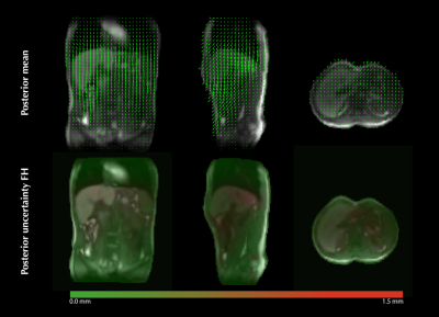 |
29 | Real-time quality assurance for volumetric motion estimation during MR-guided radiotherapy
Niek R.F. Huttinga1, Tom Bruijnen1, Cornelis A.T. van den Berg1, and Alessandro Sbrizzi1
1Department of Radiotherapy, Computational Imaging Group for MR therapy & Diagnostics, University Medical Center Utrecht, Utrecht, Netherlands
Respiratory motion during radiotherapy decreases the time-efficiency of treatments. The MR-Linac can potentially improve this with real-time 3D MR-based motion estimates. Ideally, these estimates should be accompanied with estimation uncertainty to ensure patient safety and spare organs-at-risk during bulk motion or breathing pattern change, but this is typically not provided by state-of-the-art methods. In this work we extend a previously proposed probabilistic framework for real-time motion and uncertainty estimation with a rejection criterion based on the estimation uncertainty. Results show that the proposed rejection criterion preserves low end-point-errors and rejects erroneous motion estimates during bulk motion and breathing pattern changes.
|
|
2128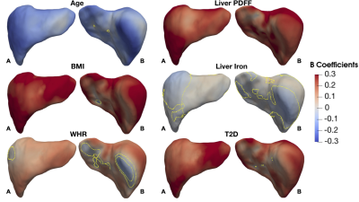 |
30 | Mass Univariate Regression Analysis on a Three-Dimensional Liver Phenotype: Associations with Disease States
Marjola Thanaj1, Nicolas Basty1, Madeleine Cule2, Elena P Sorokin2, Jimmy David Bell1, Elizabeth Louise Thomas1, and Brandon Whitcher1
1Research Centre for Optimal Health, School of Life Science, University of Westminster, London, United Kingdom, 2Calico Life Sciences LLC, South San Francisco, CA, United States
Organ MRI measurements have the potential to enhance our understanding of the precise phenotypic changes underlying many clinical conditions. Using liver mesh-based shape analysis we were able to detect significant changes in organ shape in specific anatomical regions associated with anthropometric traits and health conditions including type-2 diabetes and liver disease. We have shown that presence of T2D is associated with increase in liver shape and size. We also showed that the liver disease when accounting for liver fat showed more negative associations with the 3D liver shape phenotype.
|
||
2129 |
31 | Correlation between 18F-FDG PET/MR imaging parameters and histopathological factors from image-guided biopsy in breast
Ilaria Neri1, Carla Canevari1, Elena Venturini2, Francesca Gallivanone3, Samuele Ghezzo4, Carolina Bezzi1,4, Rosa Di Micco5, Raffaele Menichini1, Annarita Savi1, Valentino Bettinardi1, Maria Picchio1,4, Oreste Gentilini5, Pietro Panizza2, Luigi Gianolli1, and Paola Scifo1
1Nuclear Medicine, IRCCS San Raffaele Scientific Institute, Milan, Italy, 2Radiology, IRCCS San Raffaele Scientific Institute, Milan, Italy, 3Institute of Molecular Bioimaging and Physiology, National Research Council (IBFM-CNR), Milan, Italy, 4Vita-Salute San Raffaele University, Milan, Italy, 5Breast Surgery Unit, IRCCS San Raffaele Scientific Institute, Milan, Italy
This work investigates the correlations between PET/MR imaging parameters and histopathological factors of breast tumors extracted from image-guided biopsy. Results from our work show that PET and some MR parameters are statistically correlated to histopathological factors. Moreover, the integration of PET and MR (DCE-MR and ADC) parameters might help in the prediction of histopathological factors.
|
||
2130 |
32 | Feasbility of 3D MRCP for patients in both supine and prone position: a comparison study of qualitative and quantitative assessment Video Not Available
yu zhang1, yun fei zha1, and Weiyin Vivian Liu2
1Renmin Hospital of Wuhan University, wu han, China, 2GE Healthcare, Beijing, China
Increasing evidence has suggested that endoscopic retrograde cholangiopancreatography (ERCP) in the supine position is technically more technically demanding for operators than prone ERCP and leads to a greater risk of adverse cardiorespiratory events. Our results showed no differences in the CR, SNR, and CNR of the CBD between MRCP image sets. The visibility of most pancreaticobiliary ducts, motion artifact, and overall image quality were slightly better on the prone MRCP images than on the supine ones (P < 0.005). Therefore, 3D prone MRCP could provide better visualization and a roadmap directly matched with prone ERCPs for clinicians performing a surgery.
|
||
2131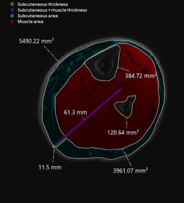 |
33 | The Subcutaneous Thickness Fraction: A new quantitative imaging metric that correlates with Lymphedema Stage.
Robert Carson Sibley1, James P. Yoon1, and Andreas M. Loening1
1Stanford University, Stanford, CA, United States
Given the dearth of quantitative metrics for staging lymphedema, we assessed subcutaneous and muscle tissue imaging metrics for patients with lower extremity lymphedema. Subcutaneous and muscle tissue thickness, subcutaneous thickness fraction (subcutaneous /sum of subcutaneous and muscle tissue thickness), and subcutaneous area fraction (subcutaneous / sum of subcutaneous and muscle tissue area) were measured. Leg subcutaneous measures strongly and significantly correlated with International Society of Lymphology lymphedema stage, while thigh subcutaneous measures demonstrated a trend to moderate correlation with stage. As lymphedema typically progresses from the distal to proximal extremity, the greater correlation with the leg is expected.
|
||
| 2132 | 34 | Preoperative Evaluation of Potential Living Liver Donors with MRI
Jitka Starekova1, Alexandra A Acher1,2, Frank A Thornton3, and Scott B Reeder1,4,5,6,7
1Radiology, University of Wisconsin-Madison, Madison, WI, United States, 2Surgery, University of Utah, Salt Lake City, Salt Lake City, UT, United States, 3University of Wisconsin-Madison, Madison, WI, United States, 4Engineering, University of Wisconsin-Madison, Madison, WI, United States, 5Medical Physics, University of Wisconsin-Madison, Madison, WI, United States, 6Medicine, University of Wisconsin-Madison, Madison, WI, United States, 7Emergency Medicine, University of Wisconsin-Madison, Madison, WI, United States
The growing need for liver transplantation has led to an increased need for living liver donors. Partial hepatectomy in donors carries significant risk, requiring rigorous preoperative evaluation. Contrast-enhanced MRI and MR cholangiography can assess liver lesions and biliary variants, while chemical shift-encoded MRI and MR elastography detect and quantify fat, iron and fibrosis in unexpected diffuse liver disease. This study summarizes our experience with an extended MRI protocol in 68 potential donors. MRI, in isolation or in combination with other test results identified more than half of patients at risk for surgical complications of liver donation.
|
||
2133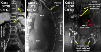 |
35 | Iron Oxide Nanoparticle Magnetic Resonance Lymphangiography (ION-MRL): A New Technique for Imaging the Peripheral and Central Lymphatic System
Robert Carson Sibley1, Maxim A. Moroz1, Shreyas S. Vasanawala1, and Andreas M. Loening1
1Stanford University, Stanford, CA, United States MR lymphangiography (MRL) utilizing the clinically available small molecular weight gadolinium contrast agents suffers from diffusion of contrast out of the lymphatic system and into the blood pool. We describe a modified MRL technique where a clinically available iron oxide nanoparticle (ferumoxytol) is used as the lymphatic contrast agent. The higher molecular weight of these nanoparticles results in retention of contrast within the lymphatic system. This simplifies peripheral lymphatic imaging by eliminating the need for vascular contamination suppression techniques and should allow better tracking of contrast through the central lymphatics following pedal injections. |
||
2134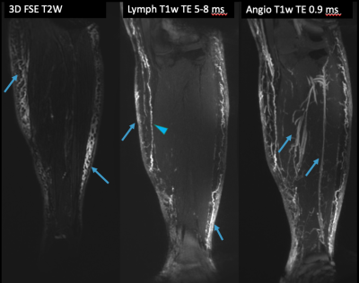 |
36 | Staging Lymphedema with Dual Agent Relaxivity Contrast Magnetic Resonance Imaging (DARC-MRL)
Robert Carson Sibley1 and Andreas M. Loening1
1Stanford University, Stanford, CA, United States
This retrospective cross-sectional study assessed different methods for staging lymphedema on Dual Agent Relaxivity Contrast MR Lymphangiography (DARC-MRL). Images from 20 patients (40 limbs) with clinically diagnosed and staged lower extremity lymphedema who had undergone DARC-MRL were assessed for dermal backflow, lymphatic vessel enhancement, and subcutaneous edema, in six regions of the lower limbs. These assessments were translated into a dermal backflow score and an edema score of our own design, as well as a recently published MRL Stage (Soga et al. 2021). All three scores demonstrated strong to excellent correlation with International Society of Lymphology (ISL) clinical stages.
|
||
2135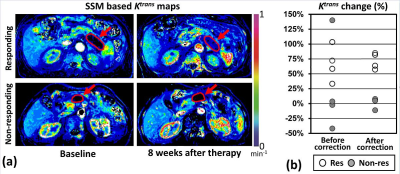 |
37 | Shutter Speed Model based Quantitative DCE-MRI for Therapeutic Response Assessment of Pancreatic Ductal Adenocarcinoma: A Pilot Study
Martin Dawson Holland1, David A Hormuth2, Xiaoyu Jiang3, Chase D Christenson4, Desiree E Morgan5, Yufeng Li5, Junzhong Xu3, Thomas E Yankeelov4, and Harrison Kim5
1Medicine, University of Alabama at Birmingham, Birmingham, AL, United States, 2The University of Texas at Austin, Austin, TX, United States, 3Vanderbilt University, Nashville, TN, United States, 4University of Texas at Austin, Austin, TX, United States, 5University of Alabama at Birmingham, Birmingham, AL, United States
Shutter Speed Model (SSM) based Ktrans, ve, kep, and τi in the pancreatic tumors before initiating chemotherapyt were 0.081±0.036 mm-1, 0.258±0.070, 0.320±0.152 mm-1, and 0.247±0.197 μs, respectively, without statistical difference between the responding and non-responding groups. The Ktrans in the tumors favorably responding to chemotherapy increased 72±14%, significantly larger than in the non-responding tumors (2±9%, p=0.0001). The ve in the responding tumors also significantly increased compared to the non-responding group (64±25% vs 3±9%, p=0.0036). However, no statistical difference was found between the responding and non-responding groups in either kep (6±11% vs 11±16%, p=0.6204) or τi (3±47% vs 147±280%, p=0.3408).
|
||
2136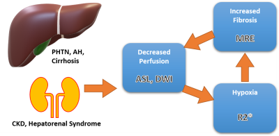 |
38 | Development of an Efficient MRE/MRI Protocol for Multiparametric Assessment of Liver, Spleen, and Kidneys to Diagnose Hepatorenal Syndrome
Nana Owusu1, Zheng Zhu1, Jiahui Li1, Kevin Glaser1, Yi Sui1, Douglas Simonetto2, Patrick Kamath2, Vijay Shah2, Sudhakar Venkatesh3, Richard Ehman1, and Meng Yin1
1Radiology, Mayo Clinic, Rochester, MN, United States, 2Gastroenterology, Mayo Clinic, Rochester, MN, United States, 3Mayo Clinic, Rochester, MN, United States
An efficient imaging protocol was developed to facilitate diagnosis of hepatorenal syndrome in patients with decompensated cirrhosis. Multiparametric MRI for quantifying perfusion, diffusion, and blood oxygenation, as well as multiparametric MRE for quantifying viscoelasticity and tissue pressure were optimized in preparation for the pilot study predicting hepatorenal syndrome. Results demonstrate the feasibility of the multiparametric protocol and provide motivation for commencing clinical testing.
|
||
2137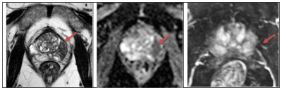 |
39 | Initial Observations on the Association of Peri-prostatic Vascular Asymmetry and Extra-prostatic Extension of Prostatic Tumors
Paul E Summers1, Alessia Minchillo2, Sarah Alessi1, Giuseppe Renne3, Ottavio De Cobelli4,5, Gennaro Musi4,5, Giulia Marvaso4,6, Barbara Alicja Jereczek-Fossa4,6, and Giuseppe Petralia4,7
1Division of Radiology, IEO, European Institute of Oncology IRCCS, Milano, Italy, 2University of Milan, Milano, Italy, 3Uropathology and Intraoperative Diagnostics, IEO, European Institute of Oncology IRCCS, Milano, Italy, 4Department of Oncology and Hemato-oncology, University of Milan, Milano, Italy, 5Division of Urology, IEO, European Institute of Oncology IRCCS, Milano, Italy, 6Division of Radiation Oncology, IEO, European Institute of Oncology IRCCS, Milano, Italy, 7Precision Imaging and Research Unit, IEO, European Institute of Oncology IRCCS, Milano, Italy
We hypothesized that the presence of asymmetry in the arterial supply of the prostate may provide a correlate of extra-prostatic tumor extension in patients with prostate cancer. In this preliminary study of 40 patients (20 with and 20 without extra-prostatic disease), a radiologist blind to the status of the patients, made a binary choice as to the presence or absence of peri-prostatic vascular asymmetry. Taking the pathological grading as the reference standard, the sensitivity, specificity, positive and negative predictive values were all 0.9. The negative likelihood ratio (LR-) was 0.11, and the positive likelihood ratio (LR+) was 9.0.
|
||
 |
2138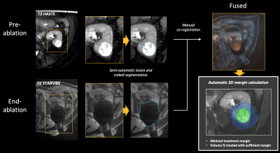 |
40 | Intraoperative MRI-derived 3D ablation margins and correlation with local outcome after MRI-guided cryoablation of small renal tumors
Nienke S. de Jager1, Tim J. van Oostenbrugge2, Torben Pätz3, Sjoerd F.M. Jenniskens1, Jurgen J. Fütterer1, Hans Langenhuijsen2, and Christiaan G. Overduin1
1Medical Imaging, Radboudumc, Nijmegen, Netherlands, 2Urology, Radboudumc, Nijmegen, Netherlands, 3Fraunhofer Institute for Digital Medicine, Bremen, Germany
MRI-guided cryoablation of small renal tumors relies on achieving sufficient ice ball coverage throughout the entire lesion for effective treatment. However, a volumetric approach to assess treatment margins intraoperatively is currently lacking. This work retrospectively derived three-dimensional ablation margins after cryoablation in 39 kidney tumors using co-registration of intraoperatively acquired pre- and post-ablation MR images. Minimal ablation margins were significantly smaller for cases with versus without local tumor progression. Radiological complete coverage (ablation margin >0 mm) was associated with local control. MRI-derived 3D ablation margins may present a useful intraprocedural tool to assess treatment success.
|
|
 Back to Meeting Home
 Back to the Program-at-a-Glance
The International Society for Magnetic Resonance in Medicine is accredited by the Accreditation Council for Continuing Medical Education to provide continuing medical education for physicians.
 Back to Meeting Home
 Back to the Program-at-a-Glance
 View the Presentation