Digital Poster
Probing the Biophysical Properties of Tumors II
Joint Annual Meeting ISMRM-ESMRMB & ISMRT 31st Annual Meeting • 07-12 May 2022 • London, UK

Digital Poster
Probing the Biophysical Properties of Tumors II
| Computer # | ||||
|---|---|---|---|---|
2581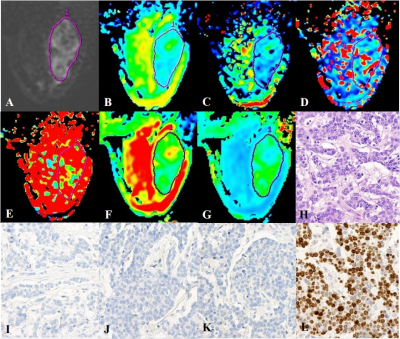 |
22 | Can combined DWI, IVIM and DKI predict prognostic factors and genotypes for patients with breast cancer?
Weiwei Wang1, Zhanguo Sun1, Yueqin Chen1, Laimin Zhu1, Zhe Zhou1, Hao Yu1, and Weiqiang Dou2
1Department of Medical Imaging, the affiliated hospital of Jining medical university, Jining,China, jining, China, 2GE Healthcare, MR Research China, Beijing, China, beijing, China
It is essential to accurately preoperatively predict genotypes and prognostic factors for patients with breast cancer in clinic treatment. Combined diffusion weighted imaging (DWI), Intravoxel incoherent motion (IVIM) and diffusion kurtosis imaging (DKI) were developed to comprehensively assess tumors at the cellular and molecular levels. In this study we aimed to explore whether the combination of these three diffusion techniques can help to comprehensively evaluate breast cancer.
|
||
2582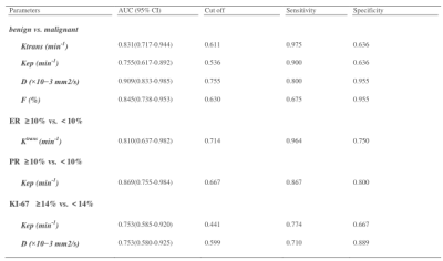 |
23 | Combination of IVIM with DCE-MRI in diagnostic and prognostic evaluation of breast cancer
Yurong zheng1, Li Li1, Rui Wang1, Tiejun Gan1, Pengfei Wang1, Jing Zhang1, and Kai Ai2
1Department of Magnetic Resonance, LanZhou University Second Hospital, Lanzhou, China, 2Philips Healthcare, Xi’an, China
Intravoxel incoherent motion (IVIM) combined with Dynamic Contrast Enhanced MRI (DCE-MRI) are meaningful MRI techniques applied to breast cancer. This study uses DCE-derived parameters (voiume translocation constant, Ktrans and rate constant, Kep) and IVIM-derived parameters (diffusion coefficient, D and perfusion fraction, f) to perform correlation analysis with prognosis of breast cancer indexes (ER, PR, her-2, Ki-67). The results show that IVIM and DCE-MRI can distinguish benign and malignant breast lesions. Therefore, there are correlation between Ktrans, Kep, D and prognostic factors of breast cancer. Our research may provide more important clinical evidence for the treatment and prognosis of breast cancer.
|
||
2583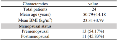 |
24 | Tumor stiffness based on 3D MR Elastography as a marker for predicting the aggressiveness in cervical cancer
Yuanqiang Xiao1, Wenying Chen1, Mengsi Li1, Jun Chen2, Meng Yin2, Richard L. Ehman2, and Jin Wang1
1Department of Radiology, The third affiliated hospital of Sun Yat-sen University, Guangzhou, China, 2Department of Radiology, Mayo Clinic College of Medicine, Mayo Clinic, Rochester, MN, United States
Preoperative prediction of aggressiveness (histologic subtype, grade of differentiation, Federation International of Gynecology and Obstetrics (FIGO) stage) in cervical cancer (CC) using a noninvasive method remains a challenge. 3D MR Elastography (MRE) is a novel functional MRI technique which can quantitatively characterize the mechanical properties of tumors. We retrospectively analyzed 24 CC patients undergoing 3D MRE examinations and found that tumor stiffness (TS) in adenocarcinoma and FIGO stage III/IV was significantly higher than that in squamous cell carcinoma and FIGO stage I/II. TS based on 3D MRE could serve as a potential noninvasive marker to assess the aggressiveness in CC.
|
||
2584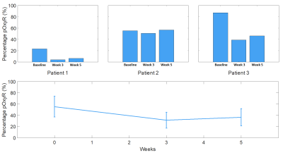 |
25 | Quantifying and mapping hypoxia modification in patients with uterine cervical cancer using oxygen-enhanced MRI
Anubhav Datta1,2, Michael Dubec1,3, David Buckley3,4, Damien McHugh3, Amal Salah5, Ross Little1, Michael Berks1, Susan Cheung1, Catharine West1, Ananya Choudhury1,6, Lisa Barraclough6, Peter Hoskin1,6,7, and James P. B. O'Connor1,2,8
1Division of Cancer Sciences, University of Manchester, Manchester, United Kingdom, 2Clinical Radiology, The Christie NHS Foundation Trust, Manchester, United Kingdom, 3Christie Medical Physics and Engineering, The Christie NHS Foundation Trust, Manchester, United Kingdom, 4School of Medicine, University of Leeds, Leeds, United Kingdom, 5Proton Beam Therapy Dept, The Christie NHS Foundation Trust, Manchester, United Kingdom, 6Clinical Oncology, The Christie NHS Foundation Trust, Manchester, United Kingdom, 7Mount Vernon Cancer Centre, Northwood, United Kingdom, 8Division of Radiotherapy and Imaging, Institute of Cancer Research, London, United Kingdom
Hypoxia is a ubiquitous negative prognostic factor in solid tumours. Oxygen-enhanced MRI can spatially map regions refractory to an oxygen challenge and, when combined with a perfusion measurement, has the potential to quantify hypoxia in vivo. We developed the technique in healthy volunteers before successfully translating into a longitudinal patient study of patients with cervical carcinoma. We present initial data to support OE-MRI quantifying and mapping hypoxia modification following therapy in this clinical dataset.
|
||
2585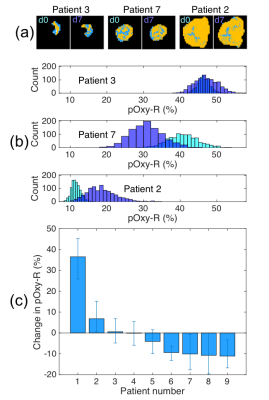 |
26 | Quantifying confidence in the OE-MRI biomarker pOxy-R using bootstrap analysis
Ross A Little1, Geoff JM Parker2,3, and James PB O'Connor1
1Division of Cancer Sciences, University of Manchester, Manchester, United Kingdom, 2Centre for Medical Image Computing, University College London, London, United Kingdom, 3Bioxydyn Limited, Manchester, United Kingdom
OE-MRI is an emerging technique for identifying, mapping and quantifying tumour hypoxia. Current analysis is based on combining data with a perfusion map and categorising each voxel absolutely as hypoxic, normoxic or necrotic. In this study we use bootstrap analysis to map the uncertainty on the biomarker pOxy-R. We investigate how this performs in synthetic tumour data before applying the method to data from 9 patients with rectal cancer undergoing chemoradiotherapy. Bootstrapping enabled estimates of confidence intervals for change, thus identifying those patients who exhibited hypoxia modification on therapy.
|
||
2586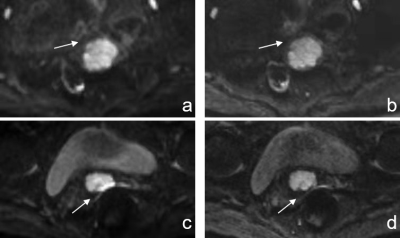 |
27 | Readout-segmented Echo-planar Imaging for Determining Cervical Cancer’s Lymph-Vascular Space Invasion and Lymph Node Metastasis Status Video Not Available
Huizhen Song1, Jiao Bai1, Yu Wang1, Juan Xie1, Mengping Huang1, Yunzhu Wu2, Shaoyu Wang3, and Jian Shu1
1Department of Radiology, Affiliated Hospital of Southwest Medical University, Luzhou,SiChuan, China, 2MR Scientific Marketing, Siemens Healthineers, Shanghai, China, 3MR Scientific Marketing, Siemens Healthineers, Xi’an, Shaanxi, China
This study compared the image quality of single-shot echo-planar imaging (ss-EPI) and readout-segmented echo-planar imaging (rs-EPI) in cervical cancer (CC). ADC values derived from these two sequences were considered as diagnostic capabilities for CC’s lymph-vascular space invasion (LVSI) and lymph node metastasis (LNM). The results showed that rs-EPI images of cervical cancer had higher subjective image quality scores, lower SNR, but no difference in CNR and ADC values. ADC was able to predict the CC’s LNM status,but couldn’t identify the LVSI status. ADC of rs-EPI sequence was superior to diagnose CC’s LNM status.
|
||
2587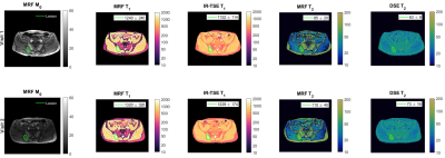 |
28 | Quantitative T1 and T2 measurements of metastatic bone lesions in prostate cancer patients: MR fingerprinting versus standard quantitative MRI
Mihaela Rata1,2, Nina Tunariu1,2, Yun Jiang3, Julie Hughes1, Georgina Hopkinson1, Erica Scurr1, Jessica M Winfield1,2, Vikas Gulani3, Dow-Mu Koh1,2, and Matthew R Orton1,2
1Radiology/MRI Unit, Royal Marsden NHS Foundation Trust, Sutton/London, United Kingdom, 2Division of Radiotherapy and Imaging, Institute of Cancer Research, Sutton/London, United Kingdom, 3Radiology, University of Michigan, Ann Arbor, MI, United States
Magnetic resonance fingerprinting (MRF) generates fast, co-registered quantitative maps from a single acquisition. This prospective study evaluates MRF-derived measures of treatment-induced T1 and T2 changes in prostate cancer patients with metastatic bone disease, by comparison with existing quantitative T1 and T2 measurements. This study demonstrated a good correlation of MRF-derived T1 and T2 changes with existing quantitative methods, supporting the use of MRF for faster measurements in bone lesions.
|
||
2588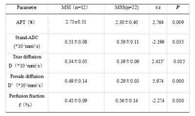 |
29 | Evaluation of microsatellite instability in endometrial cancer using APTw combined with IVIM MR imaging Video Not Available
Changjun Ma1, Ailian Liu1, Shifeng Tian1, Lihua Chen1, Nan Wang1, Qingwei Song1, and Zhiwei Shen2
1Department of Radiology, the First Affiliated Hospital of Dalian Medical University, Dalian, China, 2Philips Healthy(China), Beijing, China
Testing of Microsatellite Instability (MSI) is used to evaluate the prognosis of endometrial cancer (EC) to make the optimal treatment, and plays an important role to screen for Lynch syndrome. MSI had been detected by the traditional technology, such as PCR, IHC and Next Generation Sequencing(NGS), however, MSI also could be assessed using magnetic resonance imaging with the advantages of non-invasion and low-cost. This study aimed to investigate the clinical value in assessing the status of EC MSI with amide proton transfer weighted (APTw) imaging and intravoxel incoherent motion (IVIM) diffusion MR imaging.
|
||
 Back to Meeting Home
 Back to the Program-at-a-Glance
The International Society for Magnetic Resonance in Medicine is accredited by the Accreditation Council for Continuing Medical Education to provide continuing medical education for physicians.
 Back to Meeting Home
 Back to the Program-at-a-Glance
 View the Presentation