Digital Poster
Antenatal, Neonatal & Pediatric MRI I: Neuroimaging
Joint Annual Meeting ISMRM-ESMRMB & ISMRT 31st Annual Meeting • 07-12 May 2022 • London, UK

Digital Poster
Antenatal, Neonatal & Pediatric MRI I: Neuroimaging
| Computer # | ||||
|---|---|---|---|---|
1232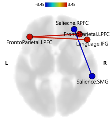 |
28 | Resting-state functional connectivity differences in preterm and term born children at school age Video Permission Withheld
Hyejin Jeong1, Hye Jung Cho2, So-Yeon Shim3, and Chan-A Park4
1Neuroscience Convergence Center, Institute of Green Manufacturing Technology, Korea University, Seoul, Korea, Republic of, 2Department of Pediatrics, Gachon University Gil Medical Center, Incheon, Korea, Republic of, 3Ewha Womans University, Seoul, Korea, Republic of, 4Biomedical Engineering Research Center, Gachon University, Seoul, Korea, Republic of
Children born preterm are at a significant risk of neurodevelopmental impairment. Alterations of functional connectivity in higher-order association cortices after preterm birth might reflect cognitive or behavior problems in school-aged children born preterm. Our results will help understanding the pathophysiology of cognitive and behavior development in children born preterm without apparent brain injury or major neurodevelopmental impairment.
|
||
1233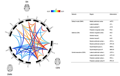 |
29 | Dynamic resting state connectivity of the default mode, salience and central executive networks in adolescents with concussion
Rachelle A. Ho1, Saurabh B Shaw2, Nicholas A Bock1, Carol DeMatteo3, Michael D Noseworthy4, and Geoffrey Hall1
1Psychology, Neuroscience & Behaviour, McMaster University, Hamilton, ON, Canada, 2Department of Psychiatry, Western University, London, ON, Canada, 3School of Rehabilitation Science, McMaster University, Hamilton, ON, Canada, 4Department of Electrical & Computer Engineering, McMaster University, Hamilton, ON, Canada
Our study evaluated the dynamic functional connectivity of adolescents with concussion in comparison to healthy controls using (1) sliding window ROI-to-ROI and (2) sliding window graph theory analyses. Adolescents with concussion exhibited higher between-network connectivity between the salience network and central executive network, but reduced between-network connectivity between the default mode network to both the salience and executive networks. This suggests lower default mode network integration and engagement during rest following concussion in adolescents.
|
||
1234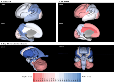 |
30 | Individualised assessment of regional brain volumes in neonates with Down syndrome reveals extreme deviation in white matter and cerebellum.
Abi Fukami - Gartner1,2, Ana A. Baburamani1, Ralica Dimitrova1,3, Prachi A. Patkee1, Olatz Ojinaga - Alfageme1,4, Alexandra F. Bonthrone1, Alena Uus1,5, Emer Hughes1, Maria Deprez1,5, Serena J. Counsell1, Joseph V. Hajnal1,5, A. David Edwards1,2, Jonathan O’Muircheartaigh1,2, and Mary A. Rutherford1,2
1Centre for the Developing Brain, School of Biomedical Engineering and Imaging Sciences, King's College London, London, United Kingdom, 2MRC Centre for Neurodevelopmental Disorders, King's College London, London, United Kingdom, 3Department of Forensic and Neurodevelopmental Science, Sackler Institute for Translational Neurodevelopment, Institute of Psychiatry, Psychology and Neuroscience, King's College London, London, United Kingdom, 4Centre for Brain and Cognitive Development, Birkbeck, University of London, London, United Kingdom, 5Biomedical Engineering Department, School of Biomedical Engineering and Imaging Sciences, King's College London, London, United Kingdom
There are relatively few early neuroimaging studies of Down syndrome (DS), despite being the most common genetic cause of intellectual disability, with characteristics present from birth. The aim of this study was to conduct a group-level analysis of volumetric differences across multiple brain regions in neonates with DS (n = 20) using individual z-scores extracted from robust normative modelling of typically developing neonatal controls (TDC; n = 493). In addition to well-documented cerebellar hypoplasia, here we have identified that neonates with DS have markedly reduced volumes in the cingulate, frontal, insular and occipital WM segments compared to TDC.
|
||
1235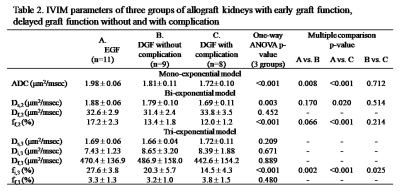 |
31 | IVIM Diffusion-Weighted MRI in Detecting Graft Function with Complications Immediately After Kidney Transplantations
JyhWen Chai1,2, Yung-Chien Chang1,3, Kuan-Jung Pan4, Mu-Chih Chung5, Hsian-Min Chen4, and Feng-Mao Chiu6
1Department of Radiology, Taichung Veterans General Hospital, Taichung, Taiwan, 2College of Medicine, National Chung Hsin University, Taichung, Taiwan, 3Department of Electrical Engineering, National Chung Hsin University, Taichung, Taiwan, 4Department of Medical Research, Taichung Veterans General Hospital, Taichung, Taiwan, 5Section of Nephrology, Taichung Veterans General Hospital, Taichung, Taiwan, 6Department of Biomedical Engineering, National Yang Ming University, Taipei, Taiwan
Delayed graft function (DGF) is a form of acute renal failure that results in post-transplantation oliguria, with various frequencies from 2% to 50%. Heretofore, there was a lack of imaging biomarkers to interpolate the DGF. The tri-exponential intravoxel incoherent motion (IVIM) model, providing three distinct signal fractions of a pure diffusion, an intermediate and an ultrafast component, is more preferable for the diffusion signal in the allograft kidneys than the mono- and bi-exponential models. Our experiment illustrates that tri-exponential IVIM model could provide a good indicator for distinguishing the early graft function, the delayed graft function without and with complications.
|
||
1236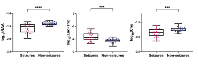 |
32 | Neonatal seizures induce significant changes in cerebral oxidative metabolism
Agnieszka Sierhej1, Alan Bainbridge2, Giles Kendall3, Magdalena Sokolska2, Kelly Pegoretti-Baruteau3, Janet Rennie3, Sean R Mathieson4, Nicola J Robertson5, Geraldine Boylan4, and Subhabrata Mitra5
1University College London, London, United Kingdom, 2Medical Physics and Bioengineering, University College London Hospitals, London, United Kingdom, 3Neonatal Unit, University College London Hospitals, London, United Kingdom, 4INFANT Research Centre, University College Cork, Cork, Ireland, 5Institute for Women's Health, University College London, London, United Kingdom
Neonatal seizures are common following neonatal encephalopathy and have the potential for themselves to cause further brain injury. Data were collected from 55 term neonates who underwent therapeutic hypothermia following neonatal encephalopathy. 25 developed seizures. Magnetic resonance Spectroscopy showed that in the seizure group, lactate was increased and Naa and Choline were decreased. Lower tNaa in neonates with seizures likely indicates further neuronal injury and may explain seizure induced neurological deficits
|
||
1237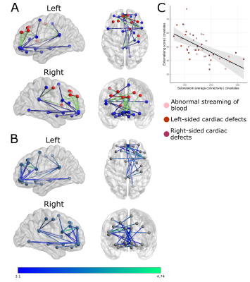 |
33 | Reduced neonatal fronto-limbic connectivity is associated with higher externalizing symptoms in toddlers with Congenital Heart Disease Video Permission Withheld
Alexandra F Bonthrone1, Andrew Chew1, Megan Ní Bhroin1,2, Christopher J Kelly1, Daan Christiaens1,3, Maximilian Pietsch1,4, J-Donald Tournier1, Lucilio Cordero-Grande1,5, Joseph V Hajnal1,6, Kuberan Pushparajah6,7, John Simpson7, A David Edwards1, Mary A Rutherford1, Chiara Nosarti1,8, Dafnis Batalle1,4, and Serena J Counsell1
1Centre for the Developing Brain, School of Biomedical Engineering and Imaging Sciences, King's College London, London, United Kingdom, 2Trinity College Institute of Neuroscience and Cognitive Systems Group, Discipline of Psychiatry, School of Medicine, Trinity College Dublin, Dublin, Ireland, 3Department of Electrical Engineering (ESAT/PSI), KU Leuven, Leuven, Belgium, 4Department for Forensic and Neurodevelopmental Sciences, Institute of Psychiatry, Psychology and Neuroscience, King's College London, London, United Kingdom, 5Biomedical Image Technologies, ETSI Telecomunicación, Universidad Politécnica de Madrid & CIBER-BBN, Madrid, Spain, 6Biomedical Engineering Department, School of Biomedical Engineering and Imaging Sciences, King's College London, London, United Kingdom, 7Paediatric Cardiology Department, Evelina London Children's Healthcare, London, United Kingdom, 8Department of Child and Adolescent Psychiatry, Institute of Psychiatry, Psychology and Neuroscience, King's College London, London, United Kingdom
Infants with Congenital Heart Disease (CHD) are at increased risk of neurodevelopmental impairments. Forty-three neonates with CHD underwent multi-shell high angular resolution diffusion MRI (HARDI) on a 3T scanner before surgery. At 22 months parents completed a questionnaire characterising internalizing and externalizing behaviours. Network-based statistics were used to characterise the relationship between internalizing and externalizing symptoms and structural connectivity. Reduced neonatal structural connectivity in fronto-limbic regions before surgery was associated with increased externalizing symptomatology in toddlers with CHD.
|
||
1238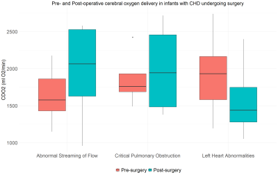 |
34 | MRI assessment of cerebral oxygen delivery before and after surgery in infants with congenital heart disease
Daniel Cromb1,2, Alexandra F Bonthrone1, Christopher Kelly1, Paul Cawley1,2, Kuberan Pushparajah3,4, Mary Rutherford1, John Simpson3,4, A. David Edwards1,2, and Serena Counsell1
1Centre for the Developing Brain, School of Biomedical Engineering and Imaging Sciences, King's College London, London, United Kingdom, 2Neonatal Intensive Care Unit, Evelina London Children's Hospital, London, United Kingdom, 3Department of Cardiovascular Imaging, School of Biomedical Engineering & Imaging Science, King's College London, London, United Kingdom, 4Department of Congenital Heart Disease, Evelina London Children's Hospital, London, United Kingdom
Reduced cerebral oxygen delivery (CDO2) in infants with congenital heart disease (CHD) is associated with abnormal brain development. We investigated how cerebral blood flow, haemoglobin, and oxygen saturations, which all contribute to CDO2, change following surgery in a cohort of 24 infants with CHD. There was no significant change in CDO2 after surgery (P = 0.62). However, both cerebral blood flow (P < 0.001) and pre-ductal oxygen saturations (P = 0.008) improved significantly, whilst haemoglobin dropped significantly (P < 0.001). Consistent with previous reports, the changes that occur in cerebral blood flow following surgery are dependent on the type of cardiac lesion.
|
||
1239 |
35 | Brain Connectivity and Autistic Traits Moderated by Olfactory Perception among Children and Adolescents with Congenital Heart Defects Video Not Available
Vanessa Jean Schmithorst1, Julia Wallace1, Pablo Polosecki2, Daryaneh Badaly3, Vincent Lee1, Sue Beers1, Pablo Meyer2, Cecilia Lo1, and Ashok Panigrahy1
1University of Pittsburgh, Pittsburgh, PA, United States, 2IBM T.J. Watson Research Center, Ossining, NY, United States, 3Child Mind Institute, New York, NY, United States
We investigate the relation between congenital heart disease (CHD), autism spectrum disorder (ASD), and olfaction in a cohort of children and young adults using fcMRI (Functional Connectivity Strength; FCS, and amygdala seed-based connectivity) and DTI (RD). ASD severity was positively correlated with FCS in widespread regions, with amygdala connectivity to the DMN, and with RD in posterior parietal white matter; however, these relations were stronger in individuals with impaired olfaction scores and individuals with CHD. The relation between impaired olfaction, CHD, and ASD may be the result of individuals with impaired olfaction and/or CHD having different brain structure-function relationships.
|
||
1240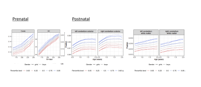 |
36 | Prenatal and Postnatal Cerebellar Development
Elizabeth Weisse1, Yaqing Chen2, Alvaro Gajardo Cataldo2, Hans-Georg Müller2, Changbo Zhu2, Jonathan O’Muircheartaigh3, Maximilian Pietsch4, Sian Wilson4, Kristofer Bouchard5,6, Sylvia Madhow5,6, James Cole7,8, Francesca Biondo7,9, Viren D’Sa10,11, Sean C. L. Deoni11,12,13, and Muriel M.K. Bruchhage1,11,14
1Department of Diagnostic Imaging, Rhode Island Hospital, Providence, RI, United States, 2Department of Statistics, UC Davis, Davis, CA, United States, 3Department of Forensic & Neurodevelopmental Sciences, King's College London, London, United Kingdom, 4Centre for the Developing Brain, Department of Perinatal Imaging and Health, King's College London, London, United Kingdom, 5Scientific Data Division & Biological Systems and Engineering Division, LBNL, Berkeley, CA, United States, 6Helen Wills Neuroscience Institute & Redwood Center for Theoretical Neuroscience, UC Berkeley, Berkeley, CA, United States, 7Centre for Medical Image Computing, Computer Science, UCL, London, United Kingdom, 8Dementia Research Centre, Queen Square Institute of Neurology, UCL, London, United Kingdom, 9Department of Neuroimaging, Institute of Psychiatry, Psychology and Neuroscience, UCL, London, United Kingdom, 10Department of Pediatrics, Rhode Island Hospital, Providence, RI, United States, 11Department of Pediatrics, Warren Alpert Medical School at Brown University, Providence, RI, United States, 12Brown's Advanced Baby Imaging Lab, Rhode Island Hospital, Providence, RI, United States, 13MRI Research, Department of Diagnostic Imaging, Rhode Island Hospital, Providence, RI, United States, 14Institute of Social Sciences, Department of Psychology, University of Stavanger, Stavanger, Norway
The cerebellum is one of the earliest structures to develop prenatally and is documented to play an important role in motor development and, more recently, cognitive development. We investigated cerebellar development and its contribution to later overall (ELC), nonverbal (NVDQ) and verbal (VDQ) cognitive development from 22 weeks of gestation to 15 years of age. Total postnatal cerebellar volume significantly increased with ELC and VDQ but decreased with NVDQ, while prenatal cerebellar volume significantly increased with ELC with age. This could be indicative of the cerebellum’s contribution to overall cognitive development refining with development and age.
|
||
| 1241 | 37 | Common Genetic Variation In IGFBP7 Is Associated With Cortical Brain Development In Preterm Infants
Daniel Cromb1, Harriet Cullen1, Christiaan De Leeuw2, Madeleine Barnett1, Jonathan O'Muircheartaigh1, Serena Counsell1, and A. David Edwards1
1Centre for the Developing Brain, School of Biomedical Engineering and Imaging Sciences, King's College London, London, United Kingdom, 2Faculty of Science, Complex Trait Genetics, Vrije Universiteit Amsterdam, Amsterdam, Netherlands
In a cohort of 185 infants born preterm we used linked MR imaging, genomic and neurodevelopmental data, to identify a significant association between common genetic variation in the IGFBP7 gene and cortical grey matter volumes as assessed at term-corrected age (p = 0.0057), as well as motor outcomes as assessed at 18-24 months of age (p = 0.044).
|
||
1242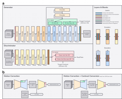 |
38 | Motion Correction of Contrast-enhanced Pediatric Brain MRI with Optional Non-contrast-enhanced Synthesis within a Single Neural Network Video Permission Withheld
Jaeuk Yi1, Sewook Kim1, Seul Lee1, Mohammed A. Al-Masni1, Sung-Min Gho2, Young Hun Choi3, and Dong-Hyun Kim1
1Department of Electrical and Electronic Engineering, Yonsei University, Seoul, Korea, Republic of, 2Collaboration & Development, GE Healthcare, Seoul, Korea, Republic of, 3Department of Radiology, Seoul National University Hospital, Seoul, Korea, Republic of
Based on the observation that motion corruption and contrast pairs do not exist in a separable way in clinically obtained pediatric contrast-enhanced scans, we developed a neural network for both motion correction and optional non-contrast-enhanced image synthesis for contrast-enhanced pediatric brain MRI. We designed a neural network architecture and training schema specific to this task. We found that motion correction performance was not degraded by doing contrast synthesis simultaneously.
|
||
1243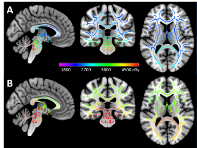 |
39 | Effect of radiation dose on the recovery of white matter integrity in children treated for medulloblastoma
John O Glass1, Jared J Sullivan1, Yian Guo2, Julie H Harreld1, Yimei Li2, Giles W Robinson3, Amar Gajjar3, Thomas E Merchant4, and Wilburn E Reddick1
1Diagnostic Imaging, St. Jude Children's Research Hospital, Memphis, TN, United States, 2Biostatistics, St. Jude Children's Research Hospital, Memphis, TN, United States, 3Oncology, St. Jude Children's Research Hospital, Memphis, TN, United States, 4Radiation Oncology, St. Jude Children's Research Hospital, Memphis, TN, United States
This study reports on the effects of pediatric medulloblastoma therapy in a population of 105 subjects. Longitudinal white matter microstructure was studied starting with the post radiation examination until approximately two years after diagnosis. The tract-based spatial statistics (TBSS) pipeline was used to combine fractional anisotropy (FA) with individual dosimetry maps before linear mixed effects models were calculated for each voxel. Significant voxels were found for increasing FA over time and a decrease of FA over time associated with higher radiation dose. These results are consistent with a recovery in FA that is attenuated by higher doses of radiation.
|
||
 Back to Meeting Home
 Back to the Program-at-a-Glance
The International Society for Magnetic Resonance in Medicine is accredited by the Accreditation Council for Continuing Medical Education to provide continuing medical education for physicians.
 Back to Meeting Home
 Back to the Program-at-a-Glance
 View the Presentation