Digital Poster
Preclinical Body
Joint Annual Meeting ISMRM-ESMRMB & ISMRT 31st Annual Meeting • 07-12 May 2022 • London, UK

| Computer # | ||||
|---|---|---|---|---|
2312 |
48 | Peri-conceptional Alcohol Exposure Shapes Placental Functions in Fetal Alcohol Spectrum Disorder Video Permission Withheld
Margaret Caroline Stapleton 1, John Richard Chaillet 2, Eric Goetzman3, Devin R. E. Cortes4, Kristina Elsa Schwab1, Amanda Sferruzzi-Perri 5, Michal Neeman6, Anthony Christodoulou 7, and Yijen L Wu1
1Developmental Biology, University of Pittsburgh, Pittsburgh, PA, United States, 2Obstetrics, Gynecology & Reproductive Sciences, University of Pittsburgh, Pittsburgh, PA, United States, 3Pediatrics, University of Pittsburgh, Pittsburgh, PA, United States, 4Biomedical Engineering, University of Pittsburgh, Pittsburgh, PA, United States, 5Physiology, University of Cambridge, Cambridge, United Kingdom, 6Biological Regulation, Weizmann Institute, Rehovot, Israel, 7Cedars Sinai Medical Center, Los Angeles, CA, United States
Peri-conceptional alcohol (PCA) exposure leading to fetal alcohol spectrum disorder continues to a significant public health concern because alcohol use ceases only after recognition of pregnancy weeks after conception, but the teratogenic damages have already occurred. Our study showed that PCA exposure in mice resulted in compromised placental perfusion and placental capability to adapt to acute hypoxia challenges. Placental abnormalities due to PCA may further exacerbate fetal neurodevelopmental deficits.
|
||
2313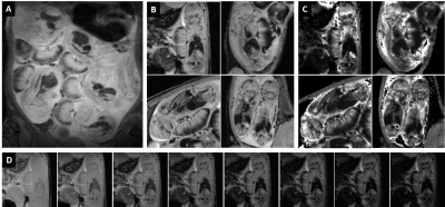 |
49 | Evaluation of Placenta Oxygenation and Perfusion in A Rat Model of Fetal Growth Restriction Using Quantitative T2* Mapping and 3D DCE-MRI
Fatimah Al Darwish1, Bram Coolen1, Fieke Terstappen2, Caren van Kammen2, Lindy Alles1, Raymond Schiffelers2, Titia lely2, and Gustav Strijkers1
1Biomedical engineering and physics, Amsterdam University Medical Center, Amsterdam, Netherlands, 2University Medical Center Utrecht, Utrech, Netherlands
Developing preclinical imaging techniques in the field of preeclampsia is essential to test the effectiveness of new therapeutic approaches. We applied T2* weighted and three-dimensional dynamic contrast-enhanced MRI in a pregnant rat model of reduced uterine perfusion pressure (RUPP) to test their feasibility and ability to detect differences in placental oxygenation and perfusion in RUPP rats compared to control. These techniques produced high-quality data that enabled the assessment of oxygenation and perfusion parameters. Oxygenation was decreased in RUPP placenta; however, perfusion parameters did not reveal differences, which requires further investigations.
|
||
2314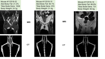 |
50 | Non-Invasive Multi-Modal Imaging of Body Composition and Bone Density in Genetically Modified Fshb Mice
Natalie Serkova1, Julia E Slack2, Jenna L Steiner3, Anna De Schutter3, Aaya AlKassi3, Jamie Henry4, and Mark S Brown3
1Radiology, University of Colorado Anschutz Medical Campus, Aurora, CO, United States, 2Emory University, Atlanta, GA, United States, 3University of Colorado Anschutz Medical Campus, Aurora, CO, United States, 4Colorado State University, Fort Collins, CO, United States
We report on a multi-modal quantitative MRI/ CT imaging protocol for characterizing the comprehensive body composition and bone health in a genetically modified mouse model. Follicle stimulating hormone (FSH) is directly involved in the regulation of estrogen production, osteoclasto-genesis, and adiposity. In this study we show that the deletion of FSH gene (-/-) in the mouse results in increased adiposity and decreased tibia density in male and female mice as they age. Multi-parametric imaging provides a unique opportunity to quantify visceral adiposity levels, muscle mass, and bone density in mutant animals, non-invasively and in real time.
|
||
2315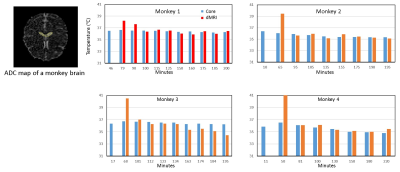 |
51 | Measuring brain temperature changes in the rhesus monkey maintained under isoflurane anesthesia using diffusion MRI
Xiaodong Zhang1 and Chun-Xia Li1
1Yerkes National Primate Research Center, Emory University, Atlanta, GA, United States Therapeutic hypothermia can improve neurological recovery and reduce mortality in acute stroke. Pharmacologically-induced hypothermia (PIH) has been investigated in preclinical and clinical studies. Usually the core temperature (rectal or esophageal) is measured to monitor the temperature changes of the subject during the treatment. However, it remains unknown how the brain temperature is affected by PIH as the brain temperature can be different from the core temperature and affected by the anesthesia. In the present study, the brain temperature changes of adult rhesus monkeys maintained under isoflurane for over 3 hours were examined and evaluated using diffusion MRI. |
||
2316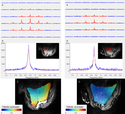 |
52 | Assessing metabolic differences in rodents on high fat diet using Deuterium Metabolic Imaging
Viktoria Ehret1, Usevalad Ustsinau2, Joachim Friske3, Thomas Scherer1, Clemens Fürnsinn1, Thomas Helbich3, Cécile Philippe2, and Martin Krššák1
1Division of Endocrinology and Metabolism, Department of Medicine III, Medical University Vienna, Vienna, Austria, 2Division of Nuclear Medicine, Department of Biomedical Imaging and Image-Guided Therapy, Medical University Vienna, Vienna, Austria, 3Division of Molecular and Structural Preclinical Imaging, Department of Biomedical Imaging and Image-Guided Therapy, Medical University Vienna, Vienna, Austria Deuterium Metabolic Imaging (DMI) is a novel method to assess metabolism in vivo using 2H MR Spectroscopic Imaging (MRSI) combined with the administration of deuterated substrates. In this pilot study, we applied 2D DMI following an intravenous injection of deuterated glucose and palmitic acid to evaluate the differences in liver metabolism of rats on standard and high fat diet at 9.4T. The spectra show a lower uptake of glucose and a higher uptake of palmitic acid after injection in high fat diet rats, indicating that liver metabolism is slowed down in rats with fatty livers. |
||
2317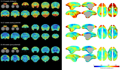 |
53 | Cortical regions in the apex transmodal network show greatest anatomical variability in infant marmosets
Stephen Sawiak1,2, Shaun Quah1, and Angela C Roberts1
1Translational Neuroimaging Laboratory, PDN, University of Cambridge, Cambridge, United Kingdom, 2Wolfson Brain Imaging Centre, University of Cambridge, Cambridge, United Kingdom
We created high-resolution paediatric templates of the marmoset brain from infancy to puberty, producing maps of anatomical variability at different ages. We show that greatest variability is present in juveniles and corresponds well with regions in a recently described transmodal apex network of higher order association areas in the marmoset (and human) brain. We hypothesise this time-period of high variability may indicate a period of heightened vulnerability to environmental stressors which may be a causal factor in the development of mental health disorders.
|
||
2318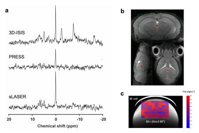 |
54 | Sequence comparison and test-retest analysis for 31P-MRS in mouse brain at 7 Tesla
Antoine Cherix1, Mohamed Tachrount1, and Jason Lerch1,2
1Wellcome Centre for Integrative Neuroimaging, WIN-FMRIB, NDCN, University of Oxford, Oxford, United Kingdom, 2Mouse Imaging Centre (MICe), Hospital for Sick Children, Toronto, ON, Canada
31P-MRS is challenging in small samples as it suffers from long T1s, short T2s and low nucleus sensitivity (γ). Thus, ultra-high field has been generally preferred for investigating the mouse brain. Less is known about its feasibility at lower field strength with more readily available scanners. Here, we assess the applicability of 31P-MRS in mouse at 7T to study neuroenergetics by comparing 3D-ISIS, PRESS and sLASER. Then, by performing a test-retest analysis and power calculations we assessed the relevance of our method for potential preclinical studies application.
|
||
2319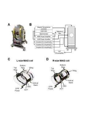 |
55 | Development of in vivo dynamic nuclear polarization (DNP)-MRI at 16 mT for large animals and redox metabolic imaging of acute hepatitis rat model
Fuminori Hyodo1, Hinako Eto2, Tatsuya Naganuma3, Abdelazim Elsayed ELHELALY1, Masaharu Murata2, Yoshifumi Noda1, Hiroki Kato1, and Masayuki Matsuo1
1Gifu University, Gifu, Japan, 2Kyushu University, Fukuoka, Japan, 3Japan REDOX Limited, Fukuoka, Japan
To use the current methods in clinical practice, the development of a prototype in vivo DNP-MRI system for preclinical examinations of large animals is indispensable for clarifying the problems peculiar to the increase in size of the DNP-MRI device. Therefore, we developed a in vivo DNP-MRI (Overhauser MRI) system with a sample bore size of 20 cm and a 16-mT magnetic field using a U-shaped permanent magnet. The in vivo DNP-MRI system developed was used to non-invasively image the redox reaction of a carbamoyl-PROXYL probe in the livers of large rats weighing 800 g and hepatitis-model rats.
|
||
2320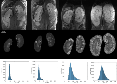 |
56 | Tracking disease progression in mouse models of polycystic kidney disease with high resolution MRI and automated postprocessing
Florian Schmid1, Georgios Koukos1, Matt Sooknah1, Sanam Assili1, and Johannes Riegler1
1Calico Life Sciences, South San Francisco, CA, United States
We present an improved MRI acquisition and data processing pipeline to assess disease progression in mouse models of polycystic kidney disease. High-resolution anatomical T2 weighted images as well as T2 mapping are used to track changes in kidney volume, cyst burden and tissue composition. We established versatile deep-learning automatic kidney segmentation, trained on a range of kidney disease stages, animal models and image resolutions.
|
||
2321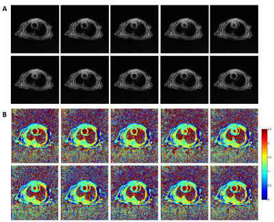 |
57 | Feasibility of Cardiovascular Magnetic Resonance Imaging of In Vivo Mouse Heart using Multitasking at 9.4T
Zhongmiao Wang1, Yang Ruan1, Qichan Gao1, Hongxia Lei1, Renkuan Zhai1, Yao Xing1, Zhongqi Zhang2, Jian Xu2, and Qi Liu2
1Wuhan United Imaging Life Science Instrument Co., Ltd., Wuhan, China, 2UIH America, Inc., Houston, TX, United States
The feasibility of cardiac T1 mapping in mouse is demonstrated at 9.4T using Multitasking, an ECG-free and free-breathing technique. High spatial- and temporal-resolution T1 maps of healthy mouse hearts without either respiratory or cardiac triggering were obtained. This technique simplifies experimental setup and opens possibilities of investigating numerous heart disease/transgenic mouse models.
|
||
2322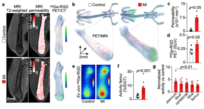 |
58 | In vivo imaging of bone marrow endothelial dysfunction promoting myeloid cell expansion in cardiovascular disease Video Permission Withheld
Katrien Vandoorne1,2, I-Hsiu Lee1, Jana Grune1, Shuang Zhang1, Cameron S. McAlpine1, Maximilian J. Schloss1, Ribhu Nayar1, Gabriel Courties1, Vanessa Frodermann1, Gregory Wojtkiewicz1, Lisa Honold1, Qi Chen3, Yoshiko Iwamoto1, Yuan Sun1, Sebastian Cremer1, Oriol Iborra-Egea4, Christian Munoz-Guijosa4, Fei Ji5, Bin Zhou6, Ralf H. Adams3,
Joshua D. Wythe7, Juan Hidalgo8, Hideto Watanabe9, Yookyung Jung10, Anja van der Laan11, Jan J. Piek11, Youmna Kfoury12,13, Pauline A. Désogère14, Claudio Vinegoni1, Partha Dutta15, Ruslan I. Sadreyev5,16, Peter Caravan14, Antoni Bayes-Genis4, Peter Libby17, David T. Scadden12,13, Charles P. Lin1,9, Kamila Naxerova1, Filip K Swirski1, Matthias Nahrendorf1, and David Rohde1,18
1Center for Systems Biology, Massachusetts General Hospital and Harvard Medical School, Boston, MA, United States, 2Biomedical Engineering, Technion, Israel Institute of Technology, Haifa, Israel, 3Max Planck Institute for Molecular Biomedicine, Muenster, Germany, 4Institut del Cor Germans Trias i Pujol, Barcelona, Spain, 5Genetics, Harvard Medical School, Boston, MA, United States, 6State Key Laboratory of Cell Biology, CAS Center for Excellence in Molecular Cell Science, Institute of Biochemistry and Cell Biology, Chinese Academic of Sciences, Shanghai, China, 7Cardiovascular Research Institute, Department of Molecular Physiology and Biophysics, Baylor College of Medicine, Houston, TX, United States, 8Institute of Neurosciences and Department of Cellular Biology, Physiology and Immunology, Universitat Autònoma de Barcelona, Barcelona, Spain, 9Institute for Molecular Science of Medicine, Aichi Medical University, Aichi, Japan, 10Wellman Center for Photomedicine, Massachusetts General Hospital and Harvard Medical School, Boston, MA, United States, 11Heart Center, Department of Cardiology, Amsterdam University Medical Center, University of Amsterdam, Amsterdam, Amsterdam, Netherlands, 12Center for Regenerative Medicine and Cancer Center, Massachusetts General Hospital, Boston, MA, United States, 13Stem Cell and Regenerative Biology, Harvard University, Cambridge, MA, United States, 14Martinos Center for Biomedical Imaging, Department of Radiolog, Massachusetts General Hospital and Harvard Medical School, Charlestown, MA, United States, 15Pittsburgh Heart, Lung, Blood and Vascular Medicine Institute, Division of Cardiology, Department of Medicine, University of Pittsburgh School of Medicine, Pittsburgh, PA, United States, 16Pathology, Massachusetts General Hospital and Harvard Medical School, Boston, MA, United States, 17Division of Cardiovascular Medicine, Department of Medicine, Brigham and Women’s Hospital and Harvard Medical School, Boston, MA, United States, 18Cardiology, Angiology and Pneumology, Heidelberg University Hospital, Heidelberg, Germany
Hematopoietic stem cells at the bone marrow (BM) niche generates excess inflammatory leukocytes harming the heart after a myocardial infarction (MI). Whether MI affects the hematopoietic organ’s microvasculature is unknown. Here intravital microscopy shows that MI triggers endothelial dysfunction, leakage, and angiogenesis in the BM, leading to systemic leukocytosis. These novel findings were imaged by noninvasive PET imaging of integrin αVβ3 activation concomitant with measuring leakiness using dynamic contrast enhanced MRI. Endothelial deletion of Vegf receptor 2 (Vegfr2) curbed emergency hematopoiesis after MI. Our findings establish that MI remodels the vascular BM niche, stimulating hematopoiesis and production of inflammatory leukocytes.
|
||
2323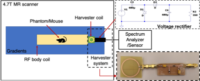 |
59 | RF Energy Harvesting System for Autonomous Respiratory Sensor in MRI
Megdouda BENAMARA1,2, Asma Bakkali2, tania-del-socorro Vergara Gomez2,3, Camille Raynard2, Christophe Vilmen3, Pierre Jomin2, Amira Trabelsi2,3, Djamel Berrahou1, Marc Dubois1, Yann Le Fur3, Frank Kober3, David Bendahan3, Emmanuel Bergeret4, Matthieu Egels4, Stefan Enoch2, and Redha Abdeddaim2
1Multiwave Imaging, Marseille, France, 2Aix Marseille Univ, CNRS, Centrale Marseille, Institut Fresnel, Marseille, France, 3Aix Marseille Univ, CNRS, CRMBM, Marseille, France, 4IM2NP - UMR 7334 CNRS, Aix-Marseille University, Marseille, France, Marseille, France
The energy harvesting system was developed for preclinical 4.7 T MRI scanners. The system is composed of a commercial birdcage coil tuned and matched at 200.1 MHz, a harvesting coil and an RF-DC rectifier. The collected energy was used to power a respiratory pressure sensor. The efficiency of the harvesting system was optimized as function of the angular positioning of the harvesting coil in the birdcage. The experiments were carried out with a water phantom and a mouse in vivo. A peak voltage of 1.5V was harvested for 45V input voltage and delivered to a load of 350 Ω.
|
||
2324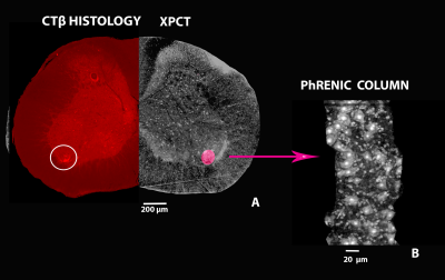 |
60 | Correlation methods between X- ray phase contrast imaging, MRI and histology for the study of the nervous central system
Laura Maugeri1, Charles Nicaise2, Maria Guidi3, Aleksandar Jankovski4, Emil Malucelli5, Alejandra Sierra6, Ali Abdollahzadeh6, Raimo A. Salo6, Irene Egidi3, Giuseppe Gigli7, Federico Giove3, Alessia Cedola8, and Michela Fratini8
1CNR -Institute of Nanotechnology, Lecce Unit & Roma Unit, LECCE, Italy, 2URPhyM – NARILIS, Université de Namur, Namur, Belgium, 3Museo Storico della Fisica e Centro Studi e Ricerche Enrico Fermi, Rome, Italy, 4NEUR division, Université catholique de Louvain (UCLouvain), Institute of NeuroScience (IoNS), Brussels, Belgium, 5Department of Pharmacy and Biotechnology, University of Bologna, Bologna, Italy, 6A. I. Virtanen Institute for Molecular Sciences, University of Eastern Finland, Kuopio, Finland, 7CNR -Institute of Nanotechnology, Lecce Unit, LECCE, Italy, 8CNR -Institute of Nanotechnology, Roma Unit, Rome, Italy
Many serious pathologies of the central nervous system (CNS) are related to anomalous development or damages to the vascular and neuronal networks. Here, we show our approach based on MRI/XPCT, XPCT/ HISTOLOGY combination in order to study neuronal and vascular alterations following a damage in mouse models.
|
||
 Back to Meeting Home
 Back to the Program-at-a-Glance
The International Society for Magnetic Resonance in Medicine is accredited by the Accreditation Council for Continuing Medical Education to provide continuing medical education for physicians.
 Back to Meeting Home
 Back to the Program-at-a-Glance
 View the Presentation