Digital Poster
Probing the Biophysical Properties of Tumors I
Joint Annual Meeting ISMRM-ESMRMB & ISMRT 31st Annual Meeting • 07-12 May 2022 • London, UK

Digital Poster
Probing the Biophysical Properties of Tumors I
| Computer # | ||||
|---|---|---|---|---|
2497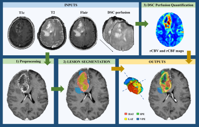 |
29 | Relative cerebral blood volume of tumor habitats can differentiate presurgical IDH-wildtype glioblastoma from IDH-mutant astrocytoma grade 4.
María del Mar Álvarez-Torres1, Elies Fuster-Garcia1,2, Carmen Balaña3, Josep Puig4, Gaspar Reynes5, Kyrre Eeg Emblem2, Enrique Mollà-Olmos6, Jose Pineda7,8, and Juan M García-Gómez1
1Biomedical Data Science Laboratory. ITACA, Universitat Politècnica de València, Valencia, Spain, 2Oslo University Hospital, Oslo, Norway, 3Institut Catala Oncologia Badalona, Barcelona, Spain, 4Institut de Diagnostic per la Image (IDI), Hospital Dr Josep Trueta, Girona, Spain, 5Instituto de Investigación Sanitaria La Fe, Valencia, Spain, 6Hospital de la Ribera, Alzira, Valencia, Spain, 7Hospital Clinic de Barcelona, Barcelona, Spain, 8void.space Lab, Facultat de Medicina, Universitat de Vic, Vic, Spain IDH-wildtype glioblastoma and IDH-mutant astrocytoma are classified as different gliomas according to WHO 2021. IDH mutations are key at clinical level, since they are associated with patient prognosis and seem to be critical for treatment selection. Despite these evidences, current protocols do not include the full sequencing for all tumors. In this sense, non-invasive and automatically calculated MRI-based biomarkers can be helpful for the clinical practice. Our results show that perfusion markers obtained in an automated, repeatable, and non-invasive manner may be candidates for being surrogate predictive markers of IDH mutation status in astrocytomas grade 4. |
||
2498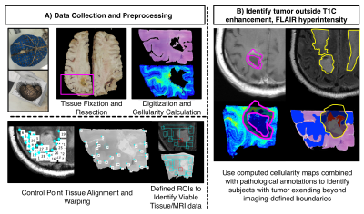 |
30 | Tumor infiltration beyond contrast enhancement and FLAIR hyperintensity at autopsy predicts survival in glioblastoma patients
Samuel Bobholz1, Allison Lowman2, Michael Brehler2, Savannah Duenweg1, John Sherman1, Fitzgerald Kyereme2, Elizabeth Cochran3, Dylan Coss3, Jennifer Connelly4, Wade Mueller5, Mohit Agarwal2, Anjishnu Banerjee6, and Peter LaViolette2,7
1Biophysics, Medical College of Wisconsin, Milwaukee, WI, United States, 2Radiology, Medical College of Wisconsin, Milwaukee, WI, United States, 3Pathology, Medical College of Wisconsin, Milwaukee, WI, United States, 4Neurology, Medical College of Wisconsin, Milwaukee, WI, United States, 5Neurosurgery, Medical College of Wisconsin, Milwaukee, WI, United States, 6Biostatistics, Medical College of Wisconsin, Milwaukee, WI, United States, 7Biomedical Engineering, Medical College of Wisconsin, Milwaukee, WI, United States
This study compared autopsy tissue samples to clinical MRI from glioblastoma (GBM) patients to assess the effects of tumor outside of T1-contrast enhancement and FLAIR hyperintensity on clinical characteristics and outcomes. Non-enhancing tumors were more frequent amongst patients who had a history of bevacizumab treatment and no history of Tumor Treating Fields treatment. Additionally, non-enhancing tumors were associated with shorter overall survival times. These results suggest that non-enhancing tumors have clinically significant correlates and may result in worse survival outcomes for GBM patients.
|
||
2499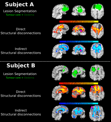 |
31 | Structural disconnections in brain tumours: investigating the similarity of Direct and Indirect Approaches
Umberto Villani1,2, Erica Silvestri1,2, Manuela Moretto1,2, Maria Colpo1,2, Alessandro Salvalaggio1,3, Maurizio Corbetta1,3,4, Diego Cecchin1,5, and Alessandra Bertoldo1,2
1Padova Neuroscience Center, University of Padova, Padova, Italy, 2Department of Information Engineering, University of Padova, Padova, Italy, 3Department of Neuroscience, University of Padova, Padova, Italy, 4Venetian Institute of Molecular Medicine, Padova, Italy, 5Department of Medicine, Unit of Nuclear Medicine, University of Padova, Padova, Italy
Gliomas are amongst the most common primary brain tumours in adults and are often associated with poor prognosis. Understanding the extent of white matter (WM) which is affected outside the tumoral lesion may be of paramount importance to explain cognitive deficits and the clinical progression of the disease. Thus, we apply both direct (i.e., tractography based) and indirect (i.e., atlas based) approaches to quantifying WM structural disconnections in a cohort of 50 glioma patients. We eventually compare the disconnections maps provided by the two methodologies in terms of spatial similarity and discuss their critical use in this field.
|
||
2500 |
32 | Chemical Exchange Saturation Transfer MR Fingerprinting (CEST-MRF) Imaging of Brain Metastases: Initial Results
Victoria Y Yu1, Kathryn R Tringale2, Robert Young3, Ricardo Otazo1, and Ouri Cohen1
1Medical Physics, Memorial Sloan Kettering Cancer Center, New York, NY, United States, 2Radiation Oncology, Memorial Sloan Kettering Cancer Center, New York, NY, United States, 3Radiology, Memorial Sloan Kettering Cancer Center, New York, NY, United States
Chemical Exchange Saturation Transfer (CEST) imaging is a promising method for cancer imaging and was recently combined with MR fingerprinting (CEST-MRF) for fast quantitative relaxation and exchange mapping. The CEST-MRF parametric maps reflect different biophysical processes, and their combination provides a comprehensive picture of complex pathologies, like brain tumors. The goal of this work is to demonstrate the potential utility of CEST-MRF in imaging brain metastases (BM). The in vivo reproducibility of the method is quantified in a healthy subject and the clinical utility is shown in subjects with metastatic brain tumors.
|
||
2501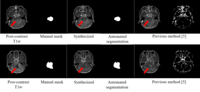 |
33 | A multi-task generative network for simultaneous post-contrast MR image synthesis and tumor segmentation: application to brainstem glioma
Yajing Zhang1, Xiangyu Xiong1, and Yaou Liu2
1MR Clinical Science, Philips Healthcare, Suzhou, China, 2Department of Radiology, Beijing Tiantan Hospital, Capital Medical University, Beijing, China
To reduce the exposure of Gadolinium-based Contrast Agents (GBCAs) in brainstem glioma detection and provide high-resolution contrast information, we propose a novel multi-task generative network for contrast-enhanced T1-weight MR synthesis on brainstem glioma images. The proposed network can simultaneously synthesize the high-resolution contrast-enhanced image and the segmentation mask of brainstem glioma lesions.
|
||
| 2502 | 34 | Inhomogeneous MT (ihMT) MRI in a glioblastoma mouse model
Gopal Varma1, Alan T Yeo2,3, Cody Callahan1,4, David C Alsop1, Aaron K Grant1, and Alain Charest2,5
1Division of MR Research, Radiology, Beth Israel Deaconess Medical Center, Harvard Medical School, Boston, MA, United States, 2Department of Medicine, Beth Israel Deaconess Medical Center, Harvard Medical School, Boston, MA, United States, 3Sackler School of Graduate Studies, Tufts University School of Medicine, Boston, MA, United States, 4Department of Pathology, Beth Israel Deaconess Medical Center, Harvard Medical School, Boston, MA, United States, 5Cancer Research Institute, Beth Israel Deaconess Medical Center, Harvard Medical School, Boston, MA, United States
Inhomogeneous MT (ihMT) MRI was applied in a genetic mouse model of glioblastoma to assess its potential to provide useful complementary information regarding the true extent of tumor infiltration. Data were acquired to produce maps of MT and ihMT ratios, which were analyzed based on regions of interest relative to brain tissue contralateral to the tumor and progression with time. The tumor was characterized by significantly lower MT and ihMT. Reduction in peripheral white matter ihMT was taken to indicate demyelinating processes associated with glioblastoma. Thus, ihMT might be used to inform on disease progression in tumor adjacent brain tissue.
|
||
2503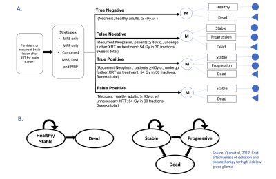 |
35 | Cost-Effectiveness Analysis of Diagnosis of Necrosis from Recurrent Tumor after RT with MRS, MRP and Combined Imaging Method for Low-Grade Glioma
Huijun Vicky Liao1, Raymond Y Huang2, Stella K Kang3, and Alexander Lin1
1Center for Clinical Spectroscopy, Brigham and Women's Hospital, Boston, MA, United States, 2Radiology, Brigham and Women's Hospital, Boston, MA, United States, 3Radiology, NYU Langone Medical Center, New York, NY, United States
Our decision-analytic model examined the cost-effectiveness of MR Spectroscopy (MRS), MR perfusion (MRP), and the combination of MRS, MRP and DWI for differentiating brain radiation necrosis from tumor progression in high-risk low-grade glioma. Our results suggested that performing combined imaging method had good diagnostic accuracy for a 3-year time horizon but not a cost-effective in the long run. In contrast, MRS and especially MRP alone were more cost-effective over all time horizons. MRS is the most cost-effective method in lifetime when its negative predictive value≥88% at the Willingness-to-Pay threshold of $100,000.
|
||
2504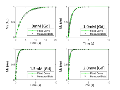 |
36 | Estimation of cellular water exchange rate, intracellular volume fraction and longitudinal relaxation time in cancer cells
Karl Kiser1, Jin Zhang1, and Gene Kim1
1Radiology, Weill Cornell Medical College, New York, NY, United States
The T1 recovery curves of cells may be sensitized to water exchange when suspended in solution with gadolinium-based contrast agent. The non-monoexponential recovery curves are fitted to a two-site exchange model to estimate the water exchange rate. This estimation process requires the inclusion of relevant parameters such as volume mol fraction and intracellular relaxation time, cannot be accurately estimated from a single curve. Here, we propose a multiple curve fitting approach, which infuses the model with additional information. By modelling multiple recovery curves of samples in a titration of GBCAs, we demonstrate an accurate and robust method for measuring invitro water exchange, volume mol fractions and intracellular relaxation rate.
The T1 recovery curves of cells may be sensitized to water exchange across the cell membrane when suspended in solution with gadolinium-based contrast agent. The two-compartment model fitting requires the inclusion of relevant parameters such as volume mol fraction and intracellular relaxation time but cannot be accurately estimated from a single curve. Here, we propose a multiple curve fitting approach, which infuses the model with additional information. By modelling multiple recovery curves of samples in a titration of GBCAs, we demonstrate an accurate and robust method for measuring invitro water exchange, cell volume and intracellular relaxation rate. |
||
2505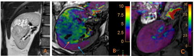 |
37 | Application of APTw imaging in clear cell renal cell carcinoma Video Not Available
Xia Wang1, Chaoqun Bu#1, Gang Tian#1, Zeliu Du1, Yu Jiang1, Na Zhao1, Chanjuan Yu1, Yuedong Han*1, Jianguang He1, Xiuzheng Yue2, and Zhiwei Shen2
1Xi'an Gaoxin Hospital, Xi'an, China, 2Philips Healthcare, Beijing, China
The application of amide proton transfer (APT) weighted imaging in molecular imaging of craniocerebral tumors has been relatively mature, but rarely reports on renal tumors. This study conducted a preliminary comparative analysis of clear cell renal cell carcinoma (ccRCC) and healthy adult renal APT imaging. The APTw values 6.40(6.26~7.75)% of tumor foci in the ccRCC group was significantly higher than that of healthy adult renal (2.05±0.24) %The preliminary results indicate that APT technology has potential clinical application value in the diagnosis of ccRCC.
|
||
2506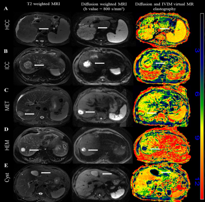 |
38 | Value of diffusion and intravoxel incoherent motion virtual MR elastography in differentiating benign and malignant focal liver lesions
Yuelang Zhang1, Xiang Li1, Chenxia Li1, Ting Cui1, Jinhan Wang1, Shuo Xing2, Xianjun Li1, and Jian Yang1
1the First Affiliated Hospital of Xi’an Jiaotong University, xi'an, China, 2Xi’an Jiaotong University, xi'an, China
Diffusion and IVIM virtual MR elastography (dMRE) provides quantitative estimates of tissue stiffness without using mechanical vibrations. It rarely applied to identify focal liver lesions (FLL). This retrospective study included 21 subjects (11 malignant lesions and 10 benign lesions). DWI were obtained to calculating dMRE. The kPa values of malignant FLLs (7.93 ± 2.37) was significantly higher than that of benign FLLs (-0.28±5.37) (P < 0.01). ROC showed an AUC of 0.93 for dMRE in differentiating maglinant FLLs and benign FLLs. The sensitivity and specificity were 91% and 90%, respectively. Malignant lesions could be distinguished from benign FLLs by dMRE.
|
||
 Back to Meeting Home
 Back to the Program-at-a-Glance
The International Society for Magnetic Resonance in Medicine is accredited by the Accreditation Council for Continuing Medical Education to provide continuing medical education for physicians.
 Back to Meeting Home
 Back to the Program-at-a-Glance
 View the Presentation