Digital Poster
Preclinical Cancer II
Joint Annual Meeting ISMRM-ESMRMB & ISMRT 31st Annual Meeting • 07-12 May 2022 • London, UK

| Computer # | ||||
|---|---|---|---|---|
2750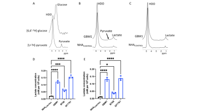 |
18 | Preclinical platform for the identification of deuterium magnetic resonance spectroscopy-based biomarkers of tumor metabolism Video Permission Withheld
Georgios Batsios1, Meryssa Tran1, Celine Taglang1, Anne Marie Gillespie1, Sabrina Ronen1, Joseph Costello2, and Pavithra Viswanath1
1Radiology and Biomedical Imaging, University of California San Francisco, San Francisco, CA, United States, 2Neurological Surgery, University of California San Francisco, San Francisco, CA, United States
Metabolic reprogramming is a fundamental hallmark of cancer, which can be exploited for non-invasive tumor imaging. Deuterium magnetic resonance spectroscopy (2H-MRS) recently emerged as a novel, clinically applicable method of non-invasively monitoring flux from 2H-labeled substrates to metabolic products. However, to date, preclinical studies have been performed in vivo, an endeavor that suffers from low-throughput and potential waste of animal lives, especially in treatment response studies. Here, we demonstrate the ability to quantify metabolism of 2H-MRS probes in live cell suspensions. Our studies will expedite the identification of novel 2H-MRS probes for imaging brain tumors and potentially other cancers.
|
||
2751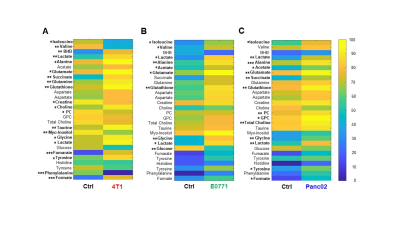 |
19 | Cancer-Induced Metabolic Reprogramming of the Spleen
James D Barnett1, Marie-France Penet1,2, Raj Kumar Sharma1, Balaji Krishnamachary1, Yelena Mironchik1, and Zaver Bhujwalla1,2,3
1Division of Cancer Imaging Research, The Russell H. Morgan Department of Radiology and Radiological Science, The Johns Hopkins University School of Medicine, Baltimore, MD, United States, 2Sidney Kimmel Comprehensive Cancer Center, The Johns Hopkins University School of Medicine, Baltimore, MD, United States, 3Department of Radiation Oncology and Molecular Radiation Sciences, The Johns Hopkins University School of Medicine, Baltimore, MD, United States
Our findings demonstrate that tumors drive metabolic alterations in the spleen. These metabolic changes may contribute to immune suppression and poor prognosis.
|
||
2752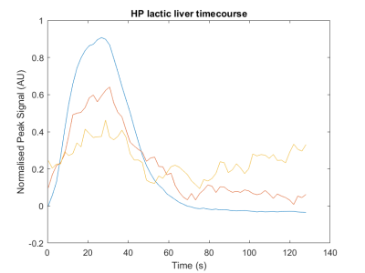 |
20 | Measuring lactate metabolism in vivo in at physiological level using [1-13C]lactate hyperpolarised with an endogenous labile radical precursor Video Not Available
Adam Philip Gaunt1, Jennifer Lewis1, Friederike Hesse1, Irene Marco-Rius1, Kevin Brindle1, and Arnaud Comment1,2
1University of Cambridge, Cambridge, United Kingdom, 2General Electric Healthcare, Chalfont St Giles, United Kingdom A hyperpolarised [1-13C]lactate solution prepared by dynamic nuclear polarization at 7T and 1.4K using photo-irradiated alpha-ketoglutarate as the polarizing agent was injected at low dose in healthy rodents to allow measuring in vivo metabolism in the rodent brain and liver at physiological concentration. As a result of the large 13C polarisation, it was possible to detect metabolic products of [1-13C]lactate in vivo, including [1-13C]pyruvate and 13C-bicarbonate. The observed pyruvate-to-lactate ratios were commensurate with in vitro values previously reported in the literature. The purely endogenous composition of the solution offers the possibility of performing similar measurements in humans. |
||
2753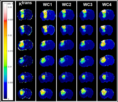 |
21 | Dynamic Contrast Enhanced MR Wavelet-Based Radiomics Analysis for Characterization of Tumor Heterogeneity of Rat Brain Tumors Video Permission Withheld
Hassan Bagher-Ebadian1, Stephen Brown1, Olivia Valadie1, Julian A Rey2, Malisa Sarntinoranont2, James R Ewing3, and Indrin J Chetty1
1Radiation Oncology, Henry Ford Cancer Institute, Detroit, MI, United States, 2Department of Mechanical & Aerospace Engineering, University of Florida, Gainesville, FL, United States, 3Neurology, Henry Ford Cancer Institute, Detroit, MI, United States
Wavelet-analysis of DCE-MR images was performed to explore the association between radiomics information and relaxivity-change (ΔR1) in human U251n tumors grown in rat brains. Sixteen DCE-MRI experiments (8 rats before- and after- radiation) were studied. Wavelet-decomposition analysis was performed using ΔR1 time trace. Frequency-based localized approximations of ΔR1 with four degrees of regularities were estimated and compared to the volume-transfer-constant (Ktrans, calculated from a modified Tofts-model pharmacokinetic analysis). Results confirm strong associations between wavelet-based radiomic information and contrast uptake/flow/leakage in the tumor vasculature. Results suggest that radiomics has potential as a biomarker of tumor physiology.
|
||
2754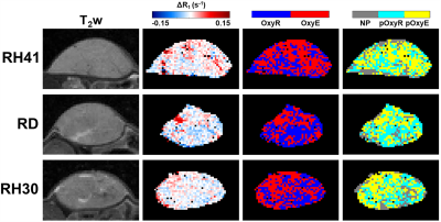 |
22 | Characterising hypoxia in rhabdomyosarcoma xenografts with oxygen-enhanced MRI
Jessica K.R. Boult1, Upasana Roy1, Carolina Bernauer2, Carol Box1, Louise Howell3, Elise Y. Lepicard1, Yann Jamin1, James P.B. O'Connor1, Janet M. Shipley2, and Simon P. Robinson1
1Division of Radiotherapy and Imaging, The Institute of Cancer Research, London, United Kingdom, 2Division of Molecular Pathology, The Institute of Cancer Research, London, United Kingdom, 3Core Research Facilities, The Institute of Cancer Research, London, United Kingdom Hypoxic gene signatures are prevalent in paediatric rhabdomyosarcomas and are important in conferring resistance to standard treatments. Oxygen-enhanced MRI (OE-MRI) and histological assessment of pimonidazole adduct formation show that xenografts derived from three human rhabdomyosarcoma cell lines exhibit high levels of hypoxia with differences in vascular perfusion evident between the models. There is much interest in hypoxia-alleviating strategies to reduce tumour hypoxia for therapeutic gain, and these will be assessed in rhabdomyosarcoma models in vivo using OE-MRI. A reduction in hypoxia in response to atovaquone in spheroids derived from the same rhabdomyosarcoma cell lines in vitro has been confirmed. |
||
2755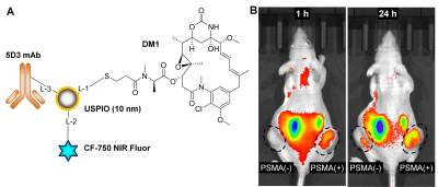 |
23 | Development of USPIO-DM1-5D3-CF750: An image-guided targeted drug delivery system for prostate cancer therapy
Sudath Hapuarachchige1,2, Ge Si1,3, Catherine Foss1, Cyril Barinka4, and Dmitri Artemov1,2
1Department of Radiology and Radiological Science, The Johns Hopkins School of Medicine, Baltimore, MD, United States, 2Department of Oncology, The Sidney Kimmel Comprehensive Cancer Center, Baltimore, MD, United States, 3Department of Chemical and Biomolecular Engineering, The Johns Hopkins University, Baltimore, MD, United States, 4Laboratory of Structural Biology, Institute of Biotechnology of the Czech Academy of Sciences, Vestec, Czech Republic
Prostate-specific membrane antigen (PSMA) is overexpressed in prostate cancers (PC) compared to normal tissues. Hence, PSMA can be used as a diagnostic biomarker and biological target to deliver drugs in PC therapy. In this study, we have conjugated ultra-small superparamagnetic iron oxide nanoparticles with mertansine (DM1), a chemotherapeutic drug, novel anti-PSMA 5D3 antibody, and NIR CF-750 fluorophore. This multimodality image-guided drug delivery system was evaluated in PC mouse models using in vivo fluorescent imaging and MRI at 9.4T. Promising results encourage further development of targeted image-guided drug delivery platforms for PC therapy.
|
||
2756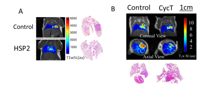 |
24 | MRI Monitoring of Therapeutic Efficacy of Novel Heme targeting drugs in Non-small cell lung cancer (NSCLC) Orthotopic Tumors in Mice
Li Liu1, Donghan Yang1, Janaka Wansapura1, Guiyang Hao1, Ralph P. Mason1, and Li Zhang2
1Radiology, The University of Texas Southwestern Medical Center, Dallas, TX, United States, 2Biological Sciences, University of Texas at Dallas, Richardson, TX, United States
The goal was to evaluate the activity of two novel heme targeting drugs in Non-Small Cell Lung Cancer models using multi-modality imaging. We found that the novel heme targeting drugs CycT (cyclopamine tartrate) and HSP2 (heme-sequestering peptide 2) were effective in suppressing NSCLC lung tumor growth as assessed by multimodality imaging and confirmed by pathology/histology. MRI detected the H1395 and H1299 orthotopic xenografts and was correlated with PET studies and histology.
|
||
 Back to Meeting Home
 Back to the Program-at-a-Glance
The International Society for Magnetic Resonance in Medicine is accredited by the Accreditation Council for Continuing Medical Education to provide continuing medical education for physicians.
 Back to Meeting Home
 Back to the Program-at-a-Glance
 View the Presentation