Digital Poster
Novel Image Reconstruction Techniques II
Joint Annual Meeting ISMRM-ESMRMB & ISMRT 31st Annual Meeting • 07-12 May 2022 • London, UK

Digital Poster
Novel Image Reconstruction Techniques II
| Computer # | ||||
|---|---|---|---|---|
2433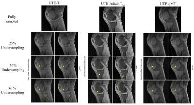 |
62 | Accelerated Quantitative 3D UTE-Cones Imaging using Compressed Sensing
Jiyo S Athertya1, Ya-Jun Ma1, Amir Masoud Afsahi1, Alicia Ji1, Eric Y Chang1,2, Jiang Du1, and Hyungseok Jang1
1Radiology, University of California San Diego, San Diego, CA, United States, 2Radiology Service, VA San Diego Healthcare System, San Diego, CA, United States
Quantitative ultrashort echo time (qUTE) imaging suffers from long acquisition time due to multiple acquisitions required for parameter estimation. In this study, feasibility of accelerated qUTE Cones imaging with compressed sensing (CS) reconstruction is investigated for fast variable flip angle UTE-T1 mapping, adiabatic UTE-T1ρ mapping, and UTE quantitative magnetization transfer (MT) modelling of macromolecular fraction (MMF). We explored these biomarkers for qUTE-Cones imaging of in vivo human knee joints at various undersampling rates. The performance of CS-reconstruction and parameter mapping was evaluated in tendons, ligaments, menisci, and cartilage.
|
||
2434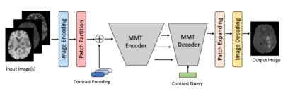 |
63 | Multi-contrast Multi-scale vision Transformers (MMT) for MRI Contrast Synthesis
Jiang Liu1, Srivathsa Pasumarthi Venkata2, and Keshav Datta2
1Department of Electrical and Computer Engineering, Johns Hopkins University, Baltimore, MD, United States, 2R&D, Subtle Medical Inc, Menlo Park, CA, United States
Complementary information from multi-modal MRI is widely used in clinical practice for disease diagnosis. Due to scan time limitations, image corruptions, and different acquisition protocols, one or more contrasts may be missing or unusable. Recently developed CNN models for contrast synthesis are unable to capture the intricate dependencies between input contrasts and are not dynamic to the varying number of inputs. This work proposes a novel Multi-contrast and Multi-scale vision Transformer (MMT) that can take any number and combination of input sequences and synthesize the missing contrasts.
|
||
2435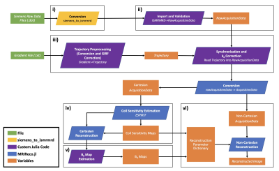 |
64 | Open-source model-based reconstruction in Julia: A pipeline for spiral diffusion imaging
Alexander Jaffray1,2, Zhe (Tim) Wu1, Kamil Uludag3,4, and Lars Kasper1
1Techna Institute, University Health Network, Toronto, ON, Canada, 2UBC MRI Research Center, University of British Columbia, Vancouver, BC, Canada, 3Techna Institute & Koerner Scientist in MR Imaging, University Health Network, Toronto, ON, Canada, 4Biomedical Engineering, Center for Neuroscience Imaging Research, Institute for Basic Science, Sungkyunkwan University, Suwon, Korea, Republic of
We investigated the use of physically-motivated corrections for non-Cartesian imaging within a fast, open-source MR reconstruction framework written in the Julia programming language. Building on existing Julia libraries for MR image reconstruction, we developed a comprehensive, open-source pipeline for non-Cartesian image reconstruction including B0 inhomogeneity correction, gradient system characterization and eddy current correction. We demonstrated that spiral images can be reconstructed using this pipeline with high geometric accuracy in a fast, accessible and efficient manner on modest hardware.
|
||
2436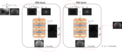 |
65 | Recurrent Variational Inference for fast and robust reconstruction of accelerated FLAIR MRI in Multiple Sclerosis
D. Karkalousos1, L. C. Liebrand1, S. Noteboom2, H. E. Hulst2,3, F. M. Vos4, and M. W. A. Caan1
1Department of Biomedical Engineering & Physics, Amsterdam UMC, University of Amsterdam, Amsterdam, Netherlands, 2Department of Anatomy & Neurosciences, MS Center Amsterdam, Amsterdam Neuroscience, Amsterdam UMC, Vrije Universiteit Amsterdam, Amsterdam, Netherlands, 3Department of Medical, Health and Neuropsychology, Leiden University, Leiden, Netherlands, 4Delft University of Technology, Delft, Netherlands Robustness when applying Deep Learning methods to clinical data is crucial for accurate high-resolution reconstructions while having fast inference times. We propose the Cascades of Independently Recurrent Variational Inference Machine (CIRVIM), targeting deep unrolled optimization and enforcing data consistency for further robustness. We quantify contrast resolution of seven and half times prospectively undersampled FLAIR MRI without fully-sampled center containing Multiple Sclerosis lesions. The proposed scheme reduces inference times by a factor of 6 compared to Compressed Sensing. Lesion contrast resolution improves by approximately 13% while preserving spatial detail with enhanced sharpness compared to more blurred results of other presented methods. |
||
2437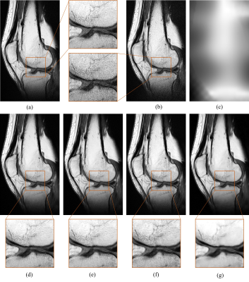 |
66 | Training a tunable, spatially-adaptive denoiser without clean targets
Laura Pfaff1,2, Julian Hossbach1,2, Elisabeth Preuhs1, Tobias Wuerfl2, Silvia Arroyo Camejo2, Dominik Nickel2, and Andreas Maier1
1Pattern Recognition Lab, Friedrich-Alexander-University Erlangen-Nuremberg, Erlangen, Germany, 2MR Application Predevelopment, Siemens Healthcare GmbH, Erlangen, Germany
Accelerating MRI is intrinsically limited by the thermal noise from the imaged object. In this work we aim to optimize MR image denoising using an unsupervised deep learning-based method. Stein's unbiased risk estimator and spatially resolved noise maps indicating the standard deviation of the noise for every pixel were incorporated into the training process. It was shown that this approach can achieve results that are equal or superior to those of state-of-the-art supervised and unsupervised methods. Furthermore, we show how to control the tradeoff between denoising and image sharpness by using a model conditioned on the noise map.
|
||
2438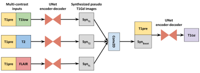 |
67 | Explainable multi-contrast deep learning model with anomaly-aware attention for reduced gadolinium dose in CE brain MRI - a feasibility study
Srivathsa Pasumarthi Venkata1, Ben Andrew Duffy1, Enhao Gong2, Greg Zaharchuk3, and Keshav Datta1
1R&D, Subtle Medical Inc, Menlo Park, CA, United States, 2R&D, Subtle Medical Inc., Menlo Park, CA, United States, 3Department of Radiology, Stanford University, Stanford, CA, United States
Complementary information from multi-contrast MRI data is used in deep learning algorithms for reducing contrast dosage in brain MRI. Though existing models produce clinically equivalent post-contrast images, they lack explainability in terms of mapping the source of contrast information from input to output. In this work we explore the feasibility of an explainable deep learning model for gadolinium dose reduction in contrast-enhanced brain MRI.
|
||
2439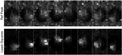 |
68 | A Least-squares Method for Improved Sensitivity Map Estimation when Imaging X-Nuclei Video Not Available
Nicholas Dwork1, Shuyu Tang1, Peder E. Z. Larson1, and Jeremy W. Gordon1
1Radiology and Biomedical Imaging, University of California San Francisco, San Francisco, CA, United States
We present a least-squares method for estimating sensitivity when imaging x-nuclei. We show results with hyperpolarized pyruvate in both the heart and the brain.
|
||
2440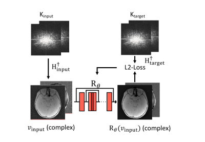 |
69 | Rapid high-resolution cranial bone MRI using deep-learning prior image reconstruction
Parna Eshraghi Boroojeni1, Paul Commean1, Cihat Eldeniz1, Weijie Gan1, Gary Skolnick1, Kamlesh Patel1, Ulugbek Kamilov1, and Hongyu An1
1Washington University in Saint Louis, Saint Louis, MO, United States
A high-resolution (HR) MRI capable of resolving the detail of bony structures at sub-millimeter resolution is desired. A short MR acquisition results in under-sampled k-space data below the Nyquist rate, leading to artifacts and high noise. We developed an HR reconstruction method regularized by a complex deep-learning prior (RECD). We achieved high-resolution MR (0.6x0.6x0.8mm3) with a one-minute acquisition time. Using images reconstructed from a 5-minute MR scan as the gold standard, we compared the peak signal to noise ratio (PSNR) and similarity index (SSIM) for 1-min RECD and 1-min compressed sensing (CS) reconstructed images. RECD outperformed CS.
|
||
2441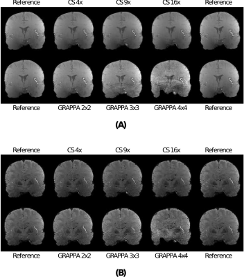 |
70 | Comparison between GRAPPA and automatic compressed sensing-based reconstruction up to 16-fold acceleration
Gabriel Varela-Mattatall1,2, Roy A.M. Haast 1,3, Omer Oran4, Ali R. Khan 1,2, and Ravi S. Menon1,2
1Centre for Functional and Metabolic Mapping (CFMM) | Robarts Research Institute | Western University, London, ON, Canada, 2Department of Medical Biophysics | Schulich School of Medicine and Dentistry | Western University, London, ON, Canada, 3Aix-Marseille Universite | CNRS | CRMBM, Marseille, France, 4Siemens Healthcare Limited, Oakville, ON, Canada
With the recent development of an automatic compressed sensing (CS)-based reconstruction method, we investigated its allowance to recover T2* maps from prospectively undersampled multi-echo, gradient echo-based acquisitions at 7T. We compared our CS method with the more conventional GRAPPA-based reconstruction using up to 16-fold acceleration. In contrast to GRAPPA, our CS-based method allows recovery of T2* maps up to 16-fold acceleration, which demonstrates its promise for ultra-fast acquisitions. However, current results also show some caveats that need to be addressed as future work.
|
||
2442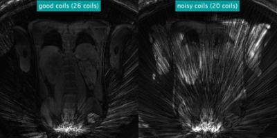 |
71 | Coil Selective Golden Angle DCE-MR Image Reconstruction using Mutual Dependence
Aziz Kocanaogullari1, Cemre Ariyurek1, Onur Afacan1, and Sila Kurugol1
1Radiology, Boston Children's Hospital, Boston, MA, United States
MRI literature shows that it is possible to increase image reconstruction quality by removing coils that cause majority of the streaking artifacts. We introduce to measure the coil quality based on mutual information between a reference and each coil image. Specifically we apply this technique to DCE-MRI reconstruction from under-sampled radial stack of stars trajectory for kidney imaging and calculate mutual dependence between a dynamic image from each coil and a reference image to assess coil contribution to reconstruction. Experiments show mutual dependence based coil selection reduces artifacts and increases reconstructed image SNR by $$$16.84\%$$$ using $$$1/3$$$ of the coils.
|
||
2443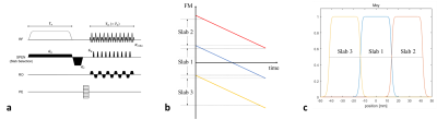 |
72 | Reconstruction for Simultaneous Multi-Slab Spatiotemporal Encoding (SMS SPEN) Using Split Slice-GRAPPA combined with CAIPIRINHA
Jaeyong Yu1,2, Sugil Kim3, and Jang-Yeon Park1,2
1Department of Biomedical Engineering, Sungkyunkwan University, Suwon, Korea, Republic of, 2Department of Intelligent Precision Healthcare Convergence, Sungkyunkwan University, Suwon, Korea, Republic of, 3Siemens Healthineers Ltd., Seoul, Korea, Republic of
To reduce acquisition time, acceleration techniques such as simultaneous multi-slice and parallel imaging techniques have been applied to various sequences. In this work, we proposed a reconstruction method for simultaneous multi-slab spatiotemporal-encoding (SMS SPEN) using split slice-GRAPPA combined with CAIPIRINHA. The proposed method was applied to the recently developed ultrafast 3D SPEN imaging sequence called ERASE (Equal-TE Rapid Acquisition with Sequential Excitation) and a case of multiband factor of 3 was demonstrated with human brain imaging at 3T.
|
||
 Back to Meeting Home
 Back to the Program-at-a-Glance
The International Society for Magnetic Resonance in Medicine is accredited by the Accreditation Council for Continuing Medical Education to provide continuing medical education for physicians.
 Back to Meeting Home
 Back to the Program-at-a-Glance
 View the Presentation