Digital Poster
Tumor Characterization & Staging
Joint Annual Meeting ISMRM-ESMRMB & ISMRT 31st Annual Meeting • 07-12 May 2022 • London, UK

Digital Poster
Tumor Characterization & Staging
| Computer # | ||||
|---|---|---|---|---|
2829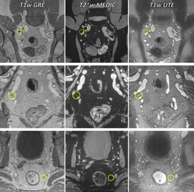 |
78 | Feasibility of T1-weighted USPIO-enhanced MR imaging of pelvic lymph nodes using stack-of-spirals UTE
Marnix C. Maas1, Patrik Zámecnik1, Moritz J. Schneider2,3, Thomas Gaass2,4, Thomas Benkert5, Julien Dinkel2,4, and Tom W.J. Scheenen1
1Medical Imaging, Radboudumc, Nijmegen, Netherlands, 2Comprehensive Pneumology Center, German Center for Lung Research (DZL), Munich, Germany, 3Antaros Medical AB, Mölndal, Sweden, 4Department of Radiology, University Hospital, LMU Munich, Munich, Germany, 5MR Application Predevelopment, Siemens Healthcare GmbH, Erlangen, Germany
Detection of lymph node (LN) metastases can be improved using Ferumoxtran-10, an ultrasmall superparamagnetic particles of iron oxide (USPIO) contrast agent . Commonly, T2* contrast is used to distinguish between normal LNs (high USPIO uptake, low signal) and metastases (low uptake, high signal). T1-weighted imaging may offer complementary information, but is challenging because of the strong T2* effects of the contrast agent. This work investigates whether ultrashort echotime (UTE) imaging with a stack-of-spirals sequence can mitigate this issue. High-resolution USPIO-enhanced T1-weighted UTE of the pelvis was achieved, and T1-mediated signal hyperintensities were indeed observed in LNs in patients with prostate cancer.
|
||
2830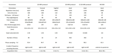 |
79 | Optimization of Compressed Sense accelerated 3D pelvis view for diagnosis of rectal cancer
Xiaoling Gong1, Daguang Wen2, Yu Shen3, Mingtian Wei3, Xiaoxiao Zhang4, Xiaoyong Zhang4, Haoyu Huang4, Ziqiang Wang3, and Bing Wu2
1Departments of Radiology, West China Hospital, Sichuan University, Cheng du, China, 2Departments of Radiology, West China Hospital, Sichuan University, Chengdu, China, 3Department of Clinical, West China Hospital, Sichuan University, Chengdu, China, 4Philips Healthcare, China, Chengdu, China Although 3D T2W imaging can make up for deficiencies, such as thick layer thickness, low spatial resolution and no retrospective post-processing, of 2D acquisitions, the long imaging time and uncertain diagnostic benefits have limited its clinical application. We compared the quality of 2D, 3D and CS sense accelerated 3D T2W imaging in patients with rectal cancer and found that 3D T2W imaging resembled CS sense accelerated 3D T2W imaging, both superior to 2D acquisitions, while the latter scanning faster than the former. It’s conclude that CS sense accelerated 3D T2W imaging can substitute the 2D acquisitions. |
||
2831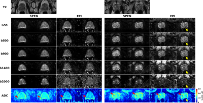 |
80 | Overcoming image distortions for DWI of prostate cancer using Spatiotemporal Encoding (SPEN)
Martins Otikovs1, Debbie Anaby2, Orith Portnoy2, Noam Nissan2, Barak Rosenzweig2, and Lucio Frydman3,4
1Weizmann Institute of Science, Rehovot, Israel, 2Sheba Medical Center, Ramat Gan, Israel, 3Department of Chemical and Biological Physics, Weizmann Institute of Science, Rehovot, Israel, 4Azrieli National Center for Brain Imaging, Weizmann Institute of Science, Rehovot, Israel
Diffusion weighted imaging (DWI) plays an important role in prostate cancer early detection and characterization. These analyses commonly rely on single shot EPI acquisitions, which can be associated with image distortions induced by field inhomogeneities induced by tissue / air interfaces. Herein we compare the faithfulness of EPI-derived DWI data, against that arising from Spatiotemporal Encoding (SPEN). In general, we find that SPEN delivers more faithful DW representations of the prostate anatomy within comparable scan times, without compromising SNR. SPEN can thus improve the contrast for prostate lesion detection, both in DW images and in calculated ADC maps.
|
||
2832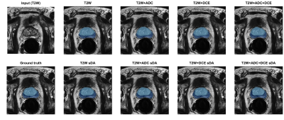 |
81 | Deep learning for prostate zonal segmentation robust to multicenter data
Simona Turco1, Hubert Blach1, Catarina Dinis Fernandes1, Jelle Barentsz2, Stijn Heijmink 3, Hessel Wijkstra1, and Massimo Mischi1
1Electrical Engineering, Eindhoven University of Technology, Eindhoven, Netherlands, 2Radiology, Radboud university medical center, Nijmegen, Netherlands, 3Radiology, Netherlands Cancer Institute, Amsterdam, Netherlands
Prostate zonal segmentation is an important step for automated PCa diagnosis, MRI-guidedradiotherapy and focal treatment planning. Here we proposed a multi-channel U-Net for automatic prostate zonal segmentation, able to include multiple MRI sequences. Using a small, multicenter, multiparametric MRI dataset, we investigated its robustness towards the acquisition protocol and whether additional imaging sequences improve segmentation performance. Our results show that T2-weighted imaging alone is sufficient for successful prostate zonal segmentation. Despite using a small multicenter dataset, the models were robust towards the acquisition protocol and the performance was comparable to that obtained with larger datasets from a single institute.
|
||
2833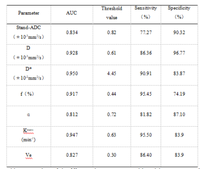 |
82 | Intravoxel Incoherent Motion Combined With DCE-MRI of Endometrial cancer:Correlations Between Multimodal Parameters and Her-2 Expression Video Not Available
Changjun Ma1, Shifeng Tian2, Lihua Chen2, Nan Wang2, Qingwei Song2, and Ailian Liu2
1Department of Radiology, the First Affiliated Hospital of Dalian Medical University, Dalian,China, China, 2Department of Radiology, the First Affiliated Hospital of Dalian Medical University, Dalian, China
At present, there are few reports on the application of imaging methods to predict the Her-2 gene expression of EC. This study will use the two functional sequences of IVIM and DCE-MRI to explore its preoperative prediction of the Her-2 gene expression of EC. Value, at the same time, discuss the correlation between the parameters of the two functional sequences.
|
||
2834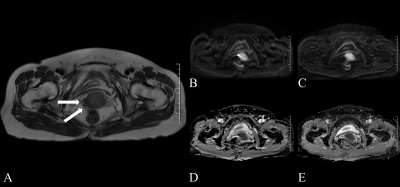 |
83 | Investigation of multiplexed sensitivity encoding diffusion-weighted imaging (DWI) in cervical carcinoma:comparison with conventional DWI Video Not Available
Tiebao Meng1, Weijing Zhang2, Haoqiang He2, Huiming Liu2, Guixiao Xu2, Fengting Zhu2, Long Qian3, and Chuanmiao Xie2
1Department of Radiology, Sun Yat-Sen University Cancer Center, Guangzhou, China, 2Sun Yat-Sen University Cancer Center, Guangzhou, China, 3GE Healthcare, Beijing, China
MUSE provides significantly better image quality in CC compared with C-DWI, with no significant difference in ADC.
|
||
2835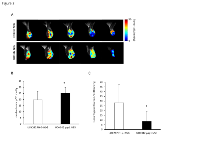 |
84 | In vivo characterization of preclinical hereditary RCC models by Electron Paramagnetic Resonance and Magnetic Resonance Imaging
Yuki Shibata1, Daniel R. Crooks1, Shun Kishimoto2, Murali C. Krishna2, and W. Marston Linehan1
1Urologic Oncology Branch, National Institute of Health, Bethesda, MD, United States, 2Radiation Biology Branch, National Institute of Health, Bethesda, MD, United States Human renal cell carcinomas (RCC) have a variety of pathologies and are known to have alterations in cellular metabolism, although many aspects remain unknown. Fumarate hydratase (FH)-deficient tumors exhibit a shift to aerobic glycolytic system due to several factors including loss and mutation of mitochondrial DNA. We investigated the partial pressure of oxygen, vascular permeability, and blood perfusion in FH-deficient UOK262 xenografts and a type 1 papillary RCC xenograft using EPRI and DCE-MRI. Despite low oxygen consumption rates in vitro, UOK262 xenografts showed a modest median pO2 and increased hypoxic fraction as compared to a type 1 papillary RCC xenografts. |
||
2836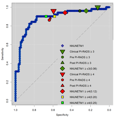 |
85 | A Follow-up Study On Prospective Para-Clinical Use by Residents of a Re-calibrating Automated Deep Learning System for Prostate Cancer Detection Video Permission Withheld
Kevin Sun Zhang1, Adrian Schrader1, Nils Netzer1, Magdalena Görtz2, Viktoria Schütz2, Constantin Schwab3, Markus Hohenfellner2, Heinz-Peter Schlemmer1, and David Bonekamp1
1Department of Radiology, German Cancer Research Center, Heidelberg, Germany, 2Department of Urology, University Hospital Heidelberg, Heidelberg, Germany, 3Institute of Pathology, University of Heidelberg Medical Center, Heidelberg, Germany
Previously validated fully-automatic detection of prostate cancer by CNNs requires further prospective validation. Para-clinical case-by-case prospective prostate MRI assessment by residents was performed both before and after review of CNN probability maps superimposed on T2w images. A previously and retrospectively validated self-parametrizing nnUNet-architecture CNN trained on more than 1000 voxel-wise annotated prostate MRIs achieved ROC AUC of 0.89. Residents did not substantially change their assessment both at PI-RADS>=3 and >=4 decisions, however achieved excellent working points, indicating success of high reading capability conveyed at an expert center.
|
||
2837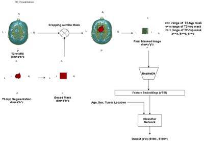 |
86 | Identification of S100 Immunopositivity on T2-weighted MRI Using Deep Learning
Abdullah Baş1, Kübra Tan2, Ayça Ersen Danyeli3,4, M.Necmettin Pamir5,6, Alp Dincer4,7, Koray Ozduman5,6, Ozge Can8, and Esin Ozturk-Isik1
1Institute of Biomedical Engineering, Bogazici University, İstanbul, Turkey, 2Health Institutes of Turkey, Istanbul, Turkey, 3Department of Medical Pathology, Acibadem Mehmet Ali Aydinlar University, İstanbul, Turkey, 4Center for Neuroradiological Applications and Reseach, Acibadem Mehmet Ali Aydinlar University, İstanbul, Turkey, 5Center for Neuroradiological Applications and Reseach, Acibadem Mehmet Ali Aydinlar University, Istanbul, Turkey, 6Department of Neurosurgery, Acibadem Mehmet Ali Aydinlar University, İstanbul, Turkey, 7Department of Radiology, Acibadem Mehmet Ali Aydinlar University, İstanbul, Turkey, 8Center for Neuroradiological Applications and Reseach Acibadem Mehmet Ali Aydinlar University, Istanbul, Turkey
S100 protein expression is a relevant indicator of prognosis in meningiomas and it is more common in benign meningiomas. To our knowledge, a clinically feasible non-invasive method that preoperatively identifies S100 protein expression is not available. In this study, we proposed registration-free deep learning models to predict S100 expression non-invasively using T2-w MRI. The proposed hybrid deep learning model could predict S100 protein expression in meningiomas using T2-w MRI, with 91% accuracy on the validation set, and 83% accuracy on the test set.
|
||
2838 |
87 | Development of a scan-specific quality control acquisition for clinical whole-body MRI (WB-MRI) protocols
Sam Keaveney1, Georgina Hopkinson1, Erica Scurr1, Maria-Alexandra Olaru2, David Collins1, Christina Messiou1, Dow-Mu Koh1, and Jessica Winfield1
1Royal Marsden Hospital & Institute of Cancer Research, Sutton, United Kingdom, 2Siemens Healthcare Ltd., Frimley, Camberley, United Kingdom A short quality control acquisition was developed to detect hardware issues and receiver coil positioning errors in whole-body MRI (WB-MRI). Individual coil element images were reconstructed to investigate potential broken elements and coil positioning was assessed by comparison with images acquired using the integral body coil. This approach was implemented in a group of healthy volunteers and patients with myeloma and successfully identified a previously undetected broken coil element. The expected signal difference between coil array and integral body coil images was defined and deviation from this expected profile was used to distinguish images acquired with deliberately imperfect coil set-ups. |
||
2839 |
88 | Influence of fat suppression and image reconstruction methods on ADC of uninvolved bone marrow in patients with myeloma.
Georgina Hopkinson1, Christina Messiou1,2, Erica Scurr1, Martin F Kaiser3,4, David J Collins1,2, and Jessica M Winfield1,2
1MRI Unit, The Royal Marsden NHS Foundation Trust, Sutton, United Kingdom, 2Division of Radiotherapy and Imaging, The Institute of Cancer Research, Sutton, United Kingdom, 3Department of Haematology, The Royal Marsden NHS Foundation Trust, Sutton, United Kingdom, 4Division of Genetics and Epidemiology, The Institute of Cancer Research, Sutton, United Kingdom
The influence of fat suppression methods and image reconstruction parameters on the apparent diffusion coefficient (ADC) in uninvolved bone marrow was assessed in 18 patients with myeloma. ADC estimates from diffusion-weighted MRI acquired using spectral adiabatic inversion recovery (SPAIR) fat suppression were significantly lower than DW-MRI with short-tau inversion recovery (STIR). ADC estimates were significantly lower in data reconstructed using sum-of-squares coil-combination mode compared with adaptive combine, but there was no difference between additional reconstruction filters (raw, elliptical). These results show that differences between imaging protocols should be considered when comparing ADC estimates with established ranges and in multi-centre studies.
|
||
 Back to Meeting Home
 Back to the Program-at-a-Glance
The International Society for Magnetic Resonance in Medicine is accredited by the Accreditation Council for Continuing Medical Education to provide continuing medical education for physicians.
 Back to Meeting Home
 Back to the Program-at-a-Glance
 View the Presentation