Digital Poster
Preclinical Cancer I
Joint Annual Meeting ISMRM-ESMRMB & ISMRT 31st Annual Meeting • 07-12 May 2022 • London, UK

| Computer # | ||||
|---|---|---|---|---|
2663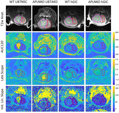 |
27 | Contrast Enhanced Imaging of Apelin Effects on Vascularization of Orthotopic Glioblastoma in Mice
Geoffrey J. Topping1, Enio Barçi2, Jiying Cheng2, Sandra Sühnel1, Rainer Glass2, Roland E. Kälin2, and Franz Schilling1
1Nuclear Medicine, School of Medicine, Klinikum rechts der Isar, Technical University of Munich, Munich, Germany, 2Neurosurgical Research, University Hospital, LMU Munich, Munich, Germany
Mice with orthotopically implanted glioblastoma were imaged during contrast injection, with the goals of establishing semi-quantitative DCE in this model and to investigate the impact of apelin-controlled tumour angiogenesis. Wild-type mice with control U87 tumours showed higher initial contrast accumulation but also faster washout compared to apelin knockout mice implanted with apelin knockdown tumour cells, consistent with apelin contributing to tumour vascularization. Control and apelin knockout mice had low contrast accumulation with genetically engineered human glioma-initiating cells.
|
||
2664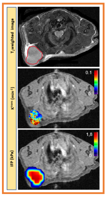 |
28 | Development of noninvasive DCE based method to estimate tumor interstitial Fluid Pressure in Mouse Models for Pancreatic Ductal Adenocarcinoma Video Permission Withheld
Ramesh Paudyal1, Eve LoCastro1, James Russell1, Ivan Wolansky1, Carl C. Lekaye1, Joseph O. Deasy1, John L. Humm1, and Amita Shukla-Dave1,2
1Medical Physics, Memorial Sloan Kettering Cancer Center, New York, NY, United States, 2Radiology, Memorial Sloan Kettering Cancer Center, New York, NY, United States
Pancreatic cancer accounts for about 3% of all cancer-related deaths in the US. An elevation of interstitial fluid pressure (IFP) is a major barrier to drug delivery in solid tumors. Noninvasive estimation of IFP as a biomarker in typically inaccessible tumors is a significant step toward assessing tumor response to therapy. We applied well-recognized fluid flow in porous media to mouse models of pancreatic ductal adenocarcinoma DCE-MRI data using extended Tofts' model-derived permeability maps with tumor-appropriate tumor geometry. Initial results suggest that after validation, IFP can be imaging biomarkers of early response to therapy.
|
||
2665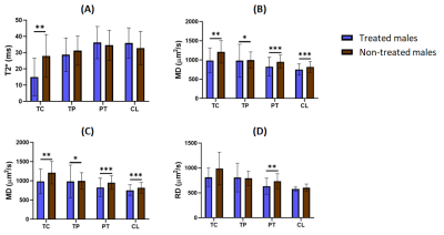 |
29 | Multiparametric MRI assessment of the sex influence in the development and response to anti-inflammatory treatment of a glioblastoma rat model
Inês Veríssimo Cabete1, Nuria Arias-Ramos1, Maria Guillén Gómez1, Margarida Catalão Almiro e Castro2, and Pilar López-Larrubia1
1Instituto de Investigaciones Biomédicas "Alberto Sols", CSIC-UAM, Madrid, Spain, 2Department of Life Sciences and Coimbra Chemistry Centre, Faculty of Science and Technology, University of Coimbra, Coimbra, Portugal
The assessment of sexual differences in glioblastoma growth and therapy response has received little attention. We assessed the influence of sex in the MRI features of an orthotropic GBM rat model submitted to anti-inflammatory therapy. Multiparametric MRI studies were acquired at an early and advanced tumor stage of development. GBMs in untreated males presented a more aggressive pattern – worsened as the tumor progressed – than females. While treated males revealed a positive response to meloxicam treatment, females did not. Our results highlighted that sex is a relevant factor to be considered in GBM outcome and anti-inflammatory therapy success.
|
||
2666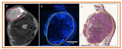 |
30 | Tumor microvasculature and microenvironment characterization in mouse models of pancreatic ductal adenocarcinoma using multiparametric MRI
Ramesh Paudyal1, James Russell1, Ivan Wolansky1, Eve LoCastro1, Carl C. Lekaye1, Joseph O. Deasy1, John L. Humm1, and Amita Shukla-Dave1,2
1Medical Physics, Memorial Sloan Kettering Cancer Center, New York, NY, United States, 2Radiology, Memorial Sloan Kettering Cancer Center, New York, NY, United States
Pancreatic ductal adenocarcinoma (PDAC) is expected to be the second cause of cancer-related deaths worldwide. Quantitative multiparametric magnetic resonance imaging (mpMRI) provides the complementary physiological properties of tumors tissue. The aim of this study was to characterize microvasculature and microenvironment in mouse models of PDAC using mpMRI. The functional status of mpMRI quantitative imaging metrics were validated with in vivo histology markers of tumor perfusion (Hoechst 33342) and tissue morphology (Hematoxylin and eosin staining).
|
||
2667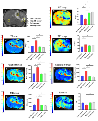 |
31 | Anatomical, functional and dynamic MRI features of the G144 GBM model developed in NOD-SCID mice
Nuria Arias-Ramos1, Ana M. Cardoso2, Amália S. Jurado2,3, M. Margarida C.A. Castro4, Maria C. Pedroso de Lima2, and Pilar López-Larrubia1
1Instituto de Investigaciones Biomédicas Alberto Sols (CSIC-UAM), Madrid, Spain, 2CNC - Center for Neuroscience and Cell Biology, CIBB - Centre for Innovative Biomedicine and Biotechnology, University of Coimbra, IIIUC - Institute for Interdisciplinary Research, Coimbra, Portugal, 3Department of Life Sciences, University of Coimbra, Coimbra, Portugal, 4Centro de Química de Coimbra, Faculdade de Ciências e Tecnologia, University of Coimbra, Coimbra, Portugal Preclinical models are needed to study glioblastoma multiforme, the most lethal malignant brain tumor. Human G144-glioblastoma stem cells have been used in in vitro studies but there are few studies in vivo, especially using MRI approaches. We aimed to evaluate in vivo, for the first time to our knowledge, a G144 glioblastoma model by multiparametric-MRI. NOD-SCID mice were orthotopically injected with G144 cells, tumor was characterized by multiparametric-MRI and dynamic contrast enhancement studies. G144 tumors presented heterogeneous imaging pattern compared to other GBM models with higher contrast uptake areas and edema related features. |
||
2668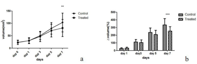 |
32 | Intravoxel incoherent motion diffusion-weighted imaging for monitoring the anti-angiogenesis efficacy in a C6 glioma rat model Video Not Available
Weishu Hou1, Xiaohu Li1, Hongli Pan1, Man Xu1, Sixing Bi1, Yujun Shen2, Yong Zhang3, Yinfeng Qian1, and Yongqiang Yu1
1the First Affiliated Hospital of Anhui Medical University, Hefei, China, 2Anhui Medical University, Hefei, China, 3GE Healthcare, Shanghai, China The purpose of this study was to investigate the feasibility of intravoxel incoherent motion (IVIM) diffusion-weighted imaging (DWI) in evaluating early effects of antiangiogenic therapy in a C6 glioma rat model. Twenty-six glioma orthotopic rat models were successfully established 14-16 days after C6 cell inoculation and randomly divided into two groups. The rats in the treated group were administered with Bev while the rats in the control group were administered with vehicle. IVIM-DWI showed significantly decreased D* values and increased ADC and D values corresponding to histological staining in Bev treated tumors. |
||
2669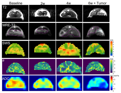 |
33 | In vivo longitudinal characterization of hepatocellular carcinoma based on viscoelasticity and water diffusivity in an orthotopic mouse model
Karolina Garczyńska1,2, Akvile Häckel1, Eyk Schellenberger1, Anja A. Kühl3, Jürgen Braun4, Lynn Jeanette Savic1, Ingolf Sack1, and Jing Guo1
1Department of Radiology, Charité Universitätsmedizin Berlin, Berlin, Germany, 2Department of Veterinary Pathology, Free University of Berlin, Berlin, Germany, 3iPATH.Berlin Core Unit of Charité - Universitätsmedizin Berlin, Charité Universitätsmedizin Berlin, Berlin, Germany, 4Institute of Medical Informatics, Charité Universitätsmedizin Berlin, Berlin, Germany The biophysical properties of hepatocellular carcinoma (HCC) and surrounding liver tissue were investigated longitudinally in a syngeneic, orthotopic mouse model using noninvasive quantitative imaging. In vivo MR elastography (MRE) and diffusion weighted imaging (DWI) were conducted prior to cancer cell implantation and three times during tumor progression. Our preliminary results suggest the involvement of the surrounding liver in terms of changes in viscoelasticity and restricted water diffusion over 6 weeks post implantation, while the HCC appeared to be stiffer and less viscous than the liver at 6 weeks. |
||
2670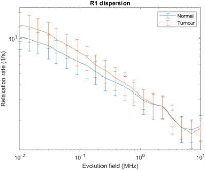 |
34 | Application of Fast Field-Cycling (FFC) NMR relaxometry in Colorectal Cancer
Amnah Alamri1, Lionel Broche1, and Leslie Samuel2
1Aberdeen Biomedical Imaging Centre, Aberdeen, Scotland, 2Aberdeen Royal Infirmary, Aberdeen, Scotland
This pilot study was performed to compare healthy and colorectal cancer samples by investigating R1 NMRD profiles acquired with Fast Field-Cycling (FFC) NMR relaxometry. We collected samples from 18 patients and found a significant difference (P=0.001) in the relaxation rates between normal and colorectal cancer tumours below 100 kHz (2.3uT), while this difference became smaller with increasing the magnetic field strengths and disappeared above 1 MHz (23 uT). This dispersion profile may lead to great potential for diagnosis, staging and monitoring treatment response.
|
||
2671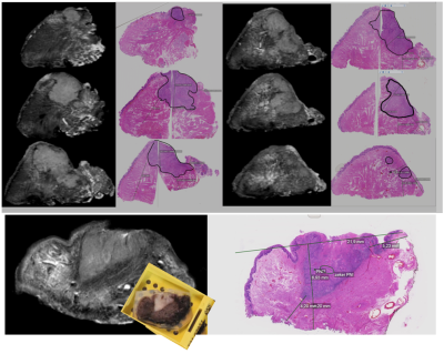 |
35 | Ex vivo 7T MRI of resection specimen of oral cancer to improve margin control.
Klijs J. de Koning1, Rob Noorlag1, Annette van der Toorn2, Gerben E. Breimer3, Jan Willem Dankbaar4, Remco de Bree1, and Marielle E.P. Philippens5
1Head and Neck Surgical Oncology, University Medical Center Utrecht, Utrecht, Netherlands, 2Biomedical MR Imaging & Spectroscopy Group, University Medical Center Utrecht, Utrecht, Netherlands, 3Pathology, University Medical Center Utrecht, Utrecht, Netherlands, 4Radiology, University Medical Center Utrecht, Utrecht, Netherlands, 5Radiotherapy, University Medical Center Utrecht, Utrecht, Netherlands
We validated the measured resection margins of the surgical specimens of tongue carcinoma on 7T ex vivo MRI with histopathology using T2 weighted MRI. In 3D T2wTSE (250 μm3) with an acquisition time of 15-20 minutes, the resection margins was undestimated by 0.5 mm (+/- 1.3 mm). This will be developed into a clinical study with immediate re-resection during surgery to prevent adjuvant (chemo)radiotherapy, which is associated with radiation induced side effects.
|
||
 Back to Meeting Home
 Back to the Program-at-a-Glance
The International Society for Magnetic Resonance in Medicine is accredited by the Accreditation Council for Continuing Medical Education to provide continuing medical education for physicians.
 Back to Meeting Home
 Back to the Program-at-a-Glance
 View the Presentation