Digital Poster
Antenatal, Neonatal & Pediatric MRI II: Translation in the Brain & Beyond
Joint Annual Meeting ISMRM-ESMRMB & ISMRT 31st Annual Meeting • 07-12 May 2022 • London, UK

Digital Poster
Antenatal, Neonatal & Pediatric MRI II: Translation in the Brain & Beyond
| Computer # | ||||
|---|---|---|---|---|
1320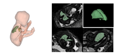 |
26 | Combined T2*-diffusion imaging of the fetal lungs in normal and preterm fetuses
Carla Lily Avena-Zampieri1,2, Lisa Story1,2, Daniel Cromb1, Mary Rutherford1, Alena Uus3, Paddy Slator4, and Jana Hutter1,3
1Perinatal Health and Imaging, Kings College London, London, United Kingdom, 2Women and Children’s Health, Kings College London, London, United Kingdom, 3Department of Biomedical Engineering, Kings College London, London, United Kingdom, 4Centre for Medical Image Computing, UCL, London, United Kingdom
Advanced functional MRI techniques were applied to the fetal lungs to increase our understanding of in-utero lung development, imperative for accurate antenatal diagnosis to identify fetuses at highest risk of morbidity. Our preliminary results show that combined T2*-diffusion imaging is a feasible technique to analyse the developing lungs in utero. A reduction in both lung volumes and T2* values associated with fetuses that subsequently delivered preterm was identified - paving the way for future more detailed in depth studies.
|
||
1321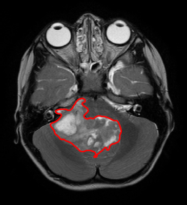 |
27 | Prediction of WHO histological grade of paediatric posterior fossa ependymoma using diagnostic MR imaging and machine learning Video Not Available
Richard J Dury1, Anbarasu Lourdusamy1, Dorothee P Auer2, Andrew Peet3, Richard G Grundy1, and Robert A Dineen2
1Children's Brain Tumour Research Centre, University of Nottingham, Nottingham, United Kingdom, 2Radiological Sciences, University of Nottingham, Nottingham, United Kingdom, 3Institute of Cancer and Genomic Sciences, University of Birmingham, Birmingham, United Kingdom
Ependymoma is the second most common paediatric malignant brain tumour and has a dismal outcome. WHO histological grade provides insight to prognosis and in most series confers a poor survival. Here we present a method to non-invasively predict the grade of paediatric posterior fossa ependymoma using diagnostic MR imaging (T2w and ADC) and machine learning. We found that WHO Grade II and III tumours can both be predicted with a sensitivity/specificity of 0.7±0.23 and 0.67±0.15 respectively. We believe these results provide the basis for a clinically important aid to decision making in the early stages of treatment.
|
||
1322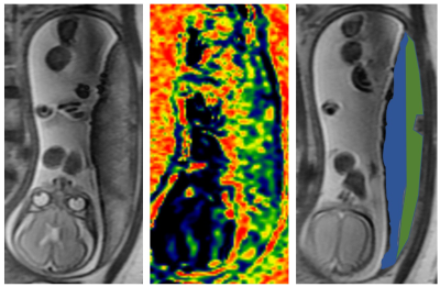 |
28 | Difference of stiffness between the fetal and the maternal part of the placenta by virtual magnetic resonance elastography Video Not Available
Ting Liu1, Jiaojiao Lu1, Junjun Li1, and Jian Yang2
1Department of Radiology, the first Affiliated Hospital of Xi'an Jiaotong University, Xi'an, China, 2the first Affiliated Hospital of Xi'an Jiaotong University, Xi'an, China
Virtual Magnetic Resonance Elastography (vMRE) is based on IVIM, which does not require any mechanical vibration, can assess the elastography of your organization. dysfunctional placenta had higher stiffness than normal placenta has been found by ultrasound shear‑wave elastography. This study intends to use this vMRE method to study the difference in elasticity between the fetus and the maternal compartment.
|
||
1323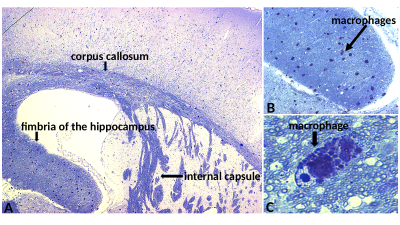 |
29 | Specific Quantification of Myelin by QSM and MWI: Comparison to DTI in Mouse of Pelizaeus Merzbacher Disease and Metachromatic Leukodystrophy
Ulrike Loebel1, Lepu Zhou2, Gabriele M Rune2, Michael Sereda3, Theresa Kungel3, Ullrich Matzner4, Jens Fiehler2, Jan Sedlacik1, and Annette Bley2
1Great Ormond Street Hospital for Children, London, United Kingdom, 2University Medical Center Hamburg-Eppendorf, Hamburg, Germany, 3Max-Planck-Institute of Experimental Medicine, Goettingen, Germany, 4Rheinische Friedrich-Wilhelms Universität Bonn, Bonn, Germany
Two different disease models, i.e., of Pelizaeus Merzbacher disease (PMD) and Metachromatic Leukodystrophy (MLD), were studied in mice using QSM, MWF, MTR and DTI as well as histopathology to assess their ability to specifically quantify myelin damage. Electron microscopy of PMD showed damaged myelin, but histological micrographs of MLD showed regular brain tissue integrity but with an abnormal presence of macrophages. Only MWF and QSM showed a difference between PMD and WT, but no difference between MLD and WT, which suggests, that MWF and QSM are highly specific to myelin damage.
|
||
1324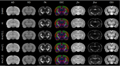 |
30 | Dose-dependent neuroprotective effects of Bovine Lactoferrin following neonatal hypoxia-ischemia in the immature rat brain
Yohan van de Looij1,2, Eduardo Sanches1, Sadou Saw1, Audrey Toulotte1, Analina Da Dilva2, Laura Modernell1, and Stéphane V Sizonenko1
1Division of Development and Growth, Department of Paediatrics and Gynaecology-Obstetrics, School of Medicine, University of Geneva, Geneva, Switzerland, 2Center for Biomedical Imaging, Animal Imaging Technology section, Federal Institute of Technology of Lausanne, Lausanne, Switzerland Injuries to the developing brain due to hypoxia–ischemia (HI) are common causes of neurological disabilities in preterm babies Lactoferrin (Lf) is an iron-binding protein shown to have antioxidant, anti-inflammatory and anti-apoptotic properties when administered to mothers as a dietary supplement during pregnancy and/or lactation in preclinical studies of developmental brain injuries. Here, we tested three different doses of dietary maternal Lf supplementation using the postnatal day 3 HI model and evaluated the acute neurochemical damage profile using 1H MRS and DTI/NODDI. In conclusion, Lf supplementation attenuates, in a dose-dependent manner, the acute and long-term cerebral injury caused by HI. |
||
1325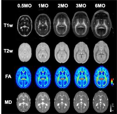 |
31 | Structural Brain Changes during Early Development in Non-human Primate, Common Marmoset: A Longitudinal MRI study
Akiko Uematsu1,2, Junichi Hata3, Makoto Fuskushima1, Noriyuki Kishi2, Ayako Muyarama2, Takuya Hayashi1, and Hideyuki Okano2,4
1BDR Laboratory for Brain Connectomics Imaging, RIKEN, Kobe, Japan, 2CBSLaboratory for Marmoset Neural Architecture, RIKEN, Wako, Japan, 3Graduate School of Human Health Sciences, Tokyo Metropolitan University, Tokyo, Japan, 4Department of Physiology, Keio University School of Medicine, Tokyo, Japan
Neuronal brain development at early life stage is critical for proper function at later life stages. Here, we delineate the maturational process of marmoset brain structure in both macro and micro level at very early life stage with longitudinal multi-modal MRI images. Integrating multi-modal MRI images provided more detailed and accurate information of changes in tissue property especially for frontal brain regions throughout development than a single-modal image analysis.
|
||
1326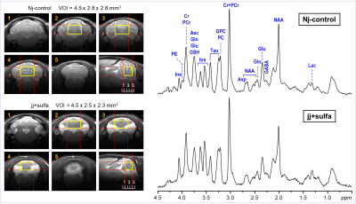 |
32 | Region-specific effects of neonatal hyperbilirubinemia on the developing brain in a preterm Gunn rat model
Ivan Tkac1, Katherine M Satrom2, and Raghavendra Rao2
1Center for Magnetic Resonance Research, University of Minnesota, Minneapolis, MN, United States, 2Department of Pediatrics, University of Minnesota, Minneapolis, MN, United States
Preterm Gunn rat model of neonatal hyperbilirubinemia (NHB) was used to investigate effects of unconjugated bilirubin (UCB) on the developing hippocampus and cerebellum. In this model, sulfadimethoxine was injected (i.p.) to Gunn rat pups on postnatal day 5 to exaggerate NHB effects at a preterm equivalent age. 1H MRS and MRI results clearly demonstrate that high levels of UCB in early postnatal age critically affects the brain development in a region-specific manner. In this model, cerebellum appears to be much more vulnerable to high levels of UCB than hippocampus resulting in abnormal postnatal development.
|
||
1327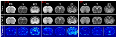 |
33 | Structural and functional brain imaging in a rat model of fetal growth restriction
Yohan van de Looij1,2, Jérôme Mairesse1,3, Julien Pansiot4, Manuela Zinni4, Stéphane V Sizonenko1, and Olivier Baud1,3
1Division of Development and Growth, Department of Paediatrics and Gynaecology-Obstetrics, School of Medicine, University of Geneva, Geneva, Switzerland, 2Center for Biomedical Imaging, Animal Imaging Technology section, Federal Institute of Technology of Lausanne, Lausanne, Switzerland, 3Division of Neonatology and Pediatric Intensive Care, Children's University Hospital of Geneva, Geneva, Switzerland, 4INSERM UMR1141 NeuroDiderot, University Paris Diderot, Sorbonne Paris Cité, Paris, France Brain injuries and subsequent neurodevelopmental impairments observed following Fetal Growth Restriction (FGR) were shown to be associated with an exacerbated neonatal neuro-inflammation. The aim of this work was to characterize a rat model of FGR by advanced diffusion MRI (DTI and NODDI) and functional ultrasounds (fUS). We show in this study that neonatal inflammation lead to transient microstructural damages and subsequent behavioral and functional disabilities at long term. This model mimics very closely clinical reality in babies born following FGR. Innovative neuroimaging tools associated with this neuro-inflammatory model pave the way for their translational use in human neonates. |
||
1328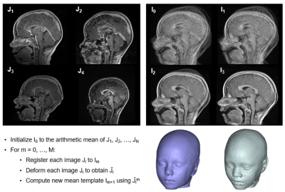 |
34 | Developing a Craniofacial Soft Tissue Anthropomorphic Database: Applying Diffeomorphic Template Building Methods to MRI Video Permission Withheld
Dillan F Villavisanis1, Pulkit Khandelwal2, Zachary D Zapatero1, Connor S Wagner2, Daniel Y Cho1, Liana Cheung1, Jessica D Blum1, Jordan W Swanson1, Jesse A Taylor1, Paul A Yushkevich 2, and Scott P Bartlett1
1Children's Hospital of Philadelphia, Philadelphia, PA, United States, 2Perelman School of Medicine at the University of Pennsylvania, Philadelphia, PA, United States
Craniofacial surgery requires reconciliation of several factors for deformity reconstruction and aesthetic enhancement. Here, we present data in developing a racially and ethnically sensitive anthropomorphic database to provide plastic and craniofacial surgeons with a set of “normal” anatomic measurements to optimize aesthetic and reconstructive outcomes. Images were used to construct composite (template) images with Advanced Normalization Tools (ANTs) and Greedy Tools. Composite templates demonstrated age-appropriate anatomic measurements as proof of concept. Application of diffeomorphic algorithms via ANTs to MRI is effective in developing composite templates representing “normal” soft tissue anatomy, which may aid in objectively quantifying anatomic abnormality.
|
||
 Back to Meeting Home
 Back to the Program-at-a-Glance
The International Society for Magnetic Resonance in Medicine is accredited by the Accreditation Council for Continuing Medical Education to provide continuing medical education for physicians.
 Back to Meeting Home
 Back to the Program-at-a-Glance
 View the Presentation