Digital Poster
Normal Development & Aging: Poster Session II
Joint Annual Meeting ISMRM-ESMRMB & ISMRT 31st Annual Meeting • 07-12 May 2022 • London, UK

Digital Poster
Normal Development & Aging: Poster Session II
| Computer # | ||||
|---|---|---|---|---|
2043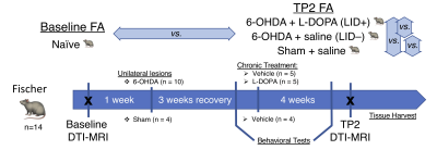 |
33 | Assessment of microstructural changes associated with dyskinesia in hemiparkinsonian rodents
Maurizio Bergamino1, Alberto Fuentes 1, David J Marmion2, Ivette M Sandoval2, Christopher Bishop3, Fredric P Manfredsson2, and Ashley M Stokes1
1Neuroimaging Division, Barrow Neurological Institute, Phoenix, AZ, United States, 2Translational Neuroscience, Barrow Neurological Institute, Phoenix, AZ, United States, 3Department of Psychology, Binghamton University, Binghamton, NY, United States
The objective of this pilot study was to assess white matter (WM) microstructural integrity in pre-clinical models of Parkinson’s disease using fractional anisotropy (FA) from diffusion tensor imaging (DTI). We assessed changes in FA in parkinsonian rats with levodopa-induced dyskinesia both over time and compared with control (sham) rats.
|
||
2044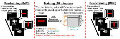 |
34 | Auditory topographic maps in the congenitally blind brain: Effects of NORDIC de-noising and sensory substitution training
Giles Hamilton-Fletcher1, Russell W. Chan1,2, Matthew C. Murphy3, Joel S. Schuman1, Amy C. Nau4,5, and Kevin C. Chan1
1Department of Ophthalmology, New York University Langone Health, New York, NY, United States, 2Neuroscience Institute, New York University Langone Health, New York City, NY, United States, 3Mayo Clinic Rochester, Rochester, MN, United States, 4Korb and associates, Boston, MA, United States, 5Department of Ophthalmology, University of Pittsburgh School of Medicine, Pittsburgh, PA, United States Sensory substitution devices (SSDs) can represent the structure of visual space through sound. To determine whether this representation is topographically organized in the congenitally blind brain, we used NORDIC de-noising and population receptive field mapping (pRF) to correlate functional brain changes to changes in sound stimulus verticality (frequency) and laterality (panning/timing) following SSD principles. We show that NORDIC significantly increased temporal signal-to-noise ratios and activation areas in subjects’ auditory cortices. PRF revealed that auditory topographic maps representing verticality and laterality emerged in occipital-parietal cortices of 3/7 congenitally blind subjects only after 10-minutes of SSD training, indicating rapid cross-modal cortical recruitment. |
||
2045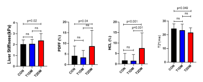 |
35 | Increased liver stiffness and shortened T2* relaxation time in people with type 2 diabetes
Yuliya Kupriyanova1,2, Pavel Bobrov2,3, Volker Burkart1,2, Vera Schrauwen-Hinderling1,2,4, and Michael Roden1,2,5
1Institute for Clinical Diabetology, German Diabetes Center, Leibniz Institute for Diabetes Research at Heinrich Heine University, Düsseldorf, Germany, 2German Center for Diabetes Research (DZD e.V.), München-Neuherberg, Germany, 3Institute for Biometrics and Epidemiology, German Diabetes Center, Leibniz Institute for Diabetes Research at Heinrich Heine University, Düsseldorf, Germany, 4Department of Radiology and Nuclear Medicine, Maastricht University Medical Center, Maastricht, Netherlands, 5Department of Endocrinology and Diabetology, Medical Faculty and University Hospital, Heinrich Heine University, Düsseldorf, Germany
This study shows the value of multiparametric magnetic resonance measurements for detecting the changes in liver tissue due to diabetes mellitus. Liver fat content (determined by mDixon MRI and 1H-MR spectroscopy), T2* relaxation time and liver stiffness were compared in humans with type 1 (T1DM), type 2 diabetes mellitus (T2DM) or normal glucose tolerance (control, CON).
|
||
2046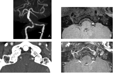 |
36 | Characterization of Symptomatic Vertebrobasilar Plaques in Diabetic Patients Using Computed Tomographic Angiography and Vessel Wall Imaging Video Not Available
Huan Yang1, Bo Liu2, Qingqing Yin3, Haipeng Wang1, Liangjie Lin4, and Ximing Wang1
1Radiology, Shandong Provincial Hospital Affiliated to Shandong First Medical University, Jinan, China, 2Radiology, Qilu Hospital of Shandong University, Jinan, China, 3Geriatric Neurology, Shandong Provincial Hospital Affiliated to Shandong First Medical University, Jinan, China, 4MSC Clinical & Technical Solutions, Philips Healthcare, Beijing, China
Diabetes mellitus is significantly associated with posterior circulation ischemic stroke. We aimed to compare the characteristics of vertebrobasilar plaques in symptomatic patients with and without diabetes using vessel wall magnetic resonance imaging and computed tomographic angiography. Characteristics of symptomatic vertebrobasilar plaques were compared between patients with and without diabetes. Multivariate analysis demonstrated differences in the presence of T1 hyperintensity and number of spotty calcifications were statistically significant. Symptomatic patients with diabetes have a higher incidence of T1 hyperintensity and larger calcification burden than those without diabetes, indicating the association of diabetes with more advanced plaque features in the posterior circulation.
|
||
2047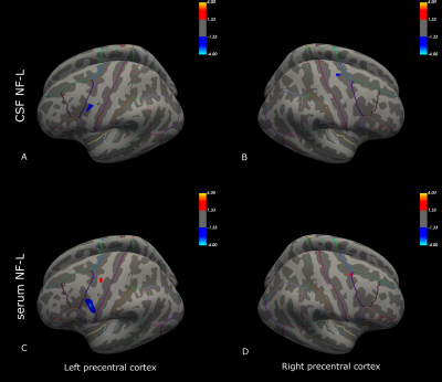 |
37 | Association between neurofilament (NF-L) levels and amyotrophic lateral sclerosis (ALS) neurodegeneration patterns in the precentral cortex
Penelope Tilsley1,2, Antoine Moutiez3, Alexandre Brodovitch4, Mohamed Mounir El Mendili1,2, Wafaa Zaaraoui1,2, Annie Verschueren1,5, Shahram Attarian5, Maxime Guye1,2, José Boucraut4,6, Aude-Marie Grapperon1,5, and Jan-Patrick Stellmann1,2,3
1Aix Marseille Univ, CNRS, CRMBM, Marseille, France, 2APHM, Hôpital de la Timone, CEMEREM, Marseille, France, 3APHM, Hôpital de la Timone, Department of Neuroradiologie, Marseille, France, 4Immunology Laboratory, Conception Hospital, AP-HM, Marseille, France, 5APHM, Hôpital de la Timone, Referral Centre for Neuromuscular Diseases and ALS, Marseille, France, 6INS, INSERM, UMR 1106, Marseille, France
Neurofilament light-chain (NF-L) levels in serum and cerebrospinal fluid (CSF) have recently been demonstrated as robust clinical biomarkers in amyotrophic lateral sclerosis (ALS). Nonetheless, the association between NF-L levels and ALS specific neurodegeneration patterns in the precentral cortex, a primarily affected area, has not been fully divulged. Combining 3T and 7T anatomical imaging with NF-L measures, clinical and electrophysiological data, we demonstrated regional-specific degradation within the precentral cortex according to affected limb areas. Further, NF-L levels negatively correlated with cortical thickness within the premotor cortex, suggesting the premotor cortex may be an important contributor to augmented NF-L levels in ALS.
|
||
2048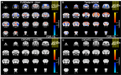 |
38 | Mild neuroanatomical phenotype in an ALS/FTD mouse model with mutation in C9orf72
Aurea B. Martins-Bach1, Carmelo Milioto2,3, Shoshana Spring4, Mireia Carcolé2,3, Thomas J. Cunningham5, Elizabeth M. C. Fisher6, Adrian M. Isaacs2,3, Brian J. Nieman4, Jason Lerch1, and Karla L. Miller1
1Wellcome Centre for Integrative Neuroimaging, FMRIB, Nuffield Department of Clinical Neurosciences, University of Oxford, Oxford, United Kingdom, 2UK Dementia Research Institute at UCL, Faculty of Brain Sciences, University College London, London, United Kingdom, 3Department of Neurodegenerative Disease, UCL Queen Square Institute of Neurology, University College London, London, United Kingdom, 4Mouse Imaging Centre, The Hospital for Sick Children, Toronto, ON, Canada, 5Mammalian Genetics Unit, MRC Harwell Institute, Oxfordshire, United Kingdom, 6Department of Neuromuscular Diseases, UCL Queen Square Institute of Neurology, University College London, London, United Kingdom
Mutations in the C9orf72 gene are the most prevalent genetic alteration in ALS/FTD. This study investigates neuroanatomical phenotypes in two C9orf72 knock-in mouse models separately expressing either poly-(PR) or poly-(GR) dipeptide-repeats. Ex-vivo structural MRI (40 μm isotropic resolution) was acquired at 7T. After registration, the deformation fields were used to estimate voxels and region-of-interest volumes for comparison between mutants and wild-type mice. Although neuroanatomical phenotypes have been previously described in C9orf72 patients and other ALS/FTD mouse models, 20-month-old poly-(PR) and poly-(GR) mice presented subtle alterations. Further investigations to assess microstructural and histological changes in these mouse models are in progress.
|
||
2049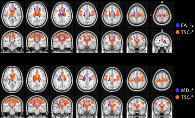 |
39 | Combined microstuctural and sodium homeostasis alterations in ALS are widespread in fast progressors: a brain DTI and sodium MRI study
Mohamed Mounir El Mendili1,2, Aude-Marie GRAPPERON2,3, Rémi DINTRICH 1,2,3, Jan patrick STELLMANN1,2, Lauriane PINI1,2, Claire COSTES1,2, Jean-Philippe RANJEVA1,2, Maxime GUYE 1,2, Annie VERSCHUEREN 3, Shahram ATTARIAN 3, and Wafaa ZAARAOUI 1,2
1Aix Marseille Univ, CNRS, CRMBM, Marseille, France, 2APHM, Hopital de la Timone, CEMEREM, Marseille, France, 3APHM, Hôpital de la Timone, Referral Centre for Neuromuscular Diseases and ALS, Marseille, France
Amyotrophic lateral sclerosis (ALS) is a heterogeneous condition showing variable progression rates. Conventional MRI lacks sensitivity and specificity to detect abnormalities in ALS and is mainly used to exclude ALS-mimics. Non-conventional MRI has gradually characterized specific neurodegeneration features in ALS.The present brain DTI and sodium MRI study evidenced combined microstructural and sodium homeostasis alterations in ALS and widespread damage in fast progressors. Our results confirm previous reports showing that non-conventional MRI technics might contribute to the diagnostic work-up of patients with different clinical profiles, especially when combined with mathematical modeling enabling predicting disease progression trajectory of a single patient.
|
||
2050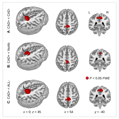 |
40 | Precuneus dysconnectivity with heart failure
Karsten Mueller1, Harald E Möller1, Birol Taskin1, Friederike Thiel1,2,3, Frank Beutner3,4, Andrej Teren3,4, Arno Arno Villringer1,2,3, and Matthias L Schroeter1,2,3
1Max Planck Institute for Human Cognitive and Brain Sciences, Leipzig, Germany, 2Clinic for Cognitive Neurology, University Hospital Leipzig, Leipzig, Germany, 3Leipzig Research Center for Civilization Diseases, Leipzig, Germany, 4Leipzig Heart Center, Leipzig, Germany
Our investigations identified the precuneus as the brain region showing a major involvement in connectivity decline in patients with heart failure. Subsequent seed-based correlation analysis showed decreased putamen connectivity related to decline in cognitive performance. In line with these findings and with current Alzheimer’s Disease models showing precuneus connectivity decline as a key feature of brain degeneration, there might be common underlying mechanisms leading to brain connectivity decrease in heart failure and dementia. This hypothesis would be also supported by specific network centrality alterations between precuneus and widely distributed cortical regions particularly in patients showing reduced cognitive performance.
|
||
2051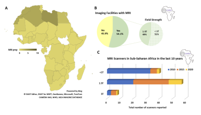 |
41 | Redefining Access to High-Value MRI: Results from the CAMERA MRI in Africa Needs Assessment Survey
Jinggang Jenny Ng1, Boaz Ehiogu2, Johnes Obungoloch3, Abiodun Fatade4, Mamadou Diop2,5, Henk-Jan Mutsaerts6, Daniel Alexander7, Mario Forjaz Secca8, Rita Nunes9, Patricia Figueiredo9, Matteo Figni7, Frank J Minja10, Vikas Gulani11, Andrew G Webb12, Iris Asllani13,14, Edward Chege Nganga15, Sola Adeleke16, Godwin Ogbole17, Farouk Dako1, and Udunna C Anazodo5,18
1Perelman School of Medicine, University of Pennsylvania, Philadelphia, PA, United States, 2Lawson Health Research Institute, London, ON, Canada, 3Mbarara University of Science and Technology, Mbarara, Uganda, 4Crestview Radiology Ltd, Lagos, Nigeria, 5Department of Medical Biophysics, Western University, London, ON, Canada, 6Department of Radiology and Nuclear Medicine, Amsterdam Neuroscience, Amsterdam University Medical Center, Amsterdam, Netherlands, 7Department of Computer Science, University College London, London, United Kingdom, 8Central Hospital of Maputo, Maputo, Mozambique, 9Department of Bioengineering, Instituto Superior, Técnico, Universidade de Lisboa, Lisbon, Portugal, 10Department of Radiology and Imaging Sciences, Emory University, Atlanta, GA, United States, 11Department of Radiology, University of Michigan, Ann Arbor, MI, United States, 12Department of Radiology, Leiden University Medical Center, Leiden, Netherlands, 13Clinical Imaging Sciences Centre, Department of Neuroscience, University of Sussex, Brighton, United Kingdom, 14Department of Biomedical Engineering, Rochester Institute of Technology, Rochester, NY, United States, 15Department of Radiology, Aga Khan University Hospital, Nairobi, Kenya, 16Department of Oncology, Guy’s & St Thomas’ Hospital, London, United Kingdom, 17Department of Radiology, University College Hospital Ibadan, Ibadan, Nigeria, 18Department of Neurology and Neurosurgery, Montreal Neurological Institute, McGill University, Montreal, QC, Canada
We examined access to MRI in Sub-Saharan Africa to provide a novel framework to address MRI needs. A 68-question needs assessment survey was distributed to collaborators, radiologists and radiographers in Africa, yielding 158 unique responses. Survey responses were analyzed to provide insight into challenges and opportunities for MRI access. Geographical information systems (GIS) were applied to responses from Nigeria to model access to high-value MRI. To our knowledge, this is the first study that used GIS mapping to estimate MRI access. This novel approach can be applied to low-resource settings globally to provide a comprehensive framework to understand MRI access.
|
||
2052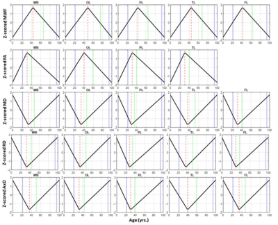 |
42 | Myelination patterns support the last-in-first-out and gain-predicts-loss paradigms in normative aging
Matthew Kiely1, Curtis Triebswetter1, Zhaoyuan Gong1, Maryam H. Alsameen1, and Mustapha Bouhrara1
1Magnetic Resonance Physics of Aging and Dementia Unit, National Institute on Aging, Baltimore, MD, United States
Using myelin water fraction (MWF), a proxy of myelin content, and diffusion tensor imaging (DTI) indices, we investigated the retrogenesis paradigm in a large cohort of cognitively unimpaired participants spanning a wide age range. Specifically, we assessed the last-in-first-out and gain-predicts-loss hypotheses, describing trends of tissue degeneration across cerebral regions and comparing maturation and degeneration rates within regions. Our MWF results support the last-in-first-out hypothesis, indicating that posterior regions are generally spared from degeneration compared to anterior regions, as well the gain-predicts-loss paradigm, suggesting that the regional maturation and degeneration rates were approximately symmetric.
|
||
 Back to Meeting Home
 Back to the Program-at-a-Glance
The International Society for Magnetic Resonance in Medicine is accredited by the Accreditation Council for Continuing Medical Education to provide continuing medical education for physicians.
 Back to Meeting Home
 Back to the Program-at-a-Glance
 View the Presentation