Digital Poster
Rare, Genetic & Metabolic Diseases I
Joint Annual Meeting ISMRM-ESMRMB & ISMRT 31st Annual Meeting • 07-12 May 2022 • London, UK

Digital Poster
Rare, Genetic & Metabolic Diseases I
| Computer # | ||||
|---|---|---|---|---|
2672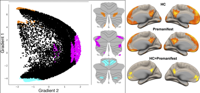 |
36 | Probing Cerebello-Cerebral Functional Connectivity in Spinocerebellar Ataxias
Sheeba Anteraper1, Ying Zhang2, Xavier Guell3, Michal Povazan4, Guita Banan5, Romain Valabregue6, Philip Ehses7, Jennifer Faber7, James M Joers2, Chiadi U Onyike4, Peter B Barker4, Jeremy D Schmahmann3, Eva-Maria Ratai3, S H Subramony5, Thomas H Mareci5, Khalaf O Bushara2, Alexandra Durr6, Thomas Klockgether7, Tetsuo Ashizawa8, Christophe Lenglet2,
and Gülin Öz2
1Carle Foundation Hospital, Champaign, IL, United States, 2University of Minnesota, Minneapolis, MN, United States, 3Massachusetts General Hospital, Boston, MA, United States, 4Johns Hopkins University, Baltimore, MD, United States, 5University of Florida, Gainesville, FL, United States, 6Sorbonne University, Paris, France, 7German Center for Neurodegenerative Diseases, Bonn, Germany, 8The Houston Methodist Research Institute, Houston, TX, United States
Spinocerebellar Ataxias (SCAs) are a group of rare, autosomal dominant diseases that result in progressive degeneration of the cerebellum. Of these, SCA1 is the fastest progressing. SCA3, the most prevalent SCA worldwide, is relentlessly progressive, disabling, and eventually fatal with no efficacious treatments other than supportive therapy. There is a strong need to improve our mechanistic understanding of the changes in the cerebello-cerebral circuitry prior to disease manifestation so that novel therapeutic strategies can be developed. Analyzing high quality magnetic resonance imaging data from the NIH-funded project, “Clinical Trial Readiness for SCA1 and SCA3 (READISCA),” will guide such efforts.
|
||
2673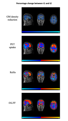 |
37 | Longitudinal hybrid PET/MRI in juvenile-onset Huntington disease (joHD): a pilot study
Maria Eugenia Caligiuri1, Patrizia Vizza2, Pierangelo Veltri3, Francesco Cicone3, Paolo Barberio3, Giuseppe Lucio Cascini3, Eugenia Scaricamazza4, Sabrina Maffi4, Simone Migliore4, Ferdinando Squitieri4,5, and Umberto Sabatini3
1Neuroscience Research Center, University "Magna Graecia", Catanzaro, Italy, 2Mater Domini University Hospital, Catanzaro, Italy, 3University "Magna Graecia", Catanzaro, Italy, 4IRCCS Casa Sollievo della Sofferenza/CSS-Mendel, San Giovanni Rotondo/Rome, Italy, 5Italian League fo Research on Huntington (LIRH Foundation), Rome, Italy
Hybrid PET-MRI is an emerging technique that allows multimodal evaluation of brain structure and function. This study evaluates longitudinal PET-MRI in one patient with stage-2 joHD, to assess changes related to disease progression. This approach might be useful to test the efficacy of disease-modifying drugs.
|
||
2674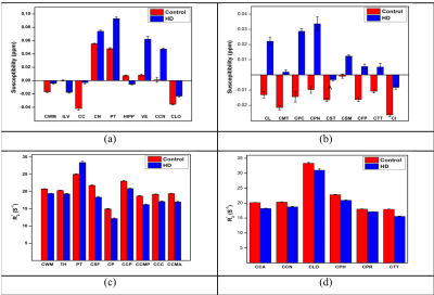 |
38 | QSM, R2* and correlation thereof as qMRI measures of iron and myelin reflect tissue changes in Huntington’s disease
Ana-Maria Oros-Peusquens1, Ravi Dadsena1, Imis Dogan2,3, Kathrin Reetz2,3, and N. Jon Shah1,2,4,5
1Institute of Neuroscience and Medicine INM-4, Research Centre Juelich, Juelich, Germany, 2Department of Neurology, RWTH Aachen University, Aachen, Germany, 3JARA-BRAIN Institute Molecular Neuroscience and Neuroimaging, Research Centre Juelich, Juelich, Germany, 4Faculty of Medicine, JARA, RWTH Aachen University, Aachen, Germany, 5Institute of Neuroscience and Medicine 11, INM-11, JARA, Research Centre Juelich, Juelich, Germany
QSM and R2* are quantitative MRI parameters sensitive to iron and myelin content, and their correlation provides further insight into the chemical form of iron. These are the tissue properties which are most affected by Huntington’s disease, besides volumetric changes. We investigate by multiparametric qMRI changes induced by HD in 18 gene-carriers by comparison to matched healthy controls. These qMRI changes are significant in several cortical and subcortical regions and correlate with clinical measures of HD, including CAG mutation length. Although the interpretation of correlations between parameters is not unambiguous, insights into tissue chemical and micro-structure are enabled.
|
||
2675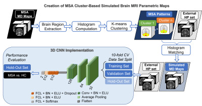 |
39 | Multiple System Atrophy Classification via 3D Convolutional Neural Network and Simulated Brain MRI Parametric Maps
Giulia Maria Mattia1, Edouard Villain1,2, Olivier Rascol3, Wassilios G. Meissner4,5,6, Xavier Franceries7, and Patrice Péran1
1ToNIC, Toulouse NeuroImaging Center, Université de Toulouse, Inserm, UPS, Toulouse, France, 2LAAS CNRS, Université de Toulouse, CNRS, INSA, UPS, Toulouse, France, 3French Reference Center for MSA, Department of Neurosciences and Clinical Pharmacology Clinical Investigation Center CIC1436, NS-Park/FCRIN Network, ToNIC, Toulouse NeuroImaging Center University Hospital of Toulouse, Inserm, Université de Toulouse 3, Toulouse, France, 4French Reference Center for MSA, Department of Neurology for Neurodegenerative Diseases, University Hospital Bordeaux, Bordeaux, France, 5Univ. Bordeaux, CNRS, IMN, UMR 5293, F-33000 Bordeaux, France, 6Dept. Medicine, University of Otago, Christchurch and New Zealand Brain Research Institute, Christchurch, New Zealand, 7CRCT, Centre de Recherche en Cancerologie de Toulouse, Inserm, Toulouse, France
As a rare neurodegenerative disorder, multiple system atrophy (MSA) can be challenging to analyse using a deep learning approach given the limited sample size. A method is submitted to produce cluster-based simulated brain MR parametric maps from healthy controls, using regional intensity distribution belonging to a set of MSA patients. This enabled to train a 3D CNN only with the simulated set. Testing on the MSA data set, the accuracy obtained was comparable to the state-of-the-art. This approach allows to deal with small samples of data in deep learning, while exploiting a-priori knowledge of the disease.
|
||
2676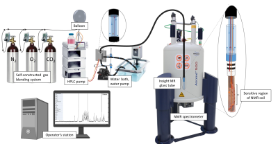 |
40 | Compartment-Specific Metabolic Investigation of 3D Cell Culture by Real-time NMR for Investigating Metabolic Diseases
Christian Urzi1,2,3, Damian Hertig1,2, Christoph Meyer1,2,3, Jean-Marc Nuoffer2,4, and Peter Vermathen1
1Magnetic Resonance Methodology, Institute of Diagnostic and Interventional Neuroradiology, University of Bern, Bern, Switzerland, 2Institute of Clinical Chemistry, University Hospital Bern, Bern, Switzerland, 3Graduate School for Cellular and Biomedical Sciences, University of Bern, Bern, Switzerland, 4Department of Pediatric Endocrinology, Diabetology and Metabolism, University Children’s Hospital of Bern, Bern, Switzerland
In this study was shown the possibility to perform compartment-specific metabolic investigations of living fibroblasts in real-time using a bioreactor system within an NMR spectrometer. Cell volume and the ratio cells to extracellular medium within the sensitive region of NMR coil were evaluated for quantification purposes. The needed sensitivity to detect metabolite changes in the extracellular footprint at different flow rates was shown. The capability of diffusion technique to distinguish between compartment-specific contributions was revealed, and to detect changes in compartment-specific metabolite pools under standard or selective culture conditions, and upon inhibitor challenges.
|
||
2677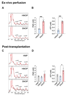 |
41 | Sub-Normothermic Ex-Vivo Perfusion of Porcine Kidney Grafts Improves Energy Metabolism: 31P-MRSI Analysis
Julien Songeon1, Thomas Agius2, Antoine Klauser1,3, Grégoire Longchamp4, Raphael Ruttimann4, Jean-Marc Corpataux2, Alban Longchamp2, and François Lazeyras1,3
1Department of Radiology and Medical Informatics, University of Geneva, Geneva, Switzerland, 2Department of Vascular Surgery, Centre Hospitalier Universitaire Vaudois and University of Lausanne, Lausanne, Switzerland, 3Center for Biomedical Imaging (CIBM), Geneva, Switzerland, 4Department of Surgery, Geneva University Hospitals and Medical School, Geneva, Switzerland Improved preservation strategies for the storage of graft collected after circulatory death could increase the number of kidneys available and improve patient survival. Warm (22 and 37°C) ex-vivo perfusion has emerged as a feasible strategy for organ recovery, but the underlying mechanism remains elusive. Using phosphorus magnetic resonance spectroscopic imaging (31P-MRSI) and histological scoring, we evaluated kidney viability and adenosine triphosphate (ATP) production during sub-normothermic ex-vivo kidney perfusion (SNOP) versus hypothermic machine perfusion (HMP) in a porcine kidney autotransplantation model. |
||
2678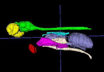 |
42 | MR imaging of Zebrafish with mitochondria diseases Video Permission Withheld
Sergey Magnitsky1, Sonal Sharma1, and Marni Falk1
1CHOP, Philadelphia, PA, United States
We created a high-resolution MR imaging atlas of wt. and three mutants (OPA1, FBXL4
|
||
2679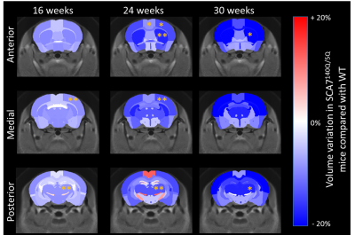 |
43 | Longitudinal metabolic and morphometric characterization of a knock-in mouse model of SpinoCerebellar Ataxia Type 7
Jean-Baptiste Pérot1, Anna Niewiadomska-Cimicka2,3,4, Emmanuel Brouillet1, Yvon Trottier2,3,4, and Julien Flament1
1Université Paris-Saclay, Commissariat à l’Energie Atomique et aux Energies Alternatives (CEA), Centre National de la Recherche Scientifique (CNRS), Molecular Imaging Research Center (MIRCen), Laboratoire des Maladies Neurodégénératives, Fontenay-aux-Roses, France, 2Université de Strasbourg, Institut de Génétique et de Biologie Moléculaire et Cellulaire, Illkirch, France, 3Centre National de la Recherche Scientifique, Unité Mixte de Recherche 7104, Illkirch, France, 4Institut National de la Santé et de la Recherche Médicale U964, Illkirch, France SpinoCerebellar Ataxia Type 7 (SCA7) is an autosomal dominant degenerative disease defined by neurodegeneration of the retina and cerebellum. While gene silencing therapeutic strategy is developing, there is a need of biomarkers for evaluation of their efficacy. Here we characterize a recent knock-in mouse model of SCA7 with in-vivo longitudinal MRI and MRS. SCA7140Q/5Q display a wide range of phenotypes, including morphological and metabolic alterations in key brain structures. Our longitudinal protocol allowed better understanding of the chronology of these alterations and offers pertinent biomarkers for evaluation of therapies using the SCA7140Q/5Q model. |
||
2680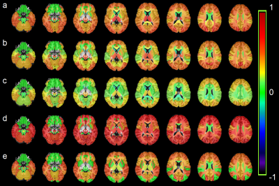 |
44 | Mapping Brain Metabolite Differences Between HIV Clade-C Infected Individuals and Healthy Subjects Using a Whole-Brain MRSI
Teddy Salan1, Sulaiman Sheriff1, Sameer Vyas2, Deepika Aggarwal2, Paramjeet Singh2, and Varan Govind1
1Radiology, University of Miami, Miami, FL, United States, 2Post Graduate Institute of Medical Education & Research, Chandigarh, India
Magnetic resonance spectroscopic imaging (MRSI) based studies investigating brain metabolite alterations due to HIV infection generally acquired MR spectra from single or multi-voxels covering regions related to HIV. In this study, we acquire whole-brain MRSI data to map regional metabolite variations in the entire brain, determine regions mostly affected by HIV clade-C infection, and identify the mechanism by which HIV damages the brain. Results show widespread increases in myo-inositol, glutamate/glutamine, choline, and creatine, and decreases in N-acetylaspartate, indicating neuronal dysfunction and astrogliosis in the white matter, as well as disruptions in synaptic transmission, neurotoxicity and inflammation throughout the brain.
|
||
2681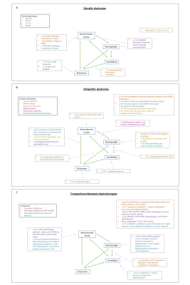 |
45 | Diffusion magnetic resonance imaging in genetic and idiopathic dystonias
Claire Louise MacIver1,2, Derek Jones1, Chantal Tax1,3, and Kathryn Peall2
1Cardiff University Brain Imaging Centre, Cardiff University, Cardiff, United Kingdom, 2Neuroscience and Mental Health Research Institute, Cardiff University, Cardiff, United Kingdom, 3Image Sciences Institute, University Medical Centre Utrecht, Utrecht, Netherlands Dystonia is a hyperkinetic movement disorder involving repetitive or sustained muscle contractions. Its pathophysiology is poorly understood, limiting therapeutic advancement. A systematic literature review on diffusion MRI in dystonia was performed to gain insight into microstructural white matter changes that may contribute to pathology. Of 403 identified records, 40 met the criteria for inclusion. The most consistent diffusion abnormalities for both genetic and idiopathic dystonia forms were lower FA values or reduced number of tractography streamlines in regions connecting brainstem, cerebellum, basal ganglia and sensorimotor cortex, with some genotype and phenotype specific differences identified. |
||
 Back to Meeting Home
 Back to the Program-at-a-Glance
The International Society for Magnetic Resonance in Medicine is accredited by the Accreditation Council for Continuing Medical Education to provide continuing medical education for physicians.
 Back to Meeting Home
 Back to the Program-at-a-Glance
 View the Presentation