Digital Poster
Rare, Genetic & Metabolic Diseases II
Joint Annual Meeting ISMRM-ESMRMB & ISMRT 31st Annual Meeting • 07-12 May 2022 • London, UK

Digital Poster
Rare, Genetic & Metabolic Diseases II
| Computer # | ||||
|---|---|---|---|---|
2757 |
25 | Liver health outcome of chronic hepatic steatosis: an interim analysis of the Dallas Heart Study cohort Video Permission Withheld
Sujoy Mukherjee1, Jarett Berry2, Carlos Duncker3, Ian Jason Neeland4, Amit Singal2, Viktoria Topper1, and Takeshi Yokoo1
1Department of Radiology and Advanced Imaging Research Center, UT Southwestern Medical Center, Dallas, TX, United States, 2Department of Internal Medicine, UT Southwestern Medical Center, Dallas, TX, United States, 3Perspectum, Oxford, United Kingdom, 4University Hospitals Cleveland Medical Center, Cleveland, OH, United States
The natural history of hepatic steatosis and risk factors for the development of advanced chronic liver disease (CLD) are unknown. This interim analysis of 191 subjects in the Dallas Heart Study longitudinal cohort shows that having hepatic steatosis (defined as proton-density fat fraction >5%) at the baseline exam was significantly associated with development of advanced CLD (defined as corrected liver T1 ≥ 800msec) 10-20 years later, with CLD prevalence of 38.8% and 16.9% in the steatosis vs. non-steatosis cohort (p<0.001). The baseline overweight or obesity status also appears to be associated with advanced CLD, independent of having steatosis at baseline.
|
||
2758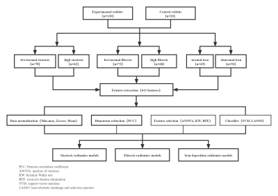 |
26 | Radiomics models based on mDixon for simultaneous liver steatosis, fibrosis and iron deposition quantification of chronic liver disease Video Permission Withheld
Qing Wang1, Ye Sheng1, Jilei Zhang2, and Weibo Chen2
1Third Affiliated Hospital of Soochow University & First People's Hospital of Changzhou, changzhou, China, 2Philips Healthcare, shanghai, China
To assess the diagnostic performance of radiomics analysis based on mDixon Quant in simultaneously quantifying liver steatosis, fibrosis and iron deposition of chronic liver disease (CLD) and eliminating the interaction of histopathological factors.
|
||
2759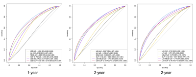 |
27 | LIA (Liver Shear Stiffness, INR, Etiology) Score Predicts Risk of Further Decompensation in Patients with Decompensated Cirrhosis
Jie Zhu1, Zhuoya Yi1, Mengsi Li1, Qilong Chen1, Yuanqiang Xiao1, Ziying Yin2, Kevin J Glaser2, Jun Chen2, Meng Yin2, Richard L. Ehman2, and Jin Wang1
1The third affiliated hospital of Sun Yat-sen University, Guangzhou, China, 2Mayo Clinic College of Medicine, Mayo Clinic, Rochester, MN, United States
In patients who have been clinically diagnosed with decompensated cirrhosis (DC), progression to more advanced decompensation is associated with high mortality. Therefore, the early detection of the risk of further decompensation is helpful to formulate individual treatment and follow-up schedules for patients with DC. Liver stiffness is a potential prognosis biomarker for patients with cirrhosis. This study evaluated MR elastography (MRE) as a tool to predict the risk of further decompensation. By integrating MRE-assessed liver stiffness with clinical information, we developed a risk score that is valuable for predicting further decompensation in patients with DC.
|
||
2760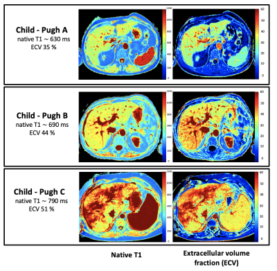 |
28 | Magnetic resonance mapping with calculation of extracellular volume fraction allows for a reliable assessment of liver cirrhosis severity
Narine Mesropyan1, Patrick A. Kupczyk1, Leona Dold2, Michael Praktiknjo2,3, Johannes Chang2,3, Alexander Isaak1, Christoph Endler1, Dmitrij Kravchenko1, Leon Bischoff1, Alois M. Sprinkart1, Claus C. Pieper1, Daniel Kuetting 1, Christian Jansen2,3, Ulrike I. Attenberger1, and Julian A. Luetkens1
1Department of Diagnostic and Interventional Radiology, University Hospital Bonn, Bonn, Germany, 2Department of Internal Medicine I, University Hospital Bonn, Bonn, Germany, 3Center for Cirrhosis and Portal Hypertension Bonn (CCB), University Hospital Bonn, Bonn, Germany
Assessment of disease severity in patients with liver cirrhosis is of great clinical importance and allows outcome prediction and overall mortality risk estimation. In the present study, we investigated the diagnostic utility of MRI-derived extracellular volume fraction (ECV) for the assessment of liver cirrhosis severity and differentiation between different Child-Pugh classes. Our study results demonstrated a high diagnostic performance of ECV for the assessment of liver cirrhosis severity and offer a reliable discrimination between different Child-Pugh classes. ECV might be a new quantitative marker for the assessment of liver cirrhosis severity, which can be calculated from quantitative liver MRI.
|
||
2761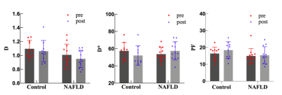 |
29 | The effect of Gd-EOB-DTPA on liver IVIM imaging of rabbits with non-alcoholic fatty liver disease Video Not Available
Xia Wang1, Yu Jiang1, Sheng Zhang1, Si Wang1, Wei Wei1, Mengzhuo Yao1, Na Zhao1, and Yuedong Han*1
1Xi’an Gaoxin Hospital, Xi’an, China
Hepatobiliary cell-specific contrast agent of Gd-EOB-DTPA hepatobiliary phase acquisition DWI sequence will not affect lesion display and ADC value [1].Few studies have been performed on the effect of IVIM on hepatic parenchyma and have focused on studies in various hepatic occupancy contexts with discrepant findings [2,3].This study modeled a healthy control group and a non-alcoholic fatty liver disease (NAFLD) group. Gd-EOB-DTPA had no significant effect on the image quality and quantitative index of liver IVIM imaging in two groups, and IVIM scanning in the hepatobiliary waiting interval after placing Gd-EOB-DTPA enhancement helped to improve the examination flow rate.
|
||
2762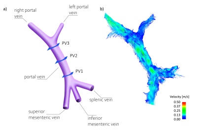 |
30 | Portal Venous 4D Flow MRI in Obese Patients without Known Liver Disease Undergoing Weight Loss Surgery
Alma Spahic1, Grant Steven Roberts1, Tanya Wolfson2, David T Harris3, Nikolaos T Panagiotopoulos3,4, Alejandro Roldán-Alzate3,5,6, Kevin M Johnson1,3, Oliver T Wieben1,3, Claude B Sirlin7, Scott B Reeder1,3,5,6,8, and Thekla Helene Oechtering3,4
1Medical Physics, University of Wisconsin- Madison, Madison, WI, United States, 2Computational and Applied Statistics Laboratory, University of California San Diego, La Jolla, CA, United States, 3Radiology, University of Wisconsin- Madison, Madison, WI, United States, 4Radiology and Nuclear Medicine, Universität zu Lübeck, Lübeck, Germany, 5Mechanical Engineering, University of Wisconsin- Madison, Madison, WI, United States, 6Biomedical Engineering, University of Wisconsin- Madison, Madison, WI, United States, 7Radiology, University of California San Diego, San Diego, CA, United States, 8Emergency Medicine, University of Wisconsin- Madison, Madison, WI, United States
4D flow MRI of the portal vein has the potential to provide diagnostic value for detecting and staging certain liver diseases. To date, it is unknown whether weight loss influences portal flow in obese patients. We aimed to investigate the effect of weight loss on portal flow rate measured by non-contrast 4D flow MRI in obese patients undergoing weight loss surgery. Preliminary data of this ongoing study detected no statistically significant change in the mean volumetric portal flow rate in subjects during 2-6 months of weight loss. However, portal flow rate normalized to body weight or BMI increased significantly.
|
||
2763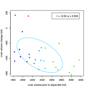 |
31 | PROGNOSTIC VALUE OF BASELINE MRI FOR LIVER VOLUME CHANGES FOLLOWING LOW CALORIE DIET
Lael K Ceriani1, Mark Barahman1, Tanya Wolfson2, Danielle N Batakis1, Robert H Lubenow1, Jake T Weeks1, Kyle A Hasenstab1, Timoteo Delgado1, Yesenia Covarrubias1, Celene Gonzalez1, Michael S Middleton1, Walter C Henderson1, David T Harris3, Nikolaos Panagiotopolous3, Scott B Reeder3, Kathryn J Fowler1, and Claude B Sirlin1
1Radiology, University of California San Diego, La Jolla, CA, United States, 2Computational and Applied Statistics Laboratory, University of California San Diego, La Jolla, CA, United States, 3University of Wisconsin Madison, Madison, WI, United States
The purpose of this study was to identify the prognostic value of baseline MRI-determined imaging biomarkers for liver volume changes following low-calorie liquid diet in bariatric surgery patients
|
||
2764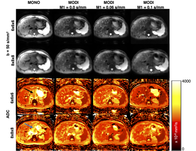 |
32 | Low Resolution Diffusion Weighted Imaging for the Assessment of Diffuse Liver Disease
Srijyotsna Volety1,2, Diego Hernando1,2, and Ali Pirasteh1,2
1Radiology, University of Wisconsin - Madison, Madison, WI, United States, 2Medical Physics, University of Wisconsin - Madison, Madison, WI, United States
We evaluated the reproducibility of liver ADC measurements across acquisition parameters using low spatial resolution DWI with M1 optimized diffusion imaging waveforms (MODI) and the routinely clinically utilized monopolar (MONO) waveforms. We also evaluated the effects of various M1 values as well as those of breath-hold and respiratory-triggering on MODI-DWI liver ADC measurements at lower resolutions than utilized in clinical practice. MONO-DWI liver ADC suffered from bias and resolution-dependence in the left lobe while MODI-DWI liver ADC did not demonstrate this effect. MODI-DWI liver ADC did not demonstrate bias with respect to resolution, M1, or breathing technique.
|
||
2765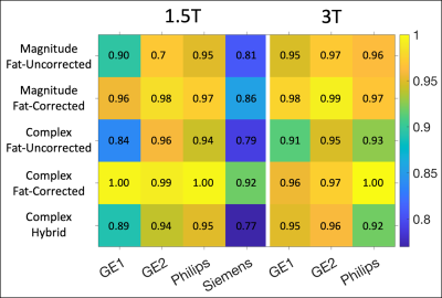 |
33 | Comparison of Centralized R2* Mapping Algorithms to Vendor R2* Mapping in a Multi-Center, Multi-Vendor Liver Iron Overload Study
Gregory Simchick1,2, Ruiyang Zhao1,2, Qing Yuan3, Mounes Aliyari Ghasabeh4, Stefan Ruschke5, Dimitrios C Karampinos5, David Harris1, Ryan J Mattison6, Michael R Jeng7, Ivan Pedrosa3,8, Ihab R Kanel3, Sheryas Vasanawala9, Takeshi Yokoo3,8, Scott B Reeder1,2,6,10,11, and Diego Hernando1,2
1Radiology, University of Wisconsin-Madison, Madison, WI, United States, 2Medical Physics, University of Wisconsin-Madison, Madison, WI, United States, 3Radiology, University of Texas Southwestern Medical Center, Dallas, TX, United States, 4Radiology, The John Hopkins University, Baltimore, MD, United States, 5Diagnostic and Interventional Radiology, Technical University of Munich, Munich, Germany, 6Medicine, University of Wisconsin-Madison, Madison, WI, United States, 7Pediatrics - Hematology & Oncology, Stanford University, Palo Alto, CA, United States, 8Advanced Imaging Research Center, University of Texas Southwestern Medical Center, Dallas, TX, United States, 9Radiology, Stanford University, Palo Alto, CA, United States, 10Biomedical Engineering, University of Wisconsin-Madison, Madison, WI, United States, 11Emergency Medicine, University of Wisconsin-Madison, Madison, WI, United States
Recently, a multi-center, multi-vendor study evaluating the reproducibility of R2* vs liver iron concentration (LIC) calibrations was completed, based on a centralized offline R2* reconstruction. However, the relationship between the offline reconstructed R2* maps and the online vendor-provided R2* maps remains unknown. In this work, subjects were recruited at four centers and imaged using 1.5T and 3T MRI scanners from three vendors. R2* maps were obtained using various offline reconstructions and compared to the online vendor reconstructions to determine R2* ranges of agreement. This may enable improved clinical interpretation of vendor R2* measurements using emerging offline R2*-LIC calibrations.
|
||
2766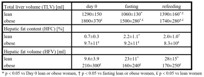 |
34 | Liver response to fasting and isocaloric high-carbohydrate refeeding in lean and obese women
Petr Sedivy1, Tereza Dusilova1, Barbora Setinova1, Monika Dezortova1, Martin Burian1, Eva Krauzova2, Michaela Siklova3, Lenka Rossmeislova3, Michal Koc3, Milan Hajek1, and Jan Kovar4
1MR unit, Department of Diagnostic and Interventional Radiology, Institute for Clinical and Experimental Medicine, Prague, Czech Republic, 2Department of Internal Medicine, Third Faculty of Medicine, Charles University, University Hospital Královské Vinohrady, Prague, Czech Republic, 3Department of Pathophysiology, Third Faculty of Medicine, Charles University, Prague, Czech Republic, 4Centre of Experimental Medicine, Institute for Clinical and Experimental Medicine, Prague, Czech Republic
Our MRS and MRI study investigates the liver response to two days of fasting followed by two days of isocaloric high carbohydrate refeeding in lean and obese women. Hepatic fat content increased after fasting in lean women whereas it did not change in obese women. The tendency to lose or accumulate fat during fasting could be related to the overall initial hepatic fat content. Moreover, liver volume significantly decreased and increased during fasting and refeeding, respectively.
|
||
2767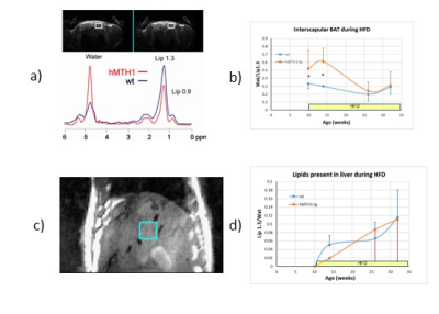 |
35 | The over-expression of human hydrolase hMTH1 modulates metabolism and fat composition in mice exposed to high fat diet: a MRI and MRS study Video Permission Withheld
Rossella Canese1, Gabriele De Luca2, Taljinder Singh1, Ambra Dell'Orso3, Egidio Iorio1, Mattea Chirico1, Maria Elena Pisanu1, Paola Fortini3, and Valeria SImonelli3
1Core Facilities, Istituto Superiore di Sanita', Rome, Italy, 2Oncology and Molecular Medicine Dept, Istituto Superiore di Sanita', Rome, Italy, 3Environmental and Health Dept, Istituto Superiore di Sanita', Rome, Italy
Oxidative stress is implicated in cancer, neurodegeneration and aging. hMTH1 is a hydrolase able to remove oxidized precursors from nucleotide’s pool, thus avoiding oxidative nucleic acids damage. Overexpression of hMTH1 in mice is protective against oxidative damage, neurodegeneration and prolongs life span. Our study showed that the overexpression of hMTH1 in mice fed with high fat diet (HFD), a dietary regimen linked to inflammation, is associated with increased brown interscapular fat (linked to protection from obesity) and with reduced perivesical fat volume (indicator of poor cardiovascular outcomes) up to four weeks. These effects seem to be reversed by prolonging HFD.
|
||
2768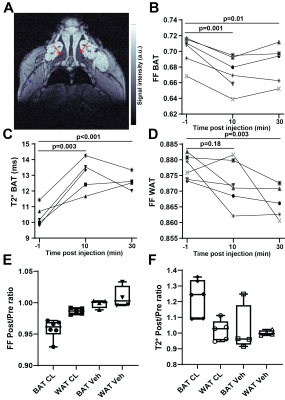 |
36 | Mouse models of diet-induced obesity allow for optimal magnetic resonance measurements of brown adipose tissue activation and heterogeneity
Michal R Tomaszewski1, Hyking Haley1, Xiangjun Meng1, and Corin O Miller1
1Translational Imaging, Merck & Co, West Point, PA, United States
Robust measurement of brown and white adipose tissue physiology in response to treatment is essential in pharmaceutical research. In this work we propose for the first time that the diet-induced obesity (DIO) mouse model can be successfully used in conjunction with both Multi Gradient Echo and Fast Spin Echo based methods for sensitive characterization of changes in brown (BAT) and white adipose tissue following pharmaceutical intervention, showing more consistent responses than lean mice. Spatial heterogeneity within BAT and response dynamics are reported, relevant for future use of the model and the proposed framework in weight loss and drug characterization studies.
|
||
 Back to Meeting Home
 Back to the Program-at-a-Glance
The International Society for Magnetic Resonance in Medicine is accredited by the Accreditation Council for Continuing Medical Education to provide continuing medical education for physicians.
 Back to Meeting Home
 Back to the Program-at-a-Glance
 View the Presentation