Digital Poster
Multiparametric MR in Cancer
Joint Annual Meeting ISMRM-ESMRMB & ISMRT 31st Annual Meeting • 07-12 May 2022 • London, UK

| Computer # | ||||
|---|---|---|---|---|
0933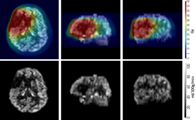 |
53 | Changes in cortical blood flow >1 year after radiation for glioma using arterial spin labelling MRI Video Permission Withheld
Magdalena Sokolska1, Meetakshi Gupta2, Björn Eiben3, Julia Markus4, Harpreet Hyare4, and Michael Kosmin2
1Medical Physics and Biomedical Engineering, University College London Hospitals, London, United Kingdom, 2Radiotheraphy, University College London Hospitals, London, United Kingdom, 3Department of Medical Physics and Biomedical Engineerin, University College London, London, United Kingdom, 4Imaging, University College London Hospitals, London, United Kingdom
This work proposes a framework for monitoring brain perfusion after radiotherapy treatment of glioma in relation to the radiotherapy dose.
|
||
0934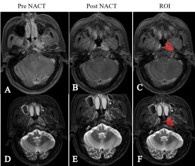 |
54 | Prediction of prognosis of nasopharyngeal carcinoma after neoadjuvant chemotherapy based on radiomics and multimodal machine learning Video Not Available
Chunmiao Hu1, Zhijian Hu2, Li Chen3, DeChun Zheng4, Yunbin Chen4, Xisheng Cao4, and Tao Lu4
1Radiology, Fujian Medical University Cancer Hospital, Fujian Cancer Hospital, Fuzhou, China, 2School of Public Health, School of Public Health, Fujian Medical University, Fuzhou Fujian 350122, China, Fuzhou Fujian, China, 3School of Arts and Sciences, School of Arts and Sciences, Fujian Medical University, Fuzhou Fujian 350122, China, Fuzhou Fujian, China, 4Fujian Medical University Cancer Hospital, Fujian Cancer Hospital, Fuzhou, China
This study is the first to adopt post-NACT MR radiomics to predict the recurrence of locally advanced NPC (stages III and IVa).
|
||
0935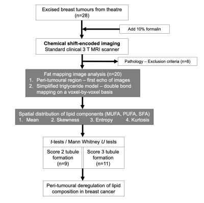 |
55 | Peri-tumoural spatial distribution of lipid composition and tubule formation in breast cancer
Sai Man Cheung1, Kwok-Shing Chan1,2, Nicholas Senn1, Ehab Husain3, Yazan Masannat4, Steven D Heys1, and Jiabao He1
1Institute of Medical Sciences, University of Aberdeen, Aberdeen, United Kingdom, 2Donders Institute for Brain, Cognition and Behaviour, Radboud University, Nijmegen, Netherlands, 3Pathology Department, Aberdeen Royal Infirmary, Aberdeen, United Kingdom, 4Breast Unit, Aberdeen Royal Infirmary, Aberdeen, United Kingdom
The spatial distribution of lipid composition in breast has a major role in breast cancer prevention, with deregulation of lipid metabolism identified in BRCA1/2 genetic mutation carriers. Neoplastic tubule formation, a key component of grading score, is directly associated with cellularity, with significant implications on prognosis. Lipid composition measurement through biochemical extraction is invasive, while conventional spectroscopic imaging demands long acquisition time. Novel chemical shift-encoded imaging (CSEI) allows lipid composition mapping of the whole breast in a clinically acceptable timeframe. We set out to examine the relationship between peri-tumoural lipid composition and tubule formation using CSEI in breast tumours.
|
||
0936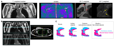 |
56 | Elevated magnetic resonance imaging measures of adipose tissue deposition in women with breast cancer treatment-related lymphedema
Rachelle Crescenzi1,2,3, Paula Donahue4, Maria Garza5, Chelsea A Lee6, Niral J Patel6, Victoria Gonzalez7, Sky Jones5, and Manus J Donahue5,8
1Department of Radiology and Radiological Sciences, Vanderbilt University Medical Center, Nashville, TN, United States, 2Vanderbilt University Institute of Imaging Science, Vanderbilt University Medical Center, Nashville, TN, United States, 3Department of Biomedical Engineering, Vanderbilt University Medical Center, Nashville, TN, United States, 4Department of Physical Medicine and Rehabilitation, Vanderbilt University Medical Center, Nashville, TN, United States, 5Department of Neurology, Vanderbilt University Medical Center, Nashville, TN, United States, 6Department of Pediatrics, Vanderbilt University Medical Center, Nashville, TN, United States, 7School of Medicine, The City College of New York, New York, NY, United States, 8Department of Psychiatry and Behavioral Sciences, Vanderbilt University Medical Center, Nashville, TN, United States
The overall goal of this work is to apply Dixon fat-water MRI to test fundamental hypotheses regarding the role of elevated adiposity in breast cancer treatment-related lymphedema (BCRL) condition severity. BCRL is a common co-morbidity of breast cancer therapies, yet factors that contribute to BCRL progression remain incompletely characterized. We observed that adiposity quantified by MRI is elevated in the affected upper extremity and torso of women with BCRL and increases with condition severity. As such, Dixon MRI may provide a surrogate marker of BCRL onset and may help to inform the varied course of lymphedema progression.
|
||
0937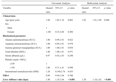 |
57 | Magnetic Resonance Elastography As a Marker for Prediction of Hepatocellular Carcinoma in Patients with HBV-Related Decompensated Cirrhosis
Haimei Chen1, Jie Zhu1, Mengsi Li1, Li Lin1, Ziying Yin2, Jun Chen2, Meng Yin2, Richard L. Ehman2, and Jin Wang1
1Department of Radiology, The Third Affiliated Hospital, Sun Yat-Sen University (SYSU), No. 600, Tianhe Road, Guangzhou, Guangdong 510630, People’s Republic of China, Guangzhou, China, 2Department of Radiology, Mayo Clinic College of Medicine, Mayo Clinic, Rochester. MN. USA, Rochester, MN, United States Hepatitis B virus (HBV)-related decompensated cirrhosis is well-known risk factor for developing hepatocellular carcinoma (HCC). Therefore, it is critical to assess the risk of developing HCC in these patients. Magnetic resonance elastography (MRE) has promise in predicting future events in many chronic liver diseases, but its prognostic role in predicting HCC development of HBV-related decompensated cirrhosis remains unclear. In our study, we evaluated the performance of baseline liver stiffness measurement (LSM) by MRE in predicting HCC development in patients with HBV-related decompensated cirrhosis. Our results showed that MRE-based LSM was associated with HCC development and is independently predictive of HCC development in patients with HBV-related decompensated cirrhosis. |
||
0938 |
58 | DTI quantitative parameter texture features for differentiating solid pseudopapillary neoplasm from pancreatic neuroendocrine tumors
Yi Wang1, Xinqi Wang2, Lu Wang2, Lizhi Xie3, Qinhe Zhang4, and Ailian Liu4
1Department of Radiology, Dalian Friendship Hospital, Dalian, China, 2School of Medical Imaging, Dalian Medical University, Dalian, China, 3GE Healthcare, MR Research China, Beijing, China, 4Department of Radiology, the First Affiliated Hospital of Dalian Medical University, Dalian, China
This work aimed for diffusion tensor imaging (DTI) quantitative parameter texture features based strategy to identify solid pseudopapillary neoplasm(SPN)and pancreatic neuroendocrine tumors(PNET),which may represent a diagnostic challenge due to many overlapping MRI features. The results showed that Large Area Emphasis (AUC: 0.737, sensitivity: 64.7%, specificity: 80%on FA signal intensity) was the optimal strategy to identify SPN and PNET.
|
||
0939 |
59 | Predictive value of T1 mapping in cervical cancer recurrence after treatment Video Permission Withheld
Jie Liu1, Shujian Li1, Qinchen Cao1, Marcel Dominik Nickel2, Jingliang Cheng1, and Jinxia Zhu3
1the First Affiliated Hospital of Zhengzhou University, Zheng zhou, China, 2MR Application Predevelopment, Siemens Healthcare GmbH, Erlangen, Germany, 3MR Collaboration, Siemens Healthineers Ltd., Beijing, China
We investigated the feasibility of T1-mapping to predict cervical cancer (CC) recurrence after treatment. Our results show that quantitative T1 values of the primary tumor could effectively predict the CC recurrence after surgery or concurrent chemoradiotherapy (CCRT). These findings suggest that the T1-mapping method could be valuable for evaluating CC recurrence after treatment, and native T1 values of the primary tumor could predict cervical cancer recurrence.
|
||
0940 |
60 | Evaluation of Multiparametric Magnetic Resonance Imaging to assist Adaptive Radiotherapy in Prostate Cancer – an interim report
Angela Turnbull1, N. Jane Taylor2, Amish Lakhani2, William McGuire2, Rachael Bowie2, Roberto Alonzi3, and Alan Mcwilliam4
1Radiotherapy Physics, Mount Vernon Cancer Centre, Northwood, United Kingdom, 2Paul Strickland Scanner Centre, Mount Vernon Cancer Centre, Northwood, United Kingdom, 3Mount Vernon Cancer Centre, Northwood, United Kingdom, 4The Christie NHS Foundation Trust and Division of Cancer Services and University of Manchester, Manchester, United Kingdom
Initial results of a prostate cancer study investigating whether multiparametric MRI (MP-MRI), involving diffusion-weighted (DW) and dynamic contrast-enhanced (DCE) MRI, can predict or assess tumour response to radiotherapy (RT) and potentially support adaptive radiotherapy for high-risk patients. Adaptive radiotherapy is a treatment technique utilised to minimise radiation related toxicity. For some cancers it is possible to adapt radiotherapy according to physical changes that occur during treatment. This is not appropriate for prostate cancer treatment since observable changes are generally due to rectal movement, however it may be possible to use multiparametric MRI (MP-MRI) to measure treatment response.
|
||
0941 |
61 | Preliminary study of amide proton transfer-weighted with intravoxel incoherent motion imaging in predicting bone metastasis of prostate cancer Video Not Available
Wenjun Hu1, Lihua Chen 1, Liangjie Lin2, Xu Dai2, Jiazheng Wang 2, and Ailian Liu1
1Department of Radiology, the First Affiliated Hospital of Dalian Medical University, Dalian, China, 2Philips Healthcare, Beijing, China
Bone metastasis is an important issue in the management of prostate cancer, and can drastically alter the treatment strategy. PET/CT has been widely used in the diagnosis of bone metastasis; however, the high cost and radiation exposure limit its extensive clinical applications. APT-weighted imaging and IVIM, recent developed function-oriented MRI sequences, allow for a non-invasive visualization of tissue composition and microscopic information without the need for contrast agents. Results of this study indicate the APT value and IVIM parameters can effectively predict bone metastases in prostate cancer. A combination of APT and D* mono can further improve the prediction performance.
|
||
0942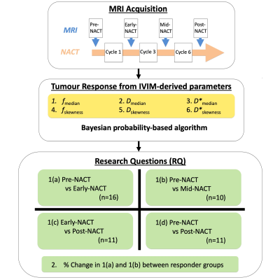 |
62 | Intravoxel incoherent motion improves diffusion-weighted imaging in detection of response towards neoadjuvant chemotherapy in breast cancer
Sai Man Cheung1, Wing Shan Wu1, Nicholas Senn1, Ravi Sharma2, Trevor McGoldrick2, Tanja Gagliardi1,3, Ehab Husain4, Yazan Masannat5, and Jiabao He1
1Institute of Medical Sciences, University of Aberdeen, Aberdeen, United Kingdom, 2Oncology Department, Aberdeen Royal Infirmary, Aberdeen, United Kingdom, 3Radiology Department, Royal Marsden Hospital, London, United Kingdom, 4Pathology Department, Aberdeen Royal Infirmary, Aberdeen, United Kingdom, 5Breast Unit, Aberdeen Royal Infirmary, Aberdeen, United Kingdom
Breast cancer is a major and expanding health challenge, and neoadjuvant chemotherapy (NACT) is increasingly prescribed to facilitate breast surgery in advanced breast cancer with an ongoing demand for improved imaging methods accurately reflecting disease load. Tissue perfusion, a sensitive marker of cancer metabolism, can be derived from intravoxel incoherent motion (IVIM) model, and recent Bayesian algorithm yields improved sensitivity and precision in breast cancer by us and pancreatic cancer. We therefore hypothesise that IVIM model powered by Bayesian algorithm is able to detect early treatment-induced changes in tumour perfusion and diffusion, with the potential to impact patient care pathway.
|
||
0943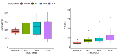 |
63 | Short- and long-term changes of ADC and rF% in patients with bone metastases from breast cancer
Alberto Colombo1, Eleonora Giardini2, Paul Eugene Summers1, Paola Pricolo1, Fabio Zugni1, Maddalena Belmonte1, and Giuseppe Petralia3,4
1Division of Radiology, IEO European Institute of Oncology IRCCS, Milan, Italy, 2Degree course in Medical radiology, imaging and radiotherapy techniques, University of Milan, Milan, Italy, 3Precision Imaging and Research Unit, Department of Medical Imaging and Radiation Sciences, IEO European Institute of Oncology IRCCS, Milan, Italy, 4Department of Oncology and Hemato-Oncology, University of Milan, Milan, Italy Evaluating short and long-term apparent diffusion coefficient (ADC) and relative fat fraction (rF%) changes could provide a better understanding of therapy response of bone metastases from breast cancer. Baseline ADC and rF% values computed from whole-body MRI, were compared with those measured at 12 and 36 week evaluations after treatment start. ADC and rF% significantly increased in responders at 12-weeks. At 36-weeks, ADC values range broadened, while rF% further increased. Short-term ADC and rF% changes are consistent with previous reports. Long-term changes highlight two patterns of response in which either high diffusion or fat repopulation prevail. |
||
 Back to Meeting Home
 Back to the Program-at-a-Glance
The International Society for Magnetic Resonance in Medicine is accredited by the Accreditation Council for Continuing Medical Education to provide continuing medical education for physicians.
 Back to Meeting Home
 Back to the Program-at-a-Glance
 View the Presentation