Digital Poster
Preclinical Neuro I
Joint Annual Meeting ISMRM-ESMRMB & ISMRT 31st Annual Meeting • 07-12 May 2022 • London, UK

| Computer # | ||||
|---|---|---|---|---|
2325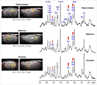 |
61 | Animal models using C57BL/6J mice are at risk of data misinterpretation due to random occurrences of animals with high levels of brain glutamine
Ivan Tkac1, Raghavendra Rao2, and Megan Paulsen2
1Center for Magnetic Resonance Research, University of Minnesota, Minneapolis, MN, United States, 2Department of Pediatrics, University of Minnesota, Minneapolis, MN, United States
Wild-type and transgenic mice derived from the C57BL/6J mouse strain are widely used in animal models of different human diseases. However, this mouse strain suffers from random appearances of sporadic congenital portosystemic shunts resulting in extremely high glutamine levels in the brain. In our recent study aimed at effects of maternal obesity on their offspring we found that in 9 out of 31 studied C57BL/6J mice had high levels of brain Gln. The purpose of this abstract is to highlight the awareness of this persisting problem in animal models based on C57BL/6J background strain.
|
||
2326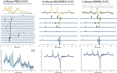 |
62 | Preclinical MR spectroscopy of GABA: conventional PRESS versus spectral editing with MEGAPRESS and HERMES
Diana Rotaru1, Steve Sawiak2,3, Camilla Simmons1,4, Eugene Kim1,4, Maria Elisa Serrano Navacerrada1,4, Davide Di Censo1,4, Adrien Le Guennec5, Richard Edden6,7, David Lythgoe1, and Diana Cash1,4
1Neuroimaging, King's College London, London, United Kingdom, 2Behavioural and Clinical Neuroscience Institute, University of Cambridge, Cambridge, United Kingdom, 3Wolfson Brain Imaging Centre, University of Cambridge, Cambridge, United Kingdom, 4BRAIN Centre (Biomarker Research And Imaging for Neuroscience), King's College London, London, United Kingdom, 5NMR Facility, Wolfson CARD, Guy's Campus, King's College London, London, United Kingdom, 6Russell H. Morgan Department of Radiology and Radiological Science, The Johns Hopkins University School of Medicine, Baltimore, Baltimore, MD, United States, 7F. M. Kirby Research Center for Functional Brain Imaging, Kennedy Krieger Institute, Baltimore, MD, United States
In vivo GABA measurements with conventional (PRESS) and spectral editing methods (MEGAPRESS and HERMES) were investigated in healthy mice at 9.4 T. Animals were divided in two groups, control mice injected with saline and mice injected with Aminooxyacetic acid to intentionally elevate GABA concentration levels (n=7 per group). Our results show all three methods successfully discriminate GABA from overlapping metabolites with stronger signals. The precision of concentration estimates is more reliable for spectral editing methods given their specificity to GABA. These outcomes suggest spectral editing methods are less susceptible to systematic biases.
|
||
2327 |
63 | MR Spectroscopy of HP [1-13C]Pyruvate in an In Vitro Model of Epilepsy
Aditya Jhajharia1, Riccardo Serra2, Muznabanu Bachani2, Jemima Olu-Owotade2, Minjie Zhu1, Joshua Rogers1, Alexander Ksendzovsky2, and Dirk Mayer1
1Diagnostic Radiology and Nuclear Medicine, University of Maryland School of Medicine, Baltimore, MD, United States, 2Department of Neurosurgery, University of Maryland School of Medicine, Baltimore, MD, United States
In this study, we investigated hyperpolarized (HP) magnetic resonance spectroscopy (MRS) as a means to identify differential lactate release in an in vitro model of epilepsy. When neurons are chronically activated, they increase LDHA expression and upregulate glycolysis as a means of cellular energy metabolism, thus increasing the production of lactate. Using MRS of HP [1-13C]pyruvate, we showed conversion to lactate in a chronic model of epilepsy. These results indicate that metabolic imaging of HP [1-13C]pyruvate can potentially be used as a tool to identify lactate elevations and thus the epileptic focus in epilepsy patients.
|
||
2328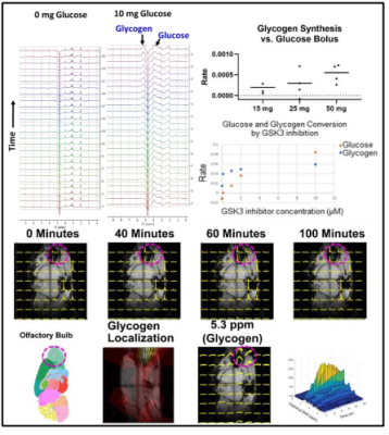 |
64 | Inhibition of GSK3 Activity Detected by Glycogen Synthase Activity from Proton MRS at 3T
Jeff Brender1, Shun Kishimoto1, Jeeva Munasinghe2, Helmutt Merkle2, Kota Yamashita1, Yasunori Otowa1, Kazutoshi Yamamoto1, and Murali Cherukuri Krishna1
1Radiation Biology Branch, NCI, Bethesda, MD, United States, 2NINDS, Bethesda, MD, United States
Glycogen synthase kinase 3 (GSK3) has been tied as a critical factor in the development of multiple major diseases from cancer to Alzheimer’s, but drug development has been stymied by a lack of options to detect GSK3 activity in vivo. We show here that GSK3 activity can be measured accurately in vivo by following the production of glycogen from a single bolus of unlabeled glucose on a standard 3T preclinical imaging system. Imaging of glycogen synthesis was possible at 9.4T by CSI, where it was found in mice to be centered on the olfactory bulb, as expected from post-mortem analysis
|
||
2329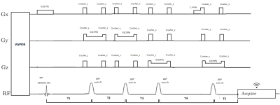 |
65 | High resolution Multi-voxel spectroscopy using CSI-semi-LASER for mouse brain preclinical studies
Zoona Javed1,2, Gary Martinez3, AnaPaula Candiota1,4,5, Miquel Cabanas2,4, and Silvia Lope2,4
1Department of Biochemistry and Molecular Biology, Universitat Autonoma de Barcelona, Bellaterra, Spain, 2Servei de Ressonància Magnètica Nuclear, Universitat Autonoma de Barcelona, Bellaterra, Spain, 3Department of Imaging Physics, University of Texas, Houston, TX, United States, 4Centro de Investigación Biomédica en Red en Bioingeniería, Biomateriales y Nanomedicina (CIBER-BBN),, Universitat Autonoma de Barcelona, Bellaterra, Spain, 5Institut de Biotecnologia i de Biomedicina (IBB), Universitat Autonoma de Barcelona, Bellaterra, Spain
This work describes an improved multi-voxel Chemical shift imaging (CSI) pulse sequence that uses the semi-LASER localization approach. Bruker CSI sequence containing PRESS localization block (CSI-PRESS) was modified by replacing the aforementioned PRESS block with a semi-LASER one resulting in a CSI-semi-LASER sequence. This work was developed on a 7T Bruker BioSpec 70/30 USR spectrometer running ParaVision 5.1 which does not contain neither built-in blocks for semi-LASER sequence nor adiabatic pulses. The sequence was tested in wt C57/BL6 mouse brain and compared with the stock CSI-PRESS sequence. The results show improved homogeneity and reduction in chemical shift displacement error (CSDE).
|
||
2330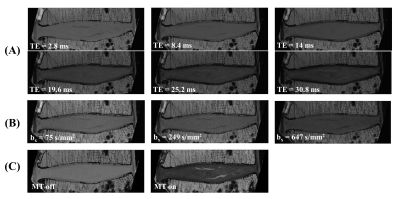 |
66 | Quantitative MR Imaging of Whole Intervertebral Disc: A Pre-Clinical Sample Study
Jiyo S Athertya1, Alecio F Lombardi2, Jonathan Wong2, Hyungseok Jang1, Saeed Jerban1, Jiang Du1, Koichi Masuda3, Eric Y Chang1,2, and Ya-Jun Ma1
1Radiology, University of California San Diego, San Diego, CA, United States, 2Radiology Service, Veterans Affairs San Diego Healthcare System, San Diego, CA, United States, 3Orthopedic Surgery, University of California San Diego, San Diego, CA, United States
Quantitative MR imaging is a powerful tool for assessing biochemical changes in tissue. In this study, we propose to measure the transverse magnetization relaxation time, diffusivity, and magnetization transfer ratio for a whole intervertebral disc (IVD), including the annulus fibrosis, cartilaginous endplate (CEP), and nucleus pulposus, on a 3T pre-clinical scanner. The sequence parameters were optimized for high resolution and high signal-to-noise ratio imaging and mapping, utilizing the high-performance gradient system on the pre-clinical scanner, and the echo times were sufficiently minimized to capture the fast-decaying CEP signals for all the quantitative imaging sequences.
|
||
2331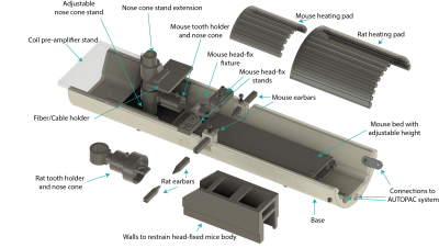 |
67 | Open-source modular 3D printed platform for in-vivo MRI experiments in awake mice and anesthetized mice and rats
Zakia Gironda1, Mihály Vöröslakos2, Youssef Wadghiri3, Omid Yaghmazadeh4, and Leeor Alon3
1Radiology, NYU School of Medicine, NEW YORK, NY, United States, 2Neuroscience Institute, NYU School of Medicine, New york, NY, United States, 3NYU School of Medicine, New York, NY, United States, 4Neuroscience Institute, NYU School of Medicine, NEW YORK, NY, United States
Despite their costly nature, the inexactitude of design in off-the-shelf MRI animal holders translates to a fastidious setup, iso-center misalignment, and incorrect animal body positioning. Our open-source 3D-print design provides precise iso-center alignment and minimizes motion artifact using its various head fixing mechanisms. It ensures comfortable positioning for rats and mice during in-vivo anesthetized and awake animal scan acquisition. The modular holder parts enable the same design to be used with other imaging modalities with minimal intervention. The design is low cost, ensures reproducibility and time efficiency of setup.
|
||
2332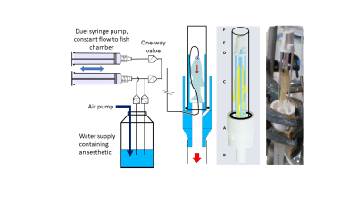 |
68 | The effect of gadolinium contrast agent on T1 and T2 relaxation of brain in live and fixed zebrafish MRI
Noémie Hamilton1, Claire Allen2, and Steven Reynolds3
1Neuroscience Institute, University of Sheffield, Sheffield, United Kingdom, 2The Bateson Centre, University of Sheffield, Sheffield, United Kingdom, 3Medical School, University of Sheffield, Sheffield, United Kingdom
Zebrafish have become a ubiquitous animal model for studying a range of diseases and conditions. Typically, these studies are conducted in transparent juvenal fish where optical imaging techniques can be used. However, this is more difficult in adult zebrafish, restricting their use in longitudinal studies. Brain pathology in adult zebrafish can be imaged by preclinical MRI. Administering gadolinium based contrast agents (GBCA), which can also reduce scanning time, can highlight brain abnormalities such as lesions. T1/T2 values have been reported for gadolinium treated fixed/sacrificed zebrafish, however, this has not been reported for live zebrafish.
|
||
2333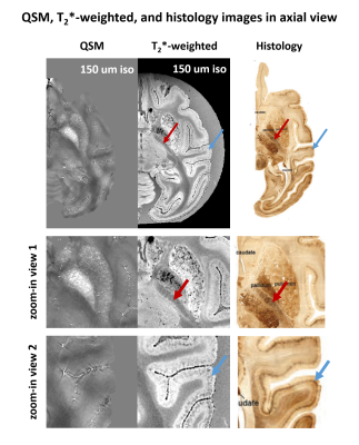 |
69 | Imaging ex-vivo cynomolgus monkey brain in 150 um isotropic resolution
Chungseok Oh1, Yu Gyeong Kim2,3, Jeongheon Park4, Youngkyu Song4, Chang-Yeop Jeon2, Kyung Seob Lim5, Jincheol Seo2, Jung Bae Seong2, Jee-Hyun Cho4, Youngjeon Lee2, Jongho Lee1, and Hyeong-Geol Shin1
1Department of Electrical and Computer Engineering, Seoul National University, Seoul, Korea, Republic of, 2National Primate Research Center, Korea Research Institute of Bioscience and Biotechnology (KRIBB), Cheongju, Korea, Republic of, 3Department of Functional Genomics, KRIBB School of Bioscience, Korea University of Science and Technology (UST), Daejeon, Korea, Republic of, 4Center for Research Equipment, Korea Basic Science Institute, Cheongju, Korea, Republic of, 5Futuristic Animal Resource & Research Center, Korea Research Institute of Bioscience and Biotechnology (KRIBB), Cheongju, Korea, Republic of
Due to the small size of the cynomolgus brain, imaging the fine structures in the brain is a challenging task. In this study, a sample preparation method is suggested for imaging the ex-vivo cynomolgus monkey brain with sub-millimeter resolution in 7T MRI. Using the proposed method, we acquired 150 um isotropic resolution MR images for whole brain, and the fine structures in the cynomolgus monkey brain was observed.
|
||
2334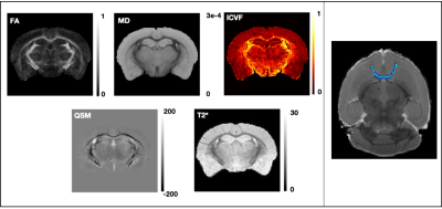 |
70 | White matter microstructure changes in a Bcan knockout mouse model
Cristiana Tisca1, Mohamed Tachrount1, Frederik J Lange1, Amy FD Howard1, Chaoyue Wang1, Lily R Qiu1, Benjamin C Tendler1, Jason P Lerch1,2,3, Aurea B Martins-Bach1, and Karla L Miller1
1Wellcome Centre for Integrative Neuroimaging, FMRIB, Nuffield Department of Clinical Neurosciences, University of Oxford, Oxford, United Kingdom, 2Mouse Imaging Centre, Hospital for Sick Children, Toronto, ON, Canada, 3Department of Medical Biophysics, University of Toronto, Toronto, ON, Canada
Recent genome-wide association studies using UK Biobank brain imaging datasets showed associations between microstructural MRI measures in white matter and genetic loci of BCAN, a gene encoding a protein implicated in neurodegeneration and synaptic transmission. To investigate these associations, we acquired ex vivo MRI data in a Bcan knockout mouse model. Our results show significant differences in FA and T2* for some tracts in wild-type males compared to homozygous males, with consistent trends of higher FA and MD across WM tracts. Future histology work in the same brains will reveal the biological changes underpinning these differences.
|
||
2335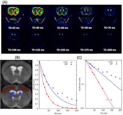 |
71 | Choroid plexus tissue perfusion and blood to CSF barrier function (BCSFB) in rats measured with continuous arterial spin labeling Video Permission Withheld
Hedok Lee1, Burhan Ozturk1, Michael Stringer2, Bradley MacIntosh3, Douglas Rothman4, and Helene Benveniste1
1Anesthesiology, Yale School of Medicine, New Haven, CT, United States, 2Centre for Clinical Brain Sciences, University of Edinburgh, Edinburgh, United Kingdom, 3Medical Biophysics, University of Toronto, Toronto, ON, Canada, 4Radiology and Biomedical Imaging, Yale School of Medicine, New Haven, CT, United States In this study choroid plexus tissue perfusion and blood to CSF barrier function in rats were studied using long and short TE continuous arterial spin labeling technique. Non-mono exponential perfusion weighted signal decay in the CP was modelled by a two-compartment perfusion model and we report that partial volume effect is significant and must be considered to derive accurate choroid plexus tissue perfusion measurements. |
||
2336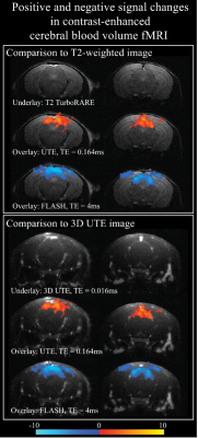 |
72 | Evaluating ultra-short echo time (UTE) cerebral blood volume (CBV)-based fMRI using an iron-oxide contrast agent in mouse visual cortex at 9.4T
Naman Jain1, Saskia Bollmann1, Jonathan R. Polimeni2,3,4, Kai-Hsiang Chuang1,5, and Markus Barth1,6,7
1Centre for Advanced Imaging, St. Lucia, Australia, 2Athinoula A. Martinos Center for Biomedical Imaging, Massachusetts General Hospital, Charlestown, MA, United States, 3Department of Radiology, Harvard Medical School, Boston, MA, United States, 4Division of Health Sciences and Technology, Massachusetts Institute of Technology, Cambridge, MA, United States, 5Queensland Brain Institute, The University of Queensland, St. Lucia, Australia, 6School of Information Technology and Electrical Engineering, St. Lucia, Australia, 7ARC Centre for Innovation in Biomedical Imaging and Technology, The University of Queensland, St. lucia, Australia
CBV-based fMRI has been demonstrated to have better spatial specificity than BOLD. CBV estimates based on iron-oxide contrast agents (CA) reflect this microvascular sensitivity, however because of strong extravascular dephasing CBV changes within large vessels are more difficult to detect. Here, we measured functional CBV changes after injecting CA with UTE-MRI and found positive signal changes (+0.5%) predominantly localized at large vessels at a TE of 0.164ms, and negative signal changes (-2%) at a TE of 4ms localized within the tissue. This indicates sensitivity to different vascular compartments at different TEs reflecting the respective T1 and T2* effects of CA.
|
||
2337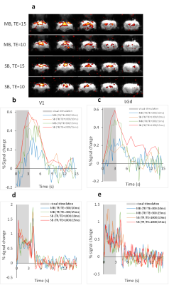 |
73 | Estimating Hemodynamic Response Function Using Simultaneous fMRI and Calcium Recording Video Permission Withheld
Shabnam khorasani gerdekoohi1, Pankaj Sah1, and Kai-Hsiang Chuang1
1Queensland Brain Institute, The University of Queensland, Brisbane, Australia · Evoked BOLD and calcium signals were recorded in the visual pathway using standard (TR=1s) and ultrafast (TR=0.3s) EPI of two TEs. · Hemodynamic response function was estimated based on neuronal calcium activity. HRF estimated from ultrafast fMRI shows faster kinetics in the lateral geniculate nucleus than the visual cortex. · BOLD signal was predicted from calcium signal convoluted by HRF. Using ultrafast fMRI significantly improved the accuracy of the predicted BOLD. |
||
2338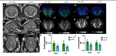 |
74 | DTI and fMRI Biomarkers in a Pig Model of Pediatric Traumatic Brain Injury
Alesa Hughson Netzley1, Andrew Badachhape2, Lauren Wade-Kleyn3, Jie Huang4, Kirk Munoz5, Aimee Colbath5, Avner Meoded2, and Galit Pelled1
1Biomedical Engineering, Michigan State University, East Lansing, MI, United States, 2Radiology, Baylor College of Medicine, Houston, TX, United States, 3Neuroscience, Michigan State University, East Lansing, MI, United States, 4Radiology, Michigan State University, East Lansing, MI, United States, 5Veterinary Medicine, Michigan State University, East Lansing, MI, United States
The goal of this work is to identify MRI biomarkers of mild traumatic brain injury (mTBI) using a pig model of pediatric concussion. We have found changes in the brain using DTI and fMRI in the days and weeks immediately following injury. We also collected data from a battery of cognitive, emotional, and motor assessments. Together, we anticipate these data will help us to identify biomarkers of injury that will improve diagnostics of children to decrease the risk of post-injury complications.
|
||
 Back to Meeting Home
 Back to the Program-at-a-Glance
The International Society for Magnetic Resonance in Medicine is accredited by the Accreditation Council for Continuing Medical Education to provide continuing medical education for physicians.
 Back to Meeting Home
 Back to the Program-at-a-Glance
 View the Presentation