Digital Poster
Preclinical Neuro II
Joint Annual Meeting ISMRM-ESMRMB & ISMRT 31st Annual Meeting • 07-12 May 2022 • London, UK

| Computer # | ||||
|---|---|---|---|---|
2411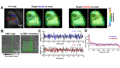 |
40 | Resting-state single-vessel cerebral blood velocity by phase contrast fMRI Video Permission Withheld
Yuanyuan Jiang1, David Christopher Hike1, and Xin Yu1
1Massachusetts General Hospital, Martinos Center for Biomedical Imaging, Charlestown, MA, United States
This research utilizes a novel high-resolution phase-contrast (PC) MRI method to measure resting-state fluctuation of cerebral blood flow-related velocity (CBFv) from penetrating vessels in the rat parietal cortex. This novel PC-based mapping scheme has identified vessel-specific correlation and anti-correlation patterns between venules and arterioles with the low-frequency oscillatory features less than 0.05Hz in anesthetized rat brains. By combining PC-based CBFv fMRI and single-vessel cerebral blood volume (CBV)-fMRI, we will further study the vasodynamic changes in diseased animal models.
|
||
2412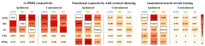 |
41 | Bilateral homotopic rs-fMRI connectivity strength is not primarily mediated by direct corticocortical interaction
Won Beom Jung1, Haiyan Jiang1,2, and Seong-Gi Kim1,2
1Center for Neuroscience Imaging Research (CNIR), Suwon, Korea, Republic of, 2Department of Biomedical Engineering, Sungkyunkwan University, Suwon, Korea, Republic of
Functional connectivity measured by rs-fMRI have generally shown a bilateral organization in homotopic cortices, presumably related to the intrinsic network of spontaneous activity. Alternatively, cortical silencing suppresses spontaneous output activity from the inactivated site and reduces input to downstream areas. Thus, the decrease in fMRI responses due to cortical silencing is related to the strength of resting-state connectivity between the stimulation site and the connected regions. To examine the contribution of spontaneous neuronal communications to bilateral homotopic connectivity of rs-MRI, we compared the somatosensory network by rs-fMRI with cortical silencing fMRI by optogenetic stimulation of interneurons and anatomical tracing data.
|
||
2413 |
42 | Hypercapnia does not change CMRO2: Data from mouse studies using multimodal NIRS-MRI at 9.4T
Mada Hashem1,2,3,4, Ying Wu2,3,4, and Jeff F. Dunn2,3,4
1Biomedical Engineering Graduate Program, University of Calgary, Calgary, AB, Canada, 2Department of Radiology, University of Calgary, Calgary, AB, Canada, 3Hotchkiss Brain Institute, University of Calgary, Calgary, AB, Canada, 4Experimental Imaging Centre, University of Calgary, Calgary, AB, Canada Non-invasive quantitative imaging of cerebral metabolic rate of oxygen (CMRO2) is essential to understand oxidative metabolism in health and disease. Hypercapnia is often used as a calibration for MRI methods associated with quantifying CMRO2. Here we show that CMRO2 remains constant during hypercapnia. We also provide a novel multimodal NIRS-MRI system that can assess absolute values of multiple metabolic correlates noninvasively in living mouse brain, and with no need for calibration with gas mixtures. Moreover, this technique is capable to monitor mitochondrial status by measuring the redox state of cytochrome oxidase, the enzyme responsible for oxygen consumption in the cell. |
||
2414 |
43 | The Digital Brain Bank, an open access platform for post-mortem datasets
Benjamin C. Tendler1, Taylor Hanayik1, Olaf Ansorge2, Sarah Bangerter-Christensen2, Gregory S. Berns3, Mads F. Bertelsen4, Katherine L. Bryant1, Sean Foxley1,5, Martijn P. van den Heuvel6,7, Amy F.D. Howard1, Istvan N. Huszar1, Alexandre A. Khrapitchev8, Anna Leonte2, Paul R. Manger9, Ricarda A.L. Menke1, Jeroen Mollink1, Duncan Mortimer1, Menuka Pallebage-Gamarallage2, Lea Roumazeilles10, Jerome Sallet10,11,
Lianne H. Scholtens6, Connor Scott2, Adele Smart1,2, Martin R. Turner1,2, Chaoyue Wang1, Saad Jbabdi1, Rogier B. Mars1,12, and Karla L. Miller1
1Wellcome Centre for Integrative Neuroimaging, FMRIB, Nuffield Department of Clinical Neurosciences, University of Oxford, Oxford, United Kingdom, 2Division of Clinical Neurology, Nuffield Department of Clinical Neurosciences, University of Oxford, Oxford, United Kingdom, 3Psychology Department, Emory University, Atlanta, GA, United States, 4Centre for Zoo and Wild Animal Health, Copenhagen Zoo, Copenhagen, Denmark, 5Department of Radiology, University of Chicago, Chicago, IL, United States, 6Department of Complex Trait Genetics, Centre for Neurogenomics and Cognitive Research, Amsterdam Neuroscience, Vrije Universiteit Amsterdam, Amsterdam, Netherlands, 7Department of Child Psychiatry, Amsterdam Neuroscience, Amsterdam UMC, Vrije Universiteit Amsterdam, Amsterdam, Netherlands, 8Medical Research Council Oxford Institute for Radiation Oncology, University of Oxford, Oxford, United Kingdom, 9School of Anatomical Sciences, Faculty of Health Sciences, University of the Witwatersrand, Johannesburg, South Africa, 10Wellcome Centre for Integrative Neuroimaging, Department of Experimental Psychology, University of Oxford, Oxford, United Kingdom, 11Stem Cell and Brain Research Institute, Université Lyon 1, INSERM, Lyon, France, 12Donders Institute for Brain, Cognition and Behaviour, Radboud University, Nijmegen, Netherlands
We introduce the Digital Brain Bank (open.win.ox.ac.uk/DigitalBrainBank), a digital platform providing open access to curated, multimodal post-mortem neuroimaging datasets. Datasets span three themes; Digital Anatomist: datasets for neuroanatomical investigations; Digital Brain Zoo: datasets for comparative neuroanatomy; Digital Pathologist: datasets for neuropathology investigations. The first release includes 21 distinctive whole-brain diffusion MRI datasets, alongside microscopy and complementary MRI modalities. This includes one of the highest-resolution whole-brain human diffusion MRI datasets ever acquired, whole-brain diffusion MRI in 14 non-human primate species, and one of the largest post-mortem whole-brain cohort imaging studies in neurodegeneration. Our resource facilitates incorporating post-mortem data into neuroimaging studies.
|
||
2415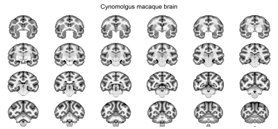 |
44 | Brain morphometric discrepancies between cynomolgus and rhesus macaques
Rakshit Dadarwal1,2 and Susann Boretius1,2
1Functional Imaging Laboratory, German Primate Center, Göttingen, Germany, 2Georg-August-University Göttingen, Göttingen, Germany
Cynomolgus and rhesus macaques are widely used in biomedical research due to their physiological proximity to humans, and both are often referred to as macaques. However, the brain morphology of cynomolgus and rhesus macaques differs. Interestingly, as shown here by a Jacobian-determinant-based approach, the smaller cynomolgus brain is not only a scaled-down version of the rhesus macaques. For instance, the gray-to-white matter ratio differs significantly between the two species.
|
||
2416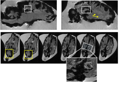 |
45 | Qualitative assessment of Ex-vivo Manganese-Enhanced MRI of gyrencephalic brains
Nathalie Just1, Martine Batailler2, Jean-Philippe Dubois2, and Martine Migaud2
1DRCMR, Copenhagen University Hospital - Amager and Hvidovre, Hvidovre, Denmark, 2INRAE, Nouzilly, France
In-vivo Manganese-Enhanced MRI (MEMRI) studies have shown important potential in rodents for the delineation of cytoarchitectural brain features and of functional details. Ex-vivo MEMRI on the other hand offers the possibility to investigate brain microstructure and function with improved spatial resolution and no motion artefacts. Here, ex-vivo lamb brains were immersed in a highly concentrated MnCl2 solution for a month and revealed interesting cytoarchitectural features already 24 hours after immersion and up to 3 months after the end of immersion on MPRAGE images at 3T. A novel ex-vivo MEMRI approach is proposed, which could benefit MEMRI translation to human studies.
|
||
2417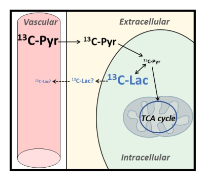 |
46 | Probing compartment-specific diffusion in the brain with 13C diffusion-weighted MRS of hyperpolarized pyruvate and lactate: initial results
Mélissa Vincent1, Xiao Gao2,3, Myriam Chaumeil2,3, and Julien Valette1
1MIRCen / Neurodegenerative Diseases Laboratory, CEA, Fontenay-aux-Roses, France, 2Department of Physical Therapy and Rehabilitation Science, UCSF, San Francisco, CA, United States, 3Department of Radiology and Biomedical Imaging, UCSF, San Francisco, CA, United States Here we propose to use 13C diffusion-weighted MRS of hyperpolarized pyruvate and lactate to probe diffusion properties in distinct compartments of the mouse brain, tentatively extracellular (ECS) and intracellular spaces (ICS). Hyperpolarized 13C-pyruvate is injected intravenously and taken up by the brain before entering cells, where it is quickly converted to lactate, so that 13C-pyruvate concentration in ICS must remain low, while newly produced 13C-lactate should remain predominantly intracellular during the relatively short measurement time. We measure faster diffusion for pyruvate even after removing IVIM effect, suggesting faster diffusion in the ECS, and slow release of lactate in the ECS. |
||
2418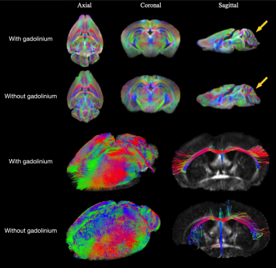 |
47 | Diffusion MRI protocol optimization for ex vivo mouse whole-brain tractography at 100μm isotropic resolution
Elise Cosenza1, Laurence Dallet1, Aurélien Trotier1, Emeline Ribot1, Laurent Petit2, and Sylvain Miraux1
1Centre de Résonance Magnétique des Systèmes Biologiques, UMR5536, CNRS, Bordeaux, France, 2Groupe dImagerie Neurofonctionnelle, Institut Des Maladies Neurodegeneratives, UMR5293, CNRS, CEA University of Bordeaux, CNRS, Bordeaux, France
A protocol, including Gadolinium-DOTA perfusion or immersion and Diffusion-Weighted multi-shot Spin-Echo EPI was developed to obtain 100μm isotropic resolution tractogram on ex vivo mouse brain. A 3mM Gd-DOTA concentration combined with 16 EPI shots at short TR (400 ms) allows to whole-brain tractograms of excellent quality and reproducible on the 15 mouse brains analyzed.
|
||
 Back to Meeting Home
 Back to the Program-at-a-Glance
The International Society for Magnetic Resonance in Medicine is accredited by the Accreditation Council for Continuing Medical Education to provide continuing medical education for physicians.
 Back to Meeting Home
 Back to the Program-at-a-Glance
 View the Presentation