Digital Poster
Preclinical: Contrast, Metabolomics & Hardware
Joint Annual Meeting ISMRM-ESMRMB & ISMRT 31st Annual Meeting • 07-12 May 2022 • London, UK

Digital Poster
Preclinical: Contrast, Metabolomics & Hardware
| Computer # | ||||
|---|---|---|---|---|
2419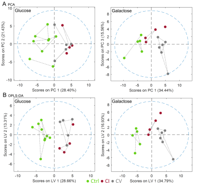 |
48 | Selective galactose culture condition reveals distinct metabolic signatures in complex I and V deficient human skin fibroblasts by 1H HR-MAS NMR
Christoph Meyer1,2,3, Damian Hertig1,2, Christian Urzi1,2,3, Janine Arnold2, Jean-Marc Nuoffer2,4, and Peter Vermathen1
1Magnetic Resonance Methodology, Institute of Diagnostic and Interventional Neuroradiology, University Bern, Bern, Switzerland, 2Institute of Clinical Chemistry, University Hospital Bern, Bern, Switzerland, 3Graduate School for Cellular and Biomedical Sciences, University of Bern, Bern, Switzerland, 4Department of Pediatric Endocrinology, Diabetology and Metabolism, University Children’s Hospital of Bern, Bern, Switzerland
Mitochondrial respiratory chain defects present as highly heterogeneous disorders which cannot be unambiguously diagnosed using standard laboratory methods. In this study we observed the biochemical consequences of complex I and complex V deficient human skin fibroblasts when cultivated under galactose stress condition compared to glucose based cell culture condition. We investigated extracellular flux using Seahorse XFe96 cell analyzer and assessed the metabolome fingerprints using High Resolution Magic Angle Spinning NMR. The selective culture method reveals CI and CV defect-specific changes in metabolites associated with the TCA cycle, malate aspartate shuttle and choline metabolism.
|
||
2420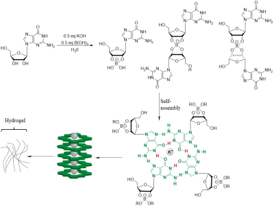 |
49 | Characterization of Self-assembled G4·K+ Hydrogel using CEST MRI Video Not Available
Xiaoling Gong1, Xiaoxiao Zhang2, Xiaoyong Zhang2, and Bing Wu1
1Department of Radiology, West China Hospital, Sichuan University, Chengdu, 610041, People’s Republic of China, Chengdu, China, 2Philips Healthcare, Beijing, China
CEST MRI is a novel molecular imaging technique in which the contrast is generated by the dynamic exchange between the exchangeable proton and water. Self-assembled G4·K+ Hydrogel can be fully characterized by its intrinsically endowed CEST contrast.
|
||
2421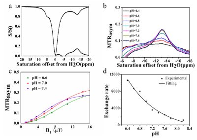 |
50 | Cross-Linked Protoporphyrin IX polymer as Efficient, Tunable CEST MRI NanoPlatform for molecular Imaging Video Permission Withheld
Xiaoxiao Zhang1, Zhigang Wu1, Peng Sun1, Zhiwei Shen1, Liangjie Lin1, Geli Hu1, Haoyu Huang1, and Jiazheng Wang1
1Philips Healthcare, Wuhan, China
CEST MRI is an novel molecular imaging technique in which the contrast is generated by the dynamic exchange between the exchangeable proton and water. Porphyrin and its analogues are a promising class of highly sensitive, diamagnetic CEST agents with highly upfield shifted protons. We synthesized a water-soluble polymeric CEST MRI agent grafted with protoporphyrin IX(PpIX). The PpIX polymeric agent resonated at -13.5 ppm from water and showed excellent CEST MRI properties.
|
||
2422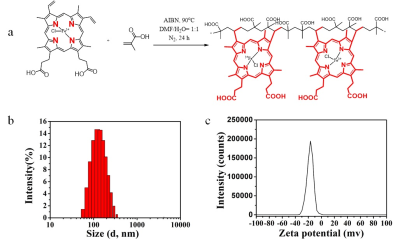 |
51 | A Highly Water-soluble Hemin-Based Polymeric Contrast Agents for Magnetic Resonance Imaging
Xiaoling Gong1, Xiaoxiao Zhang2, Xiaoyong Zhang3, Kai Ai4, and Bing Wu5
1Departments of Radiology, West China Hospital, Sichuan University, Cheng du, China, 2Department of Clinical, Philips Healthcare, China, Wuhan, China, 3Department of Clinical, Philips Healthcare, China, Chengdu, China, 4Department of Clinical, Philips Healthcare, China, Xian, China, 5Departments of Radiology, West China Hospital, Sichuan University, Chengdu, China Contrast agents play a significant role in clinical MRI and are administered to detect pathology in over a third of clinical scans. Gadolinium (Gd)-based contrast agents (GBCAs) are the mainstream MRI contrast agents used in the clinic. Although GBCAs have shown efficacy and are safe to use with most patients, some GBCAs have a small risk of adverse effects, such as nephrogenic systemic fibrosis (NSF) and gadolinium deposition in the human brain. Therefore, developing novel Gd-free MRI contrast agents are in great demand. Herein, we developed a new type of MRI contrast agent using naturally derived hemin to shorten the T1 relaxation time of water.
|
||
2423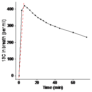 |
52 | Considering whole body metabolism in hyperpolarized MRI – an alternative way to quantification and normalization?
Steffen Houe Sejersen1, Camilla Rasmussen1, Nikolaj Bøgh1, Esben Hansen1, Rolf Schulte2, and Christoffer Laustsen1
1MR Centeret, Klinisk Institut, AU, Århus C, Denmark, 2GE Healthcare, Munich, Germany
Hyperpolarized [1-13C]pyruvate MRI is an exciting emerging clinical tool for metabolic imaging. It has the potential for absolute quantitative metabolic imaging. However, in contrast to the most abundant metabolic imaging techniques, the signal itself is less quantitative by nature and, thus the most abundant analyses are relative or semiquantitative. Here, we propose a simple normalization to the whole body metabolic oxidative metabolism to overcome this limitation.
|
||
2424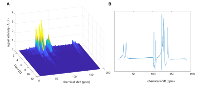 |
53 | Hyperpolarising 13C-glucose with an endogenous labile photoinduced radical Video Permission Withheld
Jennifer Lewis1, Adam Gaunt1, Irene Marco-Rius1, and Arnaud Comment1,2
1Cambridge University, Cambridge, United Kingdom, 2General Electric Healthcare, Chalfont St Giles, United Kingdom
The use of the endogenous molecule alpha-ketoglutarate (α-KG) as a non-persistent radical is examined to hyperpolarise 13C-glucose. A radical build-up curve was obtained and the maximum radical concentration was calculated to be 29 ± 3 mM. The microwave irradiation frequency was then examined to optimise the polarisation transfer between the electron on α-KG and glucose. A dissolution was successfully carried out and a hyperpolarised 13C spectrum of 13C-glucose was obtained.
|
||
2425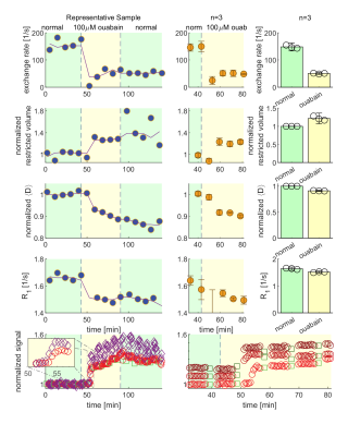 |
54 | DEXSY Reveals Water Exchange Coupled to Activity
Nathan H. Williamson1,2, Rea Ravin1, Teddy X. Cai1,3, Melanie Falgairolle4, Michael J. O'Donovan4, and Peter J. Basser1
1National Institute of Child Health and Human Development, Potomac, MD, United States, 2National Institute of General Medical Sciences, Potomac, MD, United States, 3Department of Clinical Neurosciences, Wellcome Centre for Integrative Neuroimaging, FMRIB, University of Oxford, Oxford, United Kingdom, 4National Institute of Neurological Disorders and Stroke, Potomac, MD, United States
Currently, no MRI method exists to non-invasively and absolutely measure cellular activity. One approach is to quantify steady-state transmembrane water exchange rates. Diffusion Exchange Spectroscopy (DEXSY) non-invasively and directly encodes for exchange of endogenous components. Using DEXSY-based methods implemented on a low-field, high-gradient MR system, we show in viable ex vivo neonatal mouse spinal cord samples that the rate at which water exchanges across cell membranes decreases drastically after perturbation known to induce persistent membrane depolarization. We also show that the exchange rate recovers to normal values after perturbations known to restore membrane potential.
|
||
2426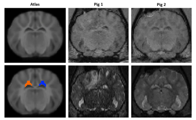 |
55 | Free Water Imaging in a Pig Model of Traumatic Brain Injury
Drew Parker1, Victoria E. Johnson2, H. Isaac Chen2,3, John A. Wolf2,3, and Ragini Verma1
1DiCIPHR (Diffusion and Connectomics in Precision Healthcare Research) Lab, University of Pennsylvania, Philadelphia, PA, United States, 2Department of Neurosurgery, Perelman School of Medicine, University of Pennsylvania, Philadelphia, PA, United States, 3Center for Neurotrauma, Neurodegeneration & Restoration, Corporal Michael J. Crescenz VA Medical Center, Philadelphia, PA, United States The way brain injury can evolve has different manifestations on imaging in multiple MRI modalities, pointing to different pathological etiologies. The true picture can be seen only by correcting for free water, as it biases all the diffusion measurements, especially the longitudinal outcome. While this has been observed in humans, it is for the first time free water analysis has been carried out in a gyrencephalic pig model of TBI. In the future, co-registration with histopathological outcomes will be undertaken to verify the specificity of the diffusion measurements, which will lay the foundation for interpretation of unbiased diffusion measurements. |
||
2427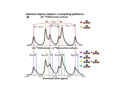 |
56 | Elucidating dynamic anaerobic metabolism in obligate anaerobes with living cells
Leo Cheng1, Aidan Pavao2, Brintha Girinathan2, Johann Peltier3, Pamela Altamirano Silva4, Bruno Dupuy5, Isabella Muti1, Craig Malloy6, and Lynn Bry2
1Radiology and Pathology, Massachusetts General HospitalMassachusetts General Hospital, Harvard Medical School, Charlestown, MA, United States, 2Massachusetts Host-Microbiome Center, Brigham and Women’s Hospital, Harvard Medical School, Boston, MA, United States, 3Institut Pasteur, Université de Paris, Paris, France, 4Centro de Investigación en Enfermedades Tropicales, Facultad de Microbiología, Universidad de Costa Rica, San Jose, Costa Rica, 5Laboratory of the Pathogenesis of Bacterial Anaerobes, Institut Pasteur, Université de Paris, Paris, France, 6Radiology, The University of Texas Southwestern Medical Center, Dallas, TX, United States
Anaerobic microbial metabolism drives critical functions within global ecosystems, host-microbiota interactions, and industrial applications, yet remains ill-defined. We resolved dynamic metabolism in living cells of the anaerobic pathogen Clostridioides difficile using High-Resolution Magic Angle Spinning (HRMAS) Nuclear Magnetic Resonance (NMR) spectroscopy to inform genome-scale predictions of cellular metabolism. Analyses leveraged the sensitivity of 13C NMR spectroscopy to simultaneously track cellular carbon and nitrogen flow from fermentable 13C and 15N-labeled substrates. We illustrate a versatile approach to elaborate complex anaerobic metabolism for clinical, scientific, and industrial applications.
|
||
2428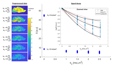 |
57 | Time dependence of the diffusion and kurtosis tensors in strongly filtered diffusion signals Video Not Available
Noam Shemesh1, Andrada Ianus1, and Sune N Jespersen2,3
1Champalimaud Centre for the Unknown, Lisbon, Portugal, 2Center for Functionally Integrative Neuroscience, Department of Clinical Medicine, Aarhus University, Aarhus, Denmark, 3Department of Physics and Astronomy, Aarhus University, Aarhus, Denmark
The sensitivity of diffusion MRI (dMRI) towards microstructural features is quite high, but its specificity is low due to the ubiquity of water in biological tissues. Here, we harness a strongly filtered diffusion kurtosis imaging (DKI) approach to (i) suppress fast diffusion spins typically associated with extracellular space and (ii) measure the time dependencies of the full filtered diffusion and kurtosis tensors in an ex-vivo mouse brain at 16.4T. Following the application of a very strong filter of b1=15ms/µm2 perpendicular to axon fibers, we find signatures for restricted diffusion that are tentatively associated with intracellular space.
|
||
2429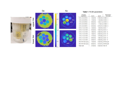 |
58 | Temperature and Concentration Dependence of Diffusion Kurtosis Parameters in a Quantitative Phantom
Dariya I. Malyarenko1, Thomas L Chenevert1, Shigeto Ono2, Ted Lynch2, and Scott D. Swanson1
1Radiology, University of Michigan, Ann Arbor, MI, United States, 2CIRS, Inc, Norfolk, VA, United States
Recently developed quantitative phantom based on lamellar-vesicles provides the range of tissue relevant diffusion kurtosis parameters for accurate evaluation of advanced multi-b DWI protocols and parametric diffusion models. This work studies temperature dependence of phantom diffusion kurtosis parameters to supply accurate nominal parameter values for typical scan room temperature range.
|
||
2430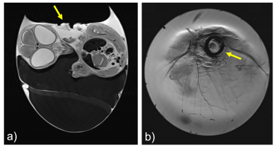 |
59 | A Transceiver Coil for improved in ovo Imaging of the Chorioallantoic Membrane at 7T
Mirsad Mahmutovic1, Manisha Shrestha1, Matthäus Poniatowski1, Jarmila Jedelská2, Udo Bakowsky3, Alexander M. König2,4, Andreas H. Mahnken2,4, and Boris Keil1
1Institute of Medical Physics and Radiation Protection, TH Mittelhessen University of Applied Sciences, Giessen, Germany, 2Center for Tumor Biology and Immunology, Core Facility for Small Animal MRI, Marburg, Germany, 3Department of Pharmaceutics and Biopharmaceutics, Philipps-University of Marburg, Marburg, Germany, 4Department of Diagnostic and Interventional Radiology, Philipps-University of Marburg, Marburg, Germany
The chorioallantoic membrane (CAM) model is a simple and low-cost model that allows screening of a large number of pharmacological samples. It is used in preclinical research as an intermediate step between in vitro and in vivo experiments and does not require approval by the animal experiment ethics committee. Latest studies show that MRI is a suitable imaging modality for evaluating drug delivering systems with the CAM-model. However, appropriate hardware for high-resolution MR imaging of the CAM model is still missing. To fill this gap, we designed, constructed, and evaluated a transceiver solenoid coil for in ovo imaging at 7T.
|
||
2431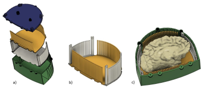 |
60 | Reusable 3D Printed Ex-vivo Brain Enclosure and Two-Piece Cutting Guide for Axial and Coronal MRI Registration with Gross Anatomy Photographs
Jacob Patrick Berardinelli1, Julia Kofler1, Noah Schweitzer1, Nadim Farhat1, Tales Santini1, Andrea Sajewski1, Joseph Mettenburg1, Milos Ikonomovic1, Howard J. Aizenstein1, and Tamer S. Ibrahim1
1University of Pittsburgh, Pittsburgh, PA, United States
A 3D printed re-usable box enclosure and two-piece cutting guide were developed to produce high quality 7T MR images that can be easily registered to gross anatomy and histology. Cuts can be made parallel or at multiple angles at 5mm intervals in both the axial and coronal orientations.
|
||
2432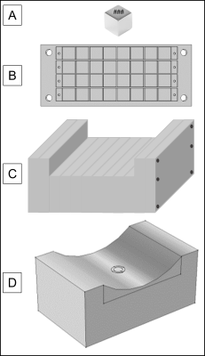 |
61 | Design and fabrication of portable, low-field MR relaxometer for clinical measurement of volemic status
Sydney Sherman1 and Michael Cima2
1HST, MIT, Cambridge, MA, United States, 2DMSC, MIT, Cambridge, MA, United States
A low-field portable MR-based sensor was fabricated for the acquisition of clinical T2 relaxometry measurements in skeletal muscle. We have previously reported methods for designing low-field permanent magnet array configurations with varying sensitive region profiles. The newly constructed array has a sensitive region 15-20mm from the surface of the magnet; field maps from the constructed magnet show good alignment with simulated fields. The exclusion of subcutaneous fat tissue in the sensitive region will improve sensitivity to fluid shifts within the skeletal muscle.
|
||
 Back to Meeting Home
 Back to the Program-at-a-Glance
The International Society for Magnetic Resonance in Medicine is accredited by the Accreditation Council for Continuing Medical Education to provide continuing medical education for physicians.
 Back to Meeting Home
 Back to the Program-at-a-Glance
 View the Presentation