Digital Poster
Advances in Diagnosis of Neurologic Disorders
Joint Annual Meeting ISMRM-ESMRMB & ISMRT 31st Annual Meeting • 07-12 May 2022 • London, UK

Digital Poster
Advances in Diagnosis of Neurologic Disorders
| Computer # | ||||
|---|---|---|---|---|
1769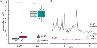 |
39 | Increased GABA in the periaqueductal grey of patients with chronic low back pain detected using 1H-magnetic resonance spectroscopy
Laura Sirucek1,2,3, Niklaus Zoelch4,5, and Petra Schweinhardt1,2
1Department of Chiropractic Medicine, Integrative Spinal Research Group, Balgrist University Hospital, University of Zurich, Zurich, Switzerland, 2University of Zurich, Zurich, Switzerland, 3Neuroscience Center Zurich, University of Zurich, Zurich, Switzerland, 4Department of Forensic Medicine and Imaging, Institute of Forensic Medicine, University of Zurich, Zurich, Switzerland, 5Department of Psychiatry, Hospital of Psychiatry, Psychotherapy and Psychosomatics, University of Zurich, Zurich, Switzerland
Preclinical studies suggest a role of altered γ-aminobutyric acid (GABA)ergic inhibitory tone in the periaqueductal grey (PAG), a key brain area for descending pain modulation, as mechanism contributing to chronic pain. The present 1H-magnetic resonance spectroscopy study investigated GABA, and glutamate/glutamine, concentrations in the PAG of patients with chronic low back pain (CLBP). Using a point resolved spectroscopy sequence with optimized frequency alignment, adequate spectral quality was achieved and increased GABA concentrations (p=0.027) identified in CLBP patients compared to pain-free controls. This finding supports dysregulations in descending pain modulation as factor contributing to chronic pain in humans.
|
||
1770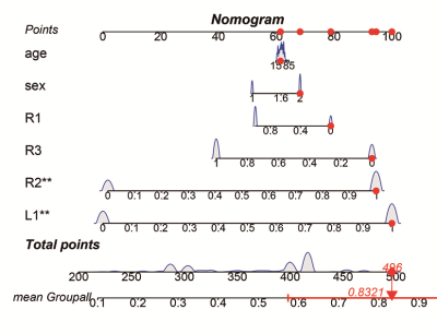 |
40 | Hippocampal subfield neurofunctional alterations in chronic insomnia Video Not Available
Yang Yang1, Yong Li1, Linwei Zhao1, Hong Chen1, Xiaoping Fan1, and Gaowu Yan1
1Suining Central Hospital, Chuanshan Suining, China
This study investigated the patterns of volume changes across neurofunctionally distinct hippocampal subfields in patients with chronic insomnia. The results suggested that L1 and R2 atrophy increased the risk of developing chronic insomnia approximately 3-fold, and negative emotion positively acted on the causal path of insomnia severity leading to R1 atrophy. In addition, we developed a practical and visual nomogram of individual competing risks for predicting chronic insomnia risk with high accurate predictive power based on these hippocampal subfields. Neurofunctional hippocampal subfield atrophy was a risk factor leading to chronic insomnia, contributing to understand of the etiology of insomnia.
|
||
1771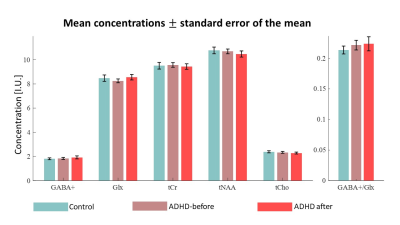 |
41 | GABA and Glutamate/Glutamine concentrations in the dACC of amphetamine treated adult ADHD patients
Yasmin Geiger1, Marganit Gonen-Shahar2, Gabriel Vainstein3, Angela Ruban2, and Assaf Tal1
1Weizmann Institute of Science, Rehovot, Israel, 2Tel Aviv University, Tel Aviv, Israel, 3Maccabi Healthcare, Kefar Sava, Israel
Attention deficit hyperactivity disorder (ADHD) is a neurodevelopmental disorder characterized by irregular levels of hyperactivity, impulsivity and inattention. It is commonly linked to dopamine and norepinephrine balance. Several studies suggested connections to γ-amino butyric acid (GABA) and glutamate (Glu) imbalance in the dorsal anterior cingulate cortex (dACC). Here we use 1H-MRS to study GABA and glutamate + glutamine (Glx) levels in the dACC of ADHD patients, before and after pharmacological treatment and compared to healthy adults. We found no metabolic difference between ADHD and control group and no change after amphetamine treatment. We found no metabolic correlation to ADHD scores.
|
||
1772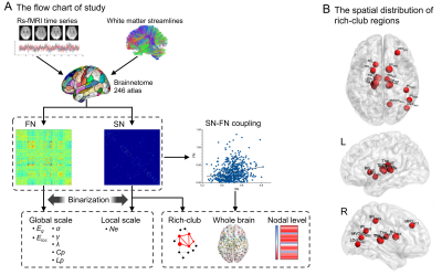 |
42 | Disruptions of functional and structural networks in classic trigeminal neuralgia patients
Pengfei Zhang1, Jun Wang1, Wanjun Hu1, Laiyang Ma1, Guangyao Liu1, Kai Ai2, and Jing Zhang1
1Department of Magnetic Resonance, Lanzhou University Second Hospital, Lanzhou, China, 2Philips Healthcare, Xi'an, China
Classic trigeminal neuralgia (CTN) has been proven related to functional or morphological alterations in critical brain regions. To test the simultaneous changes of the topological properties of structural networks (SN), functional networks (FN), as well as their couplings, we constructed SN using diffusion tensor imaging (DTI) data and FN using resting-state functional magnetic imaging (rs-fMRI) data from 29 CTN patients and 34 healthy controls (HCs). The patients showed the wide disruptions of global and local topological organizations in SN and FN, together with the abnormal coupling relationships. Our findings may provide novel perspectives for understanding the pathophysiological mechanisms of CTN.
|
||
1773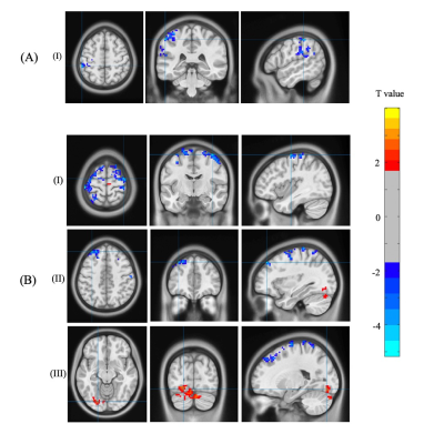 |
43 | Longitudinal resting-state functional connectivity changes with medication and nutrition treatment in chronic mild traumatic brain injury
Faezeh vedaei1, Mahdi Alizadeh1, George Zabrecky 1, Nancy Wintering 1, Anthony J. Bazzan 1, Daniel A. Monti 1, Andrew B. Newberg 1, and Feroze B. Mohamed 1
1Thomas Jefferson University, Philadelphia, PA, United States
This study provides comprehensive evaluation of functional rs-fMRI biomarkers that are associated with chronic mTBI symptoms and predicting treatment outcome and patient recovery. We evaluate the effect of medication and nutrition treatments on brain function and cognitive performance in chronic mTBI patients using resting-state fMRI metrics and neuropsychological test. We propose that treatment strategies compensate mTBI disrupt communication between brain network nodes. Our results help understanding the implication of treatment strategies in chronic mTBI patients and prediction of outcomes in brain function and cognitive recovery.
|
||
1774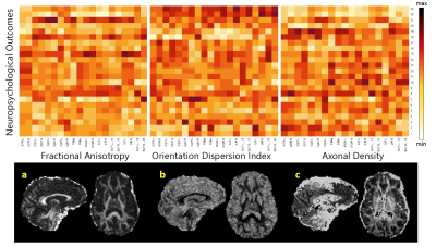 |
44 | Hybrid diffusion imaging in chronic traumatic brain injury: Longitudinal investigation and correlation with clinical outcome
Jennifer Muller1,2,3, KiChang Kang1, Devon Middleton3, Mahdi Alizadeh1, Faezeh Vedaei1, George Zabrecky4, Daniel Monti4, Nancy Wintering4, Anthony J. Bazzan4, Chengyuan Wu1, Qianhong Wu2, and Feroze B. Mohamed3
1Neurosurgery, Thomas Jefferson University Hospital, Philadelphia, PA, United States, 2Mechanical Engineering, Villanova University, Villanova, PA, United States, 3Radiology, Thomas Jefferson University Hospital, Philadelphia, PA, United States, 4Marcus Institute of Integrative Health, Villanova, PA, United States
Traumatic brain injury (TBI) is a major cause of death and disability worldwide. Currently, non-invasive and longitudinal assessment in TBI is still limited. In the current study, we applied an advanced hybrid diffusion imaging sequence (HYDI) to capture both diffusion tensor imaging and neurite orientation dispersion and density imaging to develop more sensitive biomarkers associated with longitudinal outcomes after TBI. We both contrasting and complementary correlates between diffusion metrics and clinical outcomes, suggesting HYDI to have potential clinical impact for treatment monitoring after TBI.
|
||
1775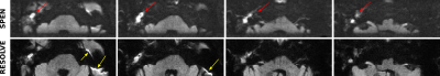 |
45 | DWI of cholesteatomas: An initial assessment based on RESOLVE and SPEN MRI
Martins Otikovs1, Debbie Anaby2, Shai Shrot2, Noam Nissan2, and Lucio Frydman1,3
1Department of Chemical and Biological Physics, Weizmann Institute of Science, Rehovot, Israel, 2Sheba Medical Center, Ramat Gan, Israel, 3Azrieli National Center for Brain Imaging, Weizmann Institute of Science, Rehovot, Israel
Cholesteatomas are destructive lesions growing in the middle ear, in need of periodic assessments. Diffusion weighted imaging (DWI) at 3T provides clear contrast of these growths, facilitating their detection. However, the close presence of air-tissue interfaces within the ear channel results in geometric distortions that have led to adoption of non-EPI based methods for these high field evaluations. Herein we present results from initial DWI experiments performed for imaging cholesteatomas, which point out the value of Spatiotemporal Encoding (SPEN) DWI methods as promising alternatives to retrieve high-contrast, high-definition images.
|
||
1776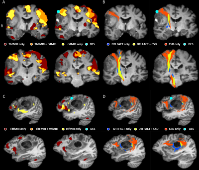 |
46 | A comparison of tractography and fMRI pre-surgical planning approaches with intraoperative mapping-based validation
Ahmed Radwan1,2, Louise Emsell1,2,3,4, Evy Cleeren5, Silvia Kovacs6, Anais Van Hoylandt5, Ronald Peeters6, Steven De Vleeschouwer2,5,7, Tom Theys2,5,7, Patrick Dupont2,8, and Stefan Sunaert1,2,6
1Imaging and pathology, Translational MRI, KU Leuven, Leuven, Belgium, 2Department of Neurosciences, KU Leuven, Leuven Brain Institute, Leuven, Belgium, 3Department of Neurosciences, Neuropsychiatry, KU Leuven, Leuven, Belgium, 4Geriatric Psychiatry, University Psychiatric Center (UPC) - Leuven, Leuven, Belgium, 5Department of Neurosurgery, University Hospitals Leuven, Leuven, Belgium, 6Department of Radiology, University Hospitals Leuven, Leuven, Belgium, 7Department of Neurosciences, Research Group Experimental Neurosurgery and Neuroanatomy, KU Leuven, Leuven, Belgium, 8Department of Neurosciences, Laboratory for cognitive neurology, KU Leuven, Leuven, Belgium
Accurate presurgical brain mapping enables preoperative risk assessment and intraoperative guidance to minimize postoperative deficits. Here we compare mapping accuracy of task-based fMRI (tbfMRI), BOLD and Functionnectome resting state fMRI (rsfMRI), DTI and constrained spherical deconvolution (CSD)-based tractography in 21 preoperative neurosurgical patients using intraoperative electrical stimulation (DES) as the ground truth for functional mapping. Accuracy was estimated based on minimum distance between MRI-based mapping and positive DES coordinates. We report that CSD outperforms DTI, and rsfMRI performs similarly to tbfMRI using DES. This demonstrates the potential benefits of using CSD and rsfMRI in clinical practice.
|
||
1777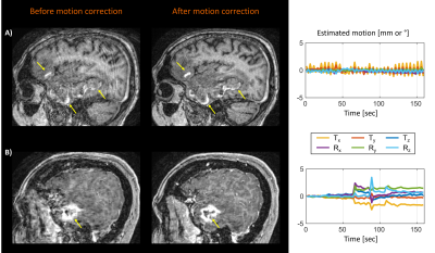 |
47 | Clinical evaluation of scout accelerated motion estimation and reduction (SAMER)
Azadeh Tabari1,2, Min Lang1,2, Daniel Polak3,4, Daniel Nicolas Splitthoff4, Bryan Clifford5, Wei-Ching Lo5, Lawrence L. Wald2,3,6, Stephen Cauley2,3, Otto Rapalino1,2, Pamela Schaefer1,2, John Conklin1,2,3, and Susie Y. Huang1,2,3,6
1Department of Radiology, Massachusetts General Hospital, Boston, MA, United States, 2Harvard Medical School, Boston, MA, United States, 3A. A. Martinos Center for Biomedical Imaging, Charlestown, MA, United States, 4Siemens Healthcare GmbH, Erlangen, Germany, 5Siemens Medical Solutions, Malvern, PA, United States, 6Harvard-MIT Health Sciences and Technology, Massachusetts Institute of Technology, Cambridge, MA, United States
Patient motion can degrade diagnostic image quality to the point that scans must be re-acquired, or patients called back. In this work, we performed a systematic clinical evaluation of SAMER, a highly efficient retrospective motion mitigation approach. 62 patients from emergency and inpatient care settings were scanned at 3T, and 14 cases were identified with a range of patient motion. Two neuro-radiologists performed blinded review of the images before and after motion correction, rating the images for motion severity. In 13 of the 14 cases, reduced motion severity and improved visualization of anatomy/pathology was obtained after SAMER motion mitigation.
|
||
1778 |
48 | The origins of BOLD signal fluctuations in non-gas-inhalation CVR mapping: an fMRI-EEG study
Parimal Pravin Joshi1, Yi Zhang2,3, Xirui Pravin Hou2, Cuimei Xu2, Paul Bottomley2, Hanzhang Lu2, and Peiying Liu1
1Department of Diagnostic Radiology and Nuclear Medicine, University of Maryland School of Medicine, Baltimore, MD, United States, 2Department of Radiology, Johns Hopkins University School of medicine, Baltimore, MD, United States, 3Department of Biomedical Engineering, College of Biomedical Engineering & Instrument Science, Zhejiang University, Hangzhou, China Cerebrovascular reactivity (CVR) is typically measured with hypercapnic challenges, which require considerable subject cooperation. Recent CVR mapping approaches using resting-state BOLD data are promising, but their sensitivity is low in subjects with minimal spontaneous changes in breathing pattern. Performed in conjunction with resting-state BOLD scans, intermittent breath modulation enhances variations in breathing pattern while requiring minimal subject compliance, and is a promising tool for CVR mapping without gas inhalation. Here we conducted simultaneous fMRI-EEG experiments to investigate origins of the global BOLD signal fluctuations during intermittent breath modulation, to verify the validity of this new approach. |
||
 Back to Meeting Home
 Back to the Program-at-a-Glance
The International Society for Magnetic Resonance in Medicine is accredited by the Accreditation Council for Continuing Medical Education to provide continuing medical education for physicians.
 Back to Meeting Home
 Back to the Program-at-a-Glance
 View the Presentation