Digital Poster
Sequences & Sampling II
Joint Annual Meeting ISMRM-ESMRMB & ISMRT 31st Annual Meeting • 07-12 May 2022 • London, UK

| Computer # | ||||
|---|---|---|---|---|
1705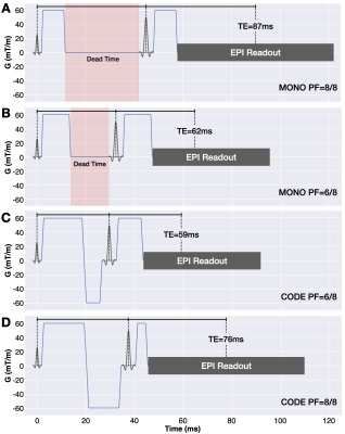 |
74 | Convex Optimized Diffusion Encoding (CODE) with Partial Fourier Imaging for EPI Diffusion Weighted Imaging
Matthew J. Middione1, Tyler E. Cork1,2, Michael Loecher1, Tim Sprenger3, Arnaud Guidon4, Shreyas S. Vasanawala1, and Daniel B. Ennis1
1Department of Radiology, Stanford University, Stanford, CA, United States, 2Department of Bioengineering, Stanford University, Stanford, CA, United States, 3GE Healthcare, Munich, Germany, 4GE Healthcare, Boston, MA, United States Diffusion weighted imaging typically uses monopolar (MONO) diffusion gradient waveforms, which may have sequence dead time that extends the TE and reduces the SNR. Partial Fourier (PF) imaging is routinely used in DWI to shorten the TE (improving SNR), but can lead to image blurring. Convex Optimized Diffusion Encoding (CODE) is a constrained optimization technique that designs time-optimal diffusion encoding gradient waveforms without sequence dead time. CODE produces a shorter TE than MONO, which leads to increased SNR. Herein, we show that CODE without PF has increased SNR and reduced image blurring compared to MONO with PF=6/8. |
||
1706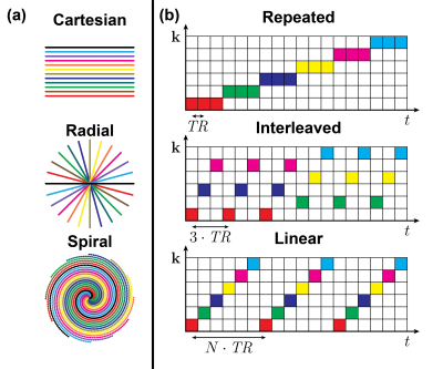 |
75 | Protocol Optimization of Spectro-Dynamic MRI
Max H.C. van Riel1, Niek R.F. Huttinga1, Tom Bruijnen1, and Alessandro Sbrizzi1
1Department of Radiotherapy, Computational Imaging Group for MR diagnostics and therapy, UMC Utrecht, Utrecht, Netherlands Inferring dynamical information from (bio)mechanical systems at a high temporal resolution can be very valuable for cardiovascular or musculoskeletal applications. Spectro-dynamic MRI is a recently proposed method that combines a measurement model and a dynamical model to characterize dynamical systems directly from k-space data. Both the displacement fields and the underlying dynamical parameters are estimated. In this work, different sampling trajectories and acquisition orderings are used to investigate the trade-off between temporal resolution and k-space coverage. A phantom experiment shows that it is possible to reconstruct a moving image from the estimated dynamics at a temporal resolution of 4.4 ms. |
||
1707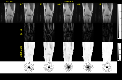 |
76 | Sampling Mask Optimization for Compressed Sensing PETRA MRI
Serhat Ilbey1, Ali Özen1, Pia Jungmann2,3, Johannes Fischer1, Matthias Jung2, and Michael Bock1
1Dept.of Radiology, Medical Physics, Medical Center University of Freiburg, Faculty of Medicine, University of Freiburg, Freiburg, Germany, 2Department of Diagnostic and Interventional Radiology, Medical Center University of Freiburg, Faculty of Medicine, University of Freiburg, Freiburg, Germany, 3Department of Radiology, Cantonal Hospital Grisons, Chur, Switzerland
Single point imaging (SPI) is intrinsically time-inefficient resulting in very long scan times in PETRA. Compressed sensing PETRA (csPETRA) shortens the acquisition time – here, various csPETRA undersampling schemes were generated and an optimal undersampling scheme was determined experimentally. It was shown in csPETRA knee imaging that in Poisson sampling combined with a spherical fully-sampled center results in minimum error compared to the fully-sampled acquisitions.
|
||
1708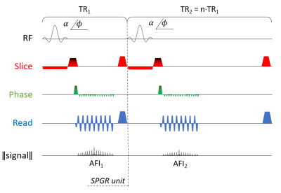 |
77 | EPIFANI: an ultrafast T1, B1 and magnetization mapping technique
Marco Andrea Zampini1,2, Jan Sijbers3, Marleen Verhoye2, and Ruslan Garipov1
1MR Solutions Ltd, Guildford, United Kingdom, 2Department of Biomedical Sciences, University of Antwerp, Wilrijk, Belgium, 3Department of Physics, University of Antwerp, Wilrijk, Belgium
Quantitative MRI still struggles to be used in clinical practice due to long acquisition times. We introduce EPIFANI, an ultrafast B1-corrected T1 mapping technique which uses EPI images acquired in two successive repetition times and a parametric map fit used in VAFI. Both a single-shot and a multi-shot version of EPIFANI were tested on phantoms and in vivo and report accurate T1 values.
|
||
1709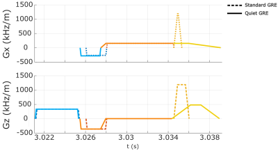 |
78 | Rapid prototyping of a motion-robust 2D Radial GRE sequence with reduced acoustic noise generation using Pulseq
Elisa Marchetto1,2, Maxim Zaitsev3, and Daniel Gallichan1,2
1School of Engineering, Cardiff University, Cardiff, United Kingdom, 2Cardiff University Brain Research Centre (CUBRIC), Cardiff University, Cardiff, United Kingdom, 3High Field MR Center, Center for Medical Physics and Biomedical Engineering, Medical University of Vienna, Vienna, Austria
A radial GRE-sequence was modified using Pulseq to reduce the acoustic noise amount produced by the rapid switching of gradient currents without excessively increasing the TR-time. Prospective motion-correction was performed using a markerless tracking device that sent the motion estimates to the scanner via the libXPACE framework, incorporated directly into Pulseq, to update gradients and RF pulses during the MR scan. The quieter sequence resulted in a acoustic noise reduction of 6 dB(A) (i.e. a factor of 2) compared to the standard one. Image quality was greatly improved using prospective motion-correction in the case of deliberate motion during the scan.
|
||
1710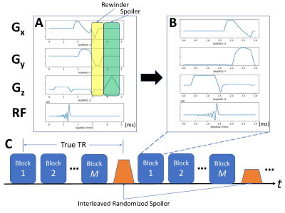 |
79 | More Efficient SPGR 3D Abdominal Imaging Using an Interleaved Randomized Spoiler
zheng zhong1, Christopher M. Sandino2, Ali Syed1, John M. Pauly2, and Shreyas S. Vasanawala1
1Radiology, Stanford University, Stanford, CA, United States, 2Electrical Engineering, Stanford University, Stanford, CA, United States
3D SPGR is commonly used clinically for contrast-enhanced body imaging, but requires time for a gradient spoiler every TR. This consumes up to 40% of scan time, even for a non-cartesian trajectories with relatively long readouts, such as cones. Removing the spoiler can save time but causes severe artifacts. In this study, we propose a novel approach of applying interleaved, randomized spoilers to a motion-robust 3D-cones sequence. The feasibility of the approach to shorten acquisition time without introducing image artifacts was validated on both healthy subjects and patients.
|
||
1711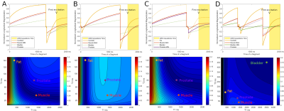 |
80 | Optimal Control of the contrast in a MP-RAGE sequence: an application on the pelvis
Benoît Vernier1,2, Eric Van Reeth1,3, Marc Lapert2, Olivier Hamelin4, Olivier Beuf1, Hélène Ratiney1, and Frank Pilleul4
1CREATIS, Lyon, France, 2Siemens Healthcare SAS, Saint-Denis, France, 3CPE, Lyon, France, 4Centre Léon Bérard, Lyon, France
MRI contrast enhancement by Optimal Control is a new approach to design optimal magnetization preparation. This allows to maximize the contrast between target tissues characterized by their relaxation times. Recent numerical implementations have made possible the optimization of such preparation in a steady state sequence without full recovery between each repetition. This abstract demonstrates the contrast flexibility offered by this approach when combined with a MPRAGE sequence on pelvis imaging at 3T.
|
||
1712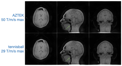 |
81 | Tennisball: slew-efficient trajectory design for 3D-radial imaging
Gopal Nataraj1 and Michael Lustig1
1University of California, Berkeley, Berkeley, CA, United States
What is the most efficient path to traverse a sphere? For a spherical gap-minimizing efficiency definition and a particular path length, the answer happens to resemble the seam lines of a tennis ball. Here we explore properties of this general class of spherical trajectories and investigate their application to slew-constrained ZTE imaging. Static and motion-resolved imaging results demonstrate that Tennisball trajectories can achieve competitive performance with existing methods but with ~30-40% lower peak slew.
|
||
1713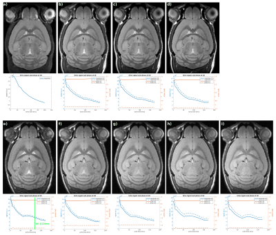 |
82 | Fast 3D Isotropic High-Resolution MRI of Mouse Brain Using a Variable Flip Angle RARE Sequence With T2 Compensation @9.4T
Lisa Y Hung1, Karl-Heinz Herrman1, and Jürgen R Reichenbach1
1Medical Physics Group, Institute of Diagnostic Radiology, Jena University Hospital, Jena, Germany
Some applications require T2-weighted mouse brain MRI with isotropic high resolution (100µm3), but at 9.4T the short T2 of mouse brain limits the echo train length of RARE sequences and exacerbates T2-decay artifacts. Adapting a RARE sequence by using variable flip-angles allows a reduction in scan time from 19min to 7-9min with similar image quality. However, long RARE echo trains and strong imaging gradients can unintentionally cause substantial diffusion weighting. To minimize the diffusion effects crusher gradients were optimized and the variable flip angles were kept above 40°.
|
||
1714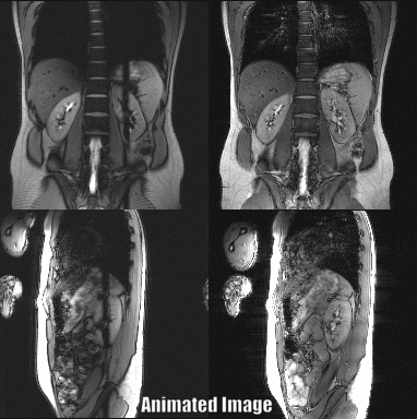 |
83 | Simultaneous T2-weighted Real-time MRI of 2 Orthogonal Slices
Samantha Hickey1, Andreas Reichert1, Thomas Bortfeld2, and Michael Bock1
1Dept. of Radiology, Medical Physics, Medical Center University of Freiburg, Faculty of Medicine, University of Freiburg, Freiburg, Germany, 2Division of Radiation Biophysics, Department of Radiation Oncology, Massachusetts General Hospital and Harvard Medical School, Boston, MA, United States
In cancer radiotherapy, MR-guidance allows for superior tumor visualization and real-time target volume tracking. Real-time pulse sequences for MR-guidance require high acquisition speeds and good tumor-to-background contrast, which is often achieved with T2-weighted acquisitions. Here, we combine a real-time sequence with orthogonal slices with a T2w echo-shifted contrast and demonstrate its use under breathhold and free-breathing conditions in a volunteer.
|
||
1715 |
84 | Bipolar Partial Echo Imaging
Holger Eggers1 and Jochen Keupp1
1Philips Research, Hamburg, Germany
Partial echo imaging is widely used in angiography to shorten the echo time and reduce flow-induced signal loss. However, sampling only an asymmetric part of k-space basically ignores high-frequency phase and thus causes signal alteration as well. In the present work, bipolar partial echo imaging is introduced to cover the full k-space sparsely with a Cartesian partial echo measurement. It involves reversing the polarity of the readout gradient during the acquisition and recovering missing data in the reconstruction without assuming smoothness of the phase. The potential of bipolar partial echo imaging is demonstrated in phantom and first volunteer experiments.
|
||
1716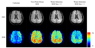 |
85 | Susceptibility weighted imaging using Stack of Spiral trajectories
Tzu-Cheng Chao1 and James G. Pipe1
1Department of Radiology, Mayo Clinic, Rochester, MN, United States
A new strategy to acquire Susceptibility Weighted Imaging was proposed with Stack of Spiral trajectories to reduce scan time while maintaining adequate SNR. Spiral encoding offers acceleration in scan speed by six times compared to conventional Cartesian methods, with similar SNR, resolution, and image quality. Due to its improved SNR efficiency, the proposed method has the potential to facilitate lesion detection with higher resolution SWI in a reasonable scan time.
|
||
 Back to Meeting Home
 Back to the Program-at-a-Glance
The International Society for Magnetic Resonance in Medicine is accredited by the Accreditation Council for Continuing Medical Education to provide continuing medical education for physicians.
 Back to Meeting Home
 Back to the Program-at-a-Glance
 View the Presentation