Digital Poster
Motion & Artifacts I
Joint Annual Meeting ISMRM-ESMRMB & ISMRT 31st Annual Meeting • 07-12 May 2022 • London, UK

| Computer # | ||||
|---|---|---|---|---|
0855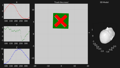 |
80 | Real time visual feedback for reproducible head motion
Adam van Niekerk1, Johan Berglund2, Henric Ryden1,3, Tim Sprenger1,4, Ola Norbeck1,3, Enrico Avventi1,3, and Stefan Skare1,3
1Clinical Neuroscience, Karolinska Institutet, Stockholm, Sweden, 2Medical Physics, Uppsala University Hospital, Uppsala, Sweden, 3Neuroradiology, Karolinska University Hospital, Stockholm, Sweden, 4MR Applied Science Laboratory Europe, GE healthcare, Stockholm, Sweden
The timing and magnitude of volunteer head motion has a significant impact on artefact level. It can therefore be difficult to reproduce motion correction results. Moreover, it is of interest to replicate patient motion retrospectively, in volunteers, to better understand the efficacy of a motion correction method. We attempt to solve this by stamping motion updates with a synchronisation “tag”. A rolling model is used to project the volunteer’s pose onto an MRI compatible display. The “tag” synchronised pose from a previous scan is then shown alongside the volunteer’s current pose, allowing them to track and hence reproduce the motion.
|
||
0856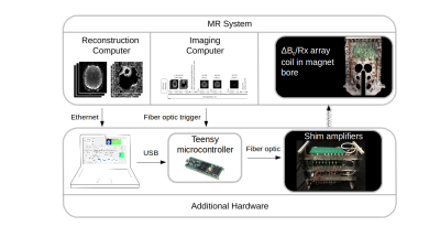 |
81 | Real-time Motion Compensated ∆B0 Shimming with an AC/DC Shim Coil and Dual-Echo vNavs
Nicolas Arango1, Robert Frost2,3, Paul Wighton2, Jason Stockmann2,3, Ovidiu C Andronesi2,3, and Andre van der Kouwe2,3
1Department of Electrical Engineering and Computer Science, Massachusetts Institute of Technology, Cambridge, MA, United States, 2A. A. Martinos Center for Biomedical Imaging, Massachusetts General Hospital, Charlestown, MA, United States, 3Department of Radiology, Harvard Medical School, Boston, MA, United States
Changes in subject position move susceptibility interfaces and therefore ∆B0 field patterns in the brain. We apply a prospective real time (TR-to-TR) shim updating scheme using dual echo EPI volume navigators to correct motion-induced changes in ∆B0 fields to reduce distortion in 2D EPI. Shim fields were produced by a 32 channel AC/DC head shim array. TR-to-TR shimming reduced EPI distortion in all head positions in in vivo experiments.
|
||
0857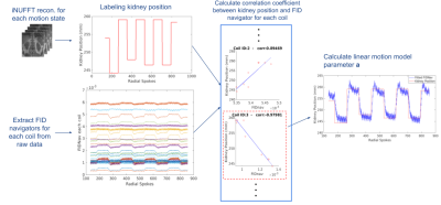 |
82 | Prospective Motion Correction in Kidney MRI Using Free Induction Decay Navigators
Cemre Ariyurek1, Tess E Wallace1, Tobias Kober2,3,4, Aziz Koçanaoğulları1, Simon K Warfield1, Sila Kurugol1, and Onur Afacan1
1Radiology, Boston Children's Hospital and Harvard Medical School, Boston, MA, United States, 2Advanced Clinical Imaging Technology, Siemens Healthcare, Lausanne, Switzerland, 3Department of Radiology, Lausanne University Hospital and University of Lausanne, Lausanne, Switzerland, 4LTS5, École Polytechnique Fédérale de Lausanne (EPFL), Lausanne, Switzerland
Abdominal MRI scans may require breath-holding to prevent image quality degradation, which can be challenging for patients. In this study, we used free induction decay navigators (FIDnavs) to correct motion prospectively. A short calibration scan was acquired prior to the prospectively corrected scan to create a linear motion model to translate the FIDnav signal into kidney displacement. Shallow, deep and continuous deep breathing scans were acquired with the proposed technique and reconstructed images were compared to those without motion correction. The proposed technique reduces blurriness and motion artifacts in the kidneys by correcting their position prospectively during the MRI acquisition.
|
||
0858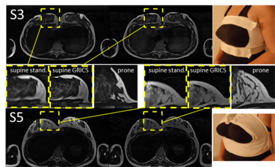 |
83 | Motion-corrected supine breast MRI with online reconstruction
Karyna Isaieva1, Pierre-André Vuissoz1, Lena Nohava1,2, Michael Obermann 2, Elmar Laistler2, Marc Fauvel3, Claire Dessale3, Nicolas Weber1, Jacques Felblinger1,3, and Freddy Odille1,3
1IADI, Université de Lorraine, INSERM, Nancy, France, 2High Field MR Center, Center for Medical Physics and Biomedical Engineering, Medical University of Vienna, Vienna, Austria, 3CIC-IT, CHRU de Nancy, INSERM, Nancy, France
A standard breast MRI protocol implies prone position. Supine position has multiple advantages; however, it requires motion correction. In this work we present our results on retrospective nonrigid motion correction of supine breast MRI on 7 healthy volunteers. A respiratory belt was used to capture the chest movements. Images were reconstructed online and fully automatically, including physiological data recording and its transfer to the reconstruction server. It was shown that application of the selected motion correction method improves image sharpness on 32-47% which was cofirmed by visual observations.
|
||
0859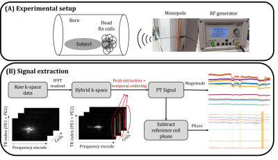 |
84 | Pilot Tone meets DISORDER: Improved data-driven motion-corrected brain MRI by leveraging Pilot Tone signal variations
Yannick Brackenier1,2,3, Thomas Wilkinson1,2,3, Lucilio Cordero-Grande1,2,4, Raphael Tomi-Tricot1,2,3,5, Philippa Bridgen1,2,3, Sharon Giles1,2,3, Enrico De Vita1,2,3, Shaihan J Malik1,2,3, and Joseph V Hajnal1,2,3
1Biomedical Engineering Department, School of Biomedical Engineering and Imaging Sciences, King's College London, London, United Kingdom, 2Centre for the Developing Brain, School of Biomedical Engineering and Imaging Sciences, King's College London, London, United Kingdom, 3London Collaborative Ultra high field System (LoCUS), London, United Kingdom, 4Biomedical Image Technologies, ETSI Telecomunicación, Universidad Politécnica de Madrid and CIBER-BNN, Madrid, Spain, 5MR Research Collaborations, Siemens Healthcare Limited, Frimley, United Kingdom
DISORDER is an established retrospective data driven motion correction approach that uses optimised phase encoding, but otherwise unmodified 3D acquisitions. It is highly effective, but requires multiple lines of k-space to be grouped together for each motion state to be estimated, and this limits temporal resolution. At 7T, head motion can also be detected by “Pilot Tone”, which is an injected RF signal picked up by each coil in the head receiver array, but a calibration step is required. Here we combine DISORDER and Pilot Tone to achieve integrated calibration and show that improved motion correction can result.
|
||
0860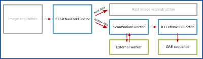 |
85 | Prospective 3D FatNav motion correction for 7T Terra
Krzysztof Klodowski1, Juraj Halabrin1, Iulius Dragonu2, and Chris Rodgers1
1Clinical Neurosciences, Wolfson Brain Imaging Centre, Cambridge, United Kingdom, 2Siemens Healthcare Limited, Frimley, United Kingdom Ultra-high field (7T) imaging can deliver images of brain structure and function at previously inaccessible spatial resolutions down to ~100um. However, subject motion, particularly for patients causes blurring which can lose some of the benefit of 7T imaging. The 3D FatNav approach to motion correction embeds short fat-excitation imaging modules in dead-time in an MRI pulse sequence. We are developing a prospective FatNav module for Siemens 7T Terra scanners. We present here a prospective FatNav module that embeds FatNav into a host pulse sequence, that separates FatNav and main image data during online reconstruction, that computes motion updates and that applies these in GRE imaging sequence in real time. |
||
0861 |
86 | Comparison of two retrospective tracking techniques in presence of fast and slow motion.
Elisa Marchetto1,2, Kevin Murphy2,3, and Daniel Gallichan1,2
1School of Engineering, Cardiff University, Cardiff, United Kingdom, 2Cardiff University Brain Research Centre (CUBRIC), Cardiff University, Cardiff, United Kingdom, 3School of Physics and Astronomy, Cardiff University, Cardiff, United Kingdom
We investigated the artifacts arising from different types of head motion during brain structural MR imaging and how well these artifacts can be compensated for using retrospective correction based on two different motion-tracking techniques: FatNavs and Tracoline systems. High image quality could be recovered in our slow-motion scenarios using both motion estimation techniques. Masking the non-rigid part of the neck during FatNav volumes registration led to higher image quality when large pitch-motion was present. The fast continuous motion scenario led to more severe image artifacts that could not be fully compensated by the retrospective motion correction techniques used.
|
||
0862 |
87 | On the limits of model-based reconstruction for retrospective motion correction in the presence of spin history effects
Aizada Nurdinova1, Stefan Ruschke1, and Dimitrios C. Karampinos1
1Department of Diagnostic and Interventional Radiology, School of Medicine, Technical University of Munich, Munich, Germany
MRI reconstruction methods comprising model-based motion correction modules often rely on the assumption of absence or piecewise constant motion patterns during signal readout and on the insignificance of spin history effects. Furthermore, the success of such methods also depends on the characteristics and motion sensitivity of the employed sequence and measured tissue properties and may therefor limit the general utility of such approaches in a clinical context. The present study demonstrates the limitations of Fourier-model-based motion correction reconstruction schemes by subtle violations of the piecewise constant motion assumption using Bloch simulations a model-based motion-corrected reconstruction approach.
|
||
0863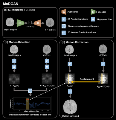 |
88 | MoDGAN: Unsupervised rigid motion detection and correction with generative adversarial networks
Mu-Yul Park1, Seul Lee1, Kyu-Jin Jung1, Jisu Yun1, and Dong-Hyun Kim1
1Department of Electrical and Electronic, Yonsei university, Seoul, Korea, Republic of Rigid motion artifacts, which are caused by subject’s motion during MRI acquisition, are a significant problem of image degradation. In this study, we propose a GAN-based method for unsupervised rigid motion detection and correction. The proposed method detects the motion-corrupted phase encoding lines using an anomaly score for motion detection. To reduce motion artifacts, we replace them with corresponding lines that generator yielded. We show that proposed method achieves high accuracy in detecting motion-corrupted line without any hardware or modification in sequence. Furthermore, the results of motion correction also showed noticeable performance. |
||
0864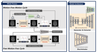 |
89 | Unsupervised Deep Learning using modified Cycle Generative Adversarial Network for rigid motion correction in pediatric brain MRI
Sewook Kim1, Seul Lee1, Jaeuk Yi1, Mohammed A. Al-Masni1, Sung-Min Gho2, Young Hun Choi3, and Dong-Hyun Kim1
1Department of Electrical and Electronic Engineering, Yonsei University, Seoul, Korea, Republic of, 2GE Healthcare, Seoul, Korea, Republic of, 3Department of Radiology, Seoul National University Hospital, Seoul, Korea, Republic of
Motion artifact that appears due to patient movement in MRI scans is a major obstacle in pediatric imaging. Since acquiring paired motion-clean and motion-corrupted dataset is difficult, unsupervised deep learning such as Cycle Generative Adversarial Network (Cycle-GAN) has been used in motion correction tasks these days. In this study, we propose a rigid motion artifact reducing strategy with modified Cycle-GAN by replacing generator that converts motion-free to motion-corrupted domain with a motion simulator. Our proposed model outperforms in contrast preservation and reducing artifacts compared to the original Cycle-GAN as well as reduces training parameters.
|
||
0865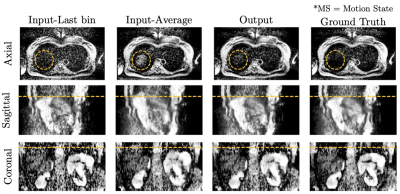 |
90 | Deep Learning-Based Respiratory Motion Correction in Free-Breathing Abdominal Diffusion-Weighted Imaging
Jinho Kim1,2,3, Fasil Gadjimuradov2,3, Thomas Benkert3, Thomas Vahle3, and Andreas Maier2
1Artificial Intelligence in Biomedical Engineering, Friedrich-Alexander Universität Erlangen-Nürnberg, Erlangen, Germany, 2Pattern Recognition Lab, Department of Computer Science, Friedrich-Alexander Universität Erlangen-Nürnberg, Erlangen, Germany, 3MR Application Pre-development, Siemens Healthcare GmbH, Erlangen, Germany
Since a single diffusion-weighted image can suffer from low SNR, multiple DWI repetitions can be averaged to improve SNR, which, however, can introduce blurring due to respiratory motion between different repetitions. Consequently, retrospective gating can be performed to overcome this problem. However, conventional retrospective gating has low SNR efficiency as it discards parts of the data and may result in certain slices to be missing for the desired motion state. This work proposes an efficient Deep Learning-based motion-correction method to improve conventional retrospective gating in free-breathing DWI, resulting in sharper images while maintaining image information from all acquired repetitions.
|
||
 Back to Meeting Home
 Back to the Program-at-a-Glance
The International Society for Magnetic Resonance in Medicine is accredited by the Accreditation Council for Continuing Medical Education to provide continuing medical education for physicians.
 Back to Meeting Home
 Back to the Program-at-a-Glance
 View the Presentation