Digital Poster
Motion & Artifacts II
Joint Annual Meeting ISMRM-ESMRMB & ISMRT 31st Annual Meeting • 07-12 May 2022 • London, UK

| Computer # | ||||
|---|---|---|---|---|
0955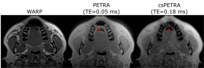 |
75 | Near Metal MRI with Compressed Sensing PETRA
Serhat Ilbey1, Michael Bock1, Matthias Jung2, Lukas Konstantinidis3, and Ali Özen1
1Dept. of Radiology, Medical Physics, Medical Center University of Freiburg, Faculty of Medicine, University of Freiburg, Freiburg, Germany, 2Department of Diagnostic and Interventional Radiology, Medical Center University of Freiburg, Faculty of Medicine, University of Freiburg, Freiburg, Germany, 3Department of Orthopaedics and Trauma Surgery, Medical Center University of Freiburg, Faculty of Medicine, University of Freiburg, Freiburg, Germany
MRI near metallic implants require high bandwidth excitation and acquisition to minimize signal void and pile-up artefacts. In this work, compressed sensing PETRA (csPETRA) was modified to have intentionally a longer TE, so that the phase-encoding part of the k-space is extended. To prevent extremely long scan times, SPI points in csPETRA sequence are pseudo-randomly undersampled to reduce the scan time below 6 minutes. csPETRA reduced artifacts near metal significantly while preserving the anatomical details. Isotropic 3D MRI of two volunteers with mouth and knee prostheses was performed with csPETRA and the artifacts are significantly reduced compared to T1w-WARP sequence.
|
||
0956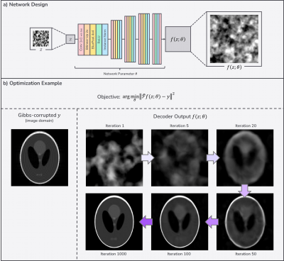 |
76 | Gibbs-ringing Removal through Anti-aliased Deep Priors Video Permission Withheld
Jaeuk Yi1, Chuanjiang Cui1, Kyu-Jin Jung1, and Dong-Hyun Kim1
1Department of Electrical and Electronic Engineering, Yonsei University, Seoul, Korea, Republic of Inspired by recent usage of Deep Image Prior (DIP) in the field of MRI that utilizes a powerful low-level image prior from a neural network architecture itself without any training dataset, we conduct k-space extrapolation using the deep prior for Gibbs-ringing removal in order to build general Gibbs-ringing correction algorithm without dependency on the dataset. We further improved the existing deep prior with the addition of anti-aliasing layers. The proposed deep prior method outperformed conventional non-learning methods quantitively and qualitatively in numerical simulations and in-vivo data. |
||
0957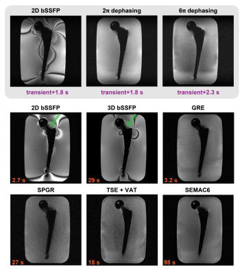 |
77 | Imaging near metal at 0.55 T using gradient-echo based sequences: Feasibility and Opportunities
Kübra Keskin1, Brian A. Hargreaves2,3,4, and Krishna S. Nayak1,5
1Electrical and Computer Engineering, University of Southern California, Los Angeles, CA, United States, 2Radiology, Stanford University, Stanford, CA, United States, 3Electrical Engineering, Stanford University, Stanford, CA, United States, 4Bioengineering, Stanford University, Stanford, CA, United States, 5Biomedical Engineering, University of Southern California, Los Angeles, CA, United States The large magnetic susceptibility difference between metallic implants and surrounding tissues causes severe MRI artifacts that scale with the B0 field strength. At conventional field strengths, spin-echo based multispectral imaging is used to mitigate this artifact and produce diagnostic images. At lower field strengths, such as 0.55 T, other strategies may be feasible. Here, we investigate gradient-echo based sequences for imaging near metal at 0.55 T, which provide high SNR efficiency and different contrast. |
||
0958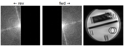 |
78 | Low Rank Off-resonance Correction for Double Half-Echo k-Space Acquisitions
Mark Bydder1, Fadil Ali1, Vahid Ghodrati1, Chencai Wang1, Akifumi Hagiwara1, and Ben Ellingson1
1UCLA, Los Angeles, CA, United States
The present study describes a model-based approach for correcting off-resonance in the context of short echo-time imaging using a double half-echo k-space acquisition. This technique employs center-out readouts in forward and reverse directions and is sensitive to off-resonance. The correction is simple and results in a marked reduction in blurring for a negligble cost in pre-processing.
|
||
0959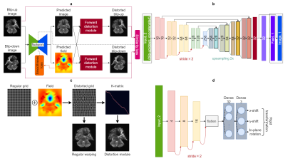 |
79 | A Deep Forward-Distortion Model for Unsupervised Correction of Susceptibility Artifacts in EPI
Abdallah Zaid Alkilani1,2, Tolga Çukur1,2,3, and Emine Ulku Saritas1,2,3
1Department of Electrical and Electronics Engineering, Bilkent University, Ankara, Turkey, 2National Magnetic Resonance Research Center (UMRAM), Bilkent University, Ankara, Turkey, 3Neuroscience Graduate Program, Bilkent University, Ankara, Turkey Echo planar imaging (EPI) requires the correction of susceptibility artifacts for further quantitative analyses. Images acquired in reversed phase-encode (PE) directions are typically used to estimate the susceptibility-induced field from EPI data. In this work, an unsupervised deep Forward-Distortion Network (FD-Net) is proposed for the correction of susceptibility artifacts in EPI: The field and underlying anatomically-correct image are predicted, subject to the constraint that forward-distortion of this image with the field explains the input warped images. This approach provides rapid correction of susceptibility artifacts, with superior performance over deep learning methods that unwarp input images based on a predicted field. |
||
0960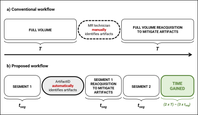 |
80 | Inline artifact identification using segmented acquisitions and deep learning
Keerthi Sravan Ravi1,2, Marina Manso Jimeno1,2, John Thomas Vaughan Jr.2, and Sairam Geethanath2
1Department of Biomedical Engineering, Columbia University, New York, NY, United States, 2Columbia Magnetic Resonance Research Center, New York, NY, United States
MR artifacts degrade image quality and affect diagnosis, requiring thorough examination by the MR technician, and reacquisition in some cases. We employ a combination of segmented acquisitions and a deep learning tool (ArtifactID) to perform more frequent updates to image quality during acquisition. ArtifactID identified wrap-around, Gibbs ringing and motion artifacts, with a mean accuracy of 99.43%. The segmented acquisitions for rescans resulted in a 12.98% time gain over the full-FOV sequence. In addition, ArtifactID alleviates burden on the MR technician via automatic artifact identification, saving image quality evaluation time and augmenting expertise.
|
||
0961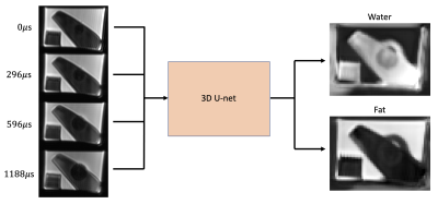 |
81 | Metal Artifact Corrected and Chemical Shift Encoded MRI
Collin J Buelo1,2, Timothy J. Colgan1, and Diego Hernando1,2
1Radiology, University of Wisconsin-Madison, Madison, WI, United States, 2Medical Physics, University of Wisconsin-Madison, Madison, WI, United States
Fat-suppressed MRI near metal remains a challenging technical problem. Chemical shift encoded (CSE) water/fat separation enables excellent SNR efficiency compared to short tau inversion recovery (STIR), but the large B0 field offsets and gradients complicate conventional CSE methods near metal. To address these challenges, we propose metal artifact correction and chemical shift encoding using optimized echo spacings, and with deep learning processing for reconstruction of water/fat separated imaging near metal. This method is able to correctly separate water and fat closer to a metallic surgical breast clip than conventional methods, without the drawbacks associated with STIR imaging.
|
||
0962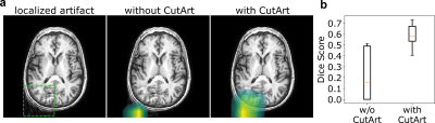 |
82 | Improved Detection and Localization of Artifacts using CutArt Augmentation
Ben A Duffy1, Greg Zaharchuk2, Enhao Gong1, and Keshav Datta1
1Subtle Medical Inc., Menlo Park, CA, United States, 2Stanford University, Stanford, CA, United States
With the aim of improving the performance of an automated quality control system, we propose to use a data augmentation technique based on cropped patches of simulated artifacts (CutArt) instead of artifacts that are distributed across the entire image. This has the advantage of improving the artifact localization and quality control classification performance, as assessed by experiments on simulated as well as real artifact affected data. Localization experiments suggested that the CutArt model learns to focus on the tissue of interest instead of the image background.
|
||
0963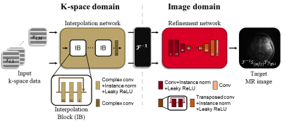 |
83 | Multi-domain Motion Correction Network for Turbo Spin-echo MRI
Jongyeon Lee1, Wonil Lee1, Namho Jeong1, and HyunWook Park1
1Korea Advanced Institute of Science and Technology, Daejeon, Korea, Republic of
Motion correction in MRI has been successfully performed by adopting deep learning techniques to correct motion artifacts with data-driven algorithms. However, many of these studies have narrowly utilized MR physics. In addition to the efficient motion correction method proposed for multi-contrast MRI, we expand our previous method to utilize data in k-space domain for a single-contrast MRI. Using the motion simulation based on MR physics, we propose a multi-domain motion correction method in both k-space and image domains. Our proposed method finely reduces motion artifacts using the multi-domain network.
|
||
0964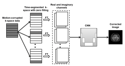 |
84 | Time-segmented and contrast-neutral motion correction using an end-to-end deep learning approach
Refaat E Gabr1, Masoud Edalati2, Lingzhi E Hu2, Adam Chandler2, Weiguo Zhang2, and Ponnada A Narayana3
1Diagnostic and Interventional Imaging, University of Texas Health Science Center at Houston, Houston, TX, United States, 2United Imaging Healthcare, Houston, TX, United States, 3University of Texas Health Science Center at Houston, Houston, TX, United States
We developed an end-to-end deep learning-based motion correction technique that utilized the time-segmented nature of MRI data acquisition. Results of computer simulations and in vivo studies show the network to be highly effective in correcting motion artifacts.
|
||
0965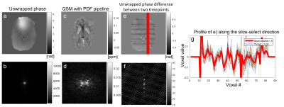 |
85 | Characterisation and Removal of Slice Dependent Artifacts in EPI Phase Images
Jannette Nassar1, Oliver Kiersnowski1, Patrick Fuchs1, and Karin Shmueli1
1Medical Physics and Biomedical Engineering, UCL, London, United Kingdom
Although EPI phase images are useful (e.g. for Quantitative Susceptibility Mapping), they often contain phase inconsistencies in the slice-select direction which persist and can degrade QSM results. Here, we analysed three EPI datasets in healthy volunteers to characterise these phase inconsistencies and understand whether they occur or interact with interleaved or sequential slice acquisition order. We characterised a ~2Hz cryogen pump artifact in sequential data and slice-to-slice phase jumps in interleaved data. We modified a previously proposed QSM processing pipeline, including 2D (VSHARP) and 3D (PDF) background field removal that removed all through-slice artifacts observed.
|
||
0966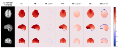 |
86 | Predicting changes in B0 field due to patient movement dynamically for MRI via Deep Learning
Stanislav Motyka1, Paul Weiser2, Beata Bachrata1, Lukas Hingerl1, Bernhard Strasser1, Dario Goranovic1, Gilbert Hangel1,3, Eva Niess1, Maxim Zaitsev4, Simon Robinson1, Georg Langs2, Siegfried Trattning1,5, and Wolfgang Bogner1
1High Field MR Center, Department of Biomedical Imaging and Image-guided Therapy, Medical University of Vienna, Vienna, Austria, 2Computational Imaging Research Lab, Department of Biomedical Imaging and Image-guided Therapy, Medical University of Vienna, Vienna, Austria, 3Department of Neurosurgery, Medical University of Vienna, Vienna, Austria, 4High Field MR Center, Center for Medical Physics and Biomedical Engineering, Medical University of Vienna, Vienna, Austria, 5Christian Doppler Laboratory for Clinical Molecular MR Imaging, Medical University of Vienna, Vienna, Austria
We proposed a U-net-based neural network to predict changes in B0 field due to patient movement. The network was trained and tested on 7T in vivo volunteer data. The method was compared against two other approaches. The estimated B0 maps were compared against the ground truth. The results suggest that prediction of B0 maps is feasible and by combination with external tracking a considerable improvement in data quality of B0-sensitive MRI methods could be achieved.
|
||
 Back to Meeting Home
 Back to the Program-at-a-Glance
The International Society for Magnetic Resonance in Medicine is accredited by the Accreditation Council for Continuing Medical Education to provide continuing medical education for physicians.
 Back to Meeting Home
 Back to the Program-at-a-Glance
 View the Presentation