Digital Poster
Strategic Acquisitions I
Joint Annual Meeting ISMRM-ESMRMB & ISMRT 31st Annual Meeting • 07-12 May 2022 • London, UK

| Computer # | ||||
|---|---|---|---|---|
2352 |
88 | Estimating B0 changes in Oscillating Steady State Imaging (OSSI) using an Artificial Neural Network
Mariama Salifu1, Melissa Haskell2, and Douglas C Noll1
1Biomedical Engineering, University of Michigan, Ann Arbor, MI, United States, 2Electrical Engineering and Computer Science, University of Michigan, Ann Arbor, MI, United States
Oscillating steady state imaging (OSSI) is a novel fMRI acquisition method which produces a high SNR T2*-weighted signal. Physiological and drift induced B0 changes cause undesired signal distortions which can significantly diminish functional contrast in OSSI fMRI. Here, we investigated a method of rapidly estimating quantitative B0 field changes from OSSI images using an artificial neural network (ANN) model. Our results demonstrated that this technique can be used to rapidly measure field changes, which has the potential to be used for prospective and retrospective B0 field correction in OSSI.
|
||
2353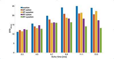 |
89 | Realtime dynamic xyz-shimming in the cervical spinal cord
Daniel Papp1, Alexandre D’Astous1, Julien Cohen-Adad1,2,3, and Eva Alonso-Ortiz4
1NeuroPoly Lab, Institute of Biomedical Engineering, Polytechnique Montreal, Montreal, QC, Canada, 2Mila - Quebec AI Institute, Montreal, Montreal, QC, Canada, 3Functional Neuroimaging Unit, Centre de recherche de l'Institut universitaire de gériatrie de Montréal, Montreal, QC, Canada, 4NeuroPoly Lab, Department of Electrical Engineering, Polytechnique Montreal, Montreal, QC, Canada
Respiration-induced changes of the magnetic field are an important source of image artefacts in spinal cord MRI. Here, we introduce realtime dynamic xyz-shimming to fully compensate for these distortions in a time-varying (‘realtime”), slicewise (‘dynamic”) fashion, using both in-plane (‘xy’) and through-plane (‘z’) gradients (‘xyz-shimming’).
|
||
2354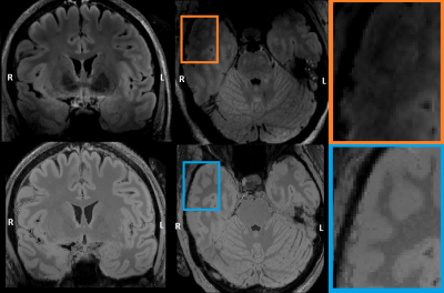 |
90 | DeepFLAIR: a neural network approach to mitigate signal loss in temporal lobe regions of 7 Tesla FLAIR images
Daniel Uher1, Jacobus F.A. Jansen1,2, Gerhard S. Drenthen1,2, Benedikt A. Poser3, Christopher J. Wiggins4, Paul A.M. Hofman2,5, Louis G. Wagner5, Rick H.G.J. van Lanen6, Christianne M. Hoeberigs2,5, Albert J. Colon5, Olaf E.M.G. Schijns1,5,6, and Walter H. Backes1,2
1School for Mental Health and Neuroscience, Maastricht University, Maastricht, Netherlands, 2Department of Radiology & Nuclear Medicine, Maastricht University Medical Centre, Maastricht, Netherlands, 3Department of Cognitive Neuroscience, Maastricht University, Maastricht, Netherlands, 4Scannexus, Maastricht, Netherlands, 5Academic Centre for Epileptology, Kempenhaeghe/Maastricht University Medical Centre, Heeze/Maastricht, Netherlands, 6Department of Neurosurgery, Maastricht University Medical Centre, Maastricht, Netherlands
In this study we aimed to improve the 7T FLAIR image quality, especially within the temporal lobe regions which are often attenuated due to field inhomogeneities. A neural network using MP2RAGE and T2-weighted images as inputs was set up to generate a new FLAIR-like image. The training was performed on the extratemporal-lobe voxels of the acquired 7T FLAIR image. The deepFLAIR showed a significant improvement in the signal-to-noise ratio and contrast-to-noise ratio in the temporal lobe regions in a number of cases. This study showed the potential to generate FLAIR-like images with reduced inhomogeneity artifacts and improved image quality.
|
||
2355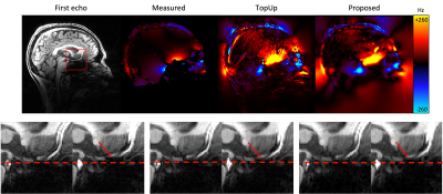 |
91 | Distortion correction in multi-echo MRI without field mapping or reverse encoding
Yael Balbastre1,2, Divya Varadarajan1,2, Klara Bas3, John Ashburner3, Robert Frost1,2, and Bruce Fischl1,2,4
1Athinoula A. Martinos Center for Biomedical Imaging, Massachusetts General Hospital, Charlestown, MA, United States, 2Department of Radiology, Harvard Medical School, Boston, MA, United States, 3Wellcome Center for Human NeuroImaging, Queen Square Institute of Neurology, University College London, London, United Kingdom, 4Computer Science and Artificial Intelligence Laboratory, Massachusetts Institute of Technology, Cambridge, MA, United States
Low-bandwidth multi-echo acquisitions suffer from spatial distortions between odd and even echoes caused by B0 homogeneity, causing blurs and loss of effective resolution in sum-of-square images or derived quantitative parameters. We show in this abstract that this curse can also be beneficial, as it allows distortions to be mapped without the need for additional field mapping data. We do so by jointly optimizing a forward model of the data with respect to exponential decay parameters and a smooth distortion map.
|
||
2356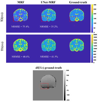 |
92 | Off-resonance correction in MRF using Deep Learning in fingerprint space
Ronal Coronado1,2, Carlos Castillo-Passi1,2, Gabriel della Maggiora1,2, Sergio Manuel Uribe1,2, Cristian Tejos1, Claudia Prieto1,2,3, Cecilia Besa4, and Pablo Irarrazaval1,2
1Biomedical Imaging Center-Universidad Católica de Chile, Santiago, Chile, 2Millennium Nucleus for Cardiovascular Magnetic Resonance, Santiago, Chile, 3King's College London, London, United Kingdom, 4Departamento de Radiologia-Universidad Católica de Chile, Santiago, Chile Magnetic Resonance Fingerprinting (MRF) acquisitions with balanced Steady State Free Precession (bSSFP) and spiral trajectories are prone to off-resonance artifacts. These artifacts affect the reconstruction of the tissue maps (T1 and T2). We propose to use a UNet CNN feed with fingerprints corrupted by off-resonance to generate corrected fingerprints with only aliasing in the bSSFP-MRF sequence. The feasibility of the proposed approach was evaluated in simulations and in-vivo brain data. Our method improved the NRMSE values for both quantitative maps T1 and T2. Considerably reducing the effects of the off-resonance by UNet-MRF in comparison to classical bSSFP-MRF. |
||
2357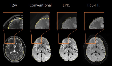 |
93 | Comparison of distortion correction with Echo Planar Imaging Correction and the 2D navigator based multi-shot SENSE EPI schemes
Yajing Zhang1, Zhigang Wu2, Xiuquan Hu3, Guangyu Jiang4, Jing Zhang3, Yan Zhao5, Marc Van Cauteren6, and Jiazheng Wang3
1BU-MR Clinical Science, Philips Healthcare, Suzhou, China, 2Philips Healthcare (China), Shenzhen, China, 3Philips Healthcare (China), Beijing, China, 4BU-MR Clinical Application, Philips Healthcare, Suzhou, China, 5BU-MR R&D, Philips Healthcare, Suzhou, China, 6BU-MR Clinical Science, Philips Healthcare, Tokyo, Japan Brain diffusion imaging by single shot EPI has remained challenging for the local B0 field variation, which leads to geometry distortion and signal inhomogeneity in the resulting images. In this study, we show that the brain single shot EPI image quality could be improved by correction of the geometric distortion and increased resolution, when compared to the traditional acquisition scheme. In-plane resolution of 1.2x1.2 mm2 was achieved and the distortion correction was effectively reduced. |
||
2358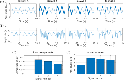 |
94 | Measurements of composite signals with MR-based neuronal current imaging: one step closer to clinical application. Video Permission Withheld
Milena Capiglioni1, Claus Kiefer2, and Roland Wiest2
1Institute for Diagnostic and Interventional Neuroradiology, Support Center for Advanced Neuroimaging, University of Bern, Bern, Switzerland, 2Institute for Diagnostic and Interventional Neuroradiology, Support Center for Advanced Neuroimaging (SCAN), Inselspital, Bern, Bern, Switzerland
We evaluated the performance of stimulus-induced rotational saturation contrast (SIRS) to assess the possibility of detecting neuronal currents. We analyzed composite signals with multiple frequencies to evaluate the ability of the technique to be used as a filter of spectral components. We conclude that the method can separate and distinguish frequency components and reconstruct the distribution of spectral components between individually acquired signals. These tests are the basis for moving towards the clinical application of this sequence to locate fields with frequencies associated with specific pathologies.
|
||
2359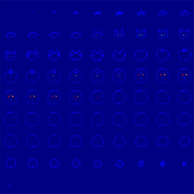 |
95 | MEDI-FM: Field Map Error Guided Regularization for Shadow Reduction
Alexandra Grace Roberts1, Pascal Spincemaille2, Thanh Nguyen2, and Yi Wang2,3
1Electrical and Computer Engineering, Cornell University, Ithaca, NY, United States, 2Radiology, Weill Cornell Medicine, New York, NY, United States, 3Biomedical Engineering, Cornell University, Ithaca, NY, United States
Morphology Enabled Dipole Inversion (MEDI) is an iterative reconstruction algorithm for Quantitative Susceptibility Mapping (QSM) that is effective in suppressing streaking artifacts by exploiting the magnitude image as a morphological prior. However, contiguous areas of dipole-incompatibility (such as noise) induce shadow artifacts whose spatial frequency components are not sufficiently regularized by the gradient based regularization in MEDI. Regularizing these spatially connected regions reduces shadow artifacts in Morphology Enabled Dipole Inversion based Quantitative Susceptibility Mapping reconstructions.
|
||
2360 |
96 | B0 field distortions monitoring and correction for 3D non-Cartesian fMRI acquisitions using a field camera: Application to 3D-SPARKLING at 7T
Zaineb Amor1, Chaithya G.R.1,2, Caroline Le Ster1, Guillaume Daval-Frérot1,2,3, Nicolas Boulant1, Franck Mauconduit1, Christian Mirkes4, Philippe Ciuciu1,2, and Alexandre Vignaud1
1Université Paris-Saclay, CEA, Neurospin, CNRS, Gif-sur-Yvette, France, 2Université Paris-Saclay, Inria, Parietal, Palaiseau, France, 3Siemens Healthcare SAS, Saint Denis, France, 4Skope Magnetic Resonance Technologies AG, Zurich, Switzerland
The benefit of retrospective correction of static and dynamic $$$B_{0}$$$ field imperfections was studied on non-Cartesian compressed sensing-based acquisition patterns by taking as an example 3D-SPARKLING encoding scheme. Long-TR dynamic acquisitions of T2*-weighted MRI (fMRI-like) volumes were acquired at 7T, retrospectively corrected, and the evaluation of the corrections was based on the image quality and the gain in temporal signal-to-noise ratio (tSNR). We found that correcting for the static and dynamic $$$0^{th}$$$ order contributions is enough to enhance the image quality and tSNR significantly (up to 22% gain in median tSNR).
|
||
 Back to Meeting Home
 Back to the Program-at-a-Glance
The International Society for Magnetic Resonance in Medicine is accredited by the Accreditation Council for Continuing Medical Education to provide continuing medical education for physicians.
 Back to Meeting Home
 Back to the Program-at-a-Glance
 View the Presentation