Digital Poster
Data Acquisition for Diffusion, Perfusion & Spectroscopy I
Joint Annual Meeting ISMRM-ESMRMB & ISMRT 31st Annual Meeting • 07-12 May 2022 • London, UK

Digital Poster
Data Acquisition for Diffusion, Perfusion & Spectroscopy I
| Computer # | ||||
|---|---|---|---|---|
1032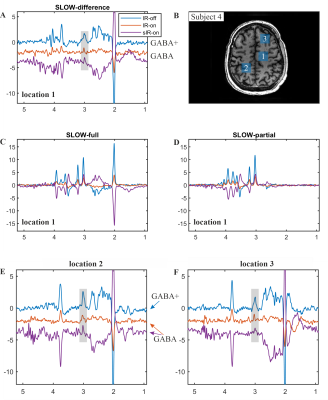 |
39 | SLOW-editing for GABA/GABA+ MRSI at 7T
Guodong Weng1,2, Piotr Radojewski1,2, and Johannes Slotboom1,2
1Institute for Diagnostic and Interventional Neuroradiology, Support Center for Advanced Neuroimaging (SCAN), University of Bern, Bern, Switzerland, 2Translational Imaging Center, sitem-insel AG, Bern, Switzerland Purpose: whole brain in vivo detection of GABA/GABA+ using SLOW-editing at 7T. Methods: An EPSI-based B0/B1+ robust SLOW-editing was applied in four healthy subjects. Pure GABA was measured by two different macromolecular-nulling inversion recovery pulses, namely a broadband- and a narrowband-inversion pulse. Results: the editing GABA/GABA+ signal at 3.01 ppm are presented. The signal intensity of GABA/GABA+ ratio is 40 – 60%. Conclusions: whole-brain in vivo GABA/GABA+-editing can be performed using SLOW-EPSI in around 10 minutes measurement time and is therefore clinically applicable. |
||
1033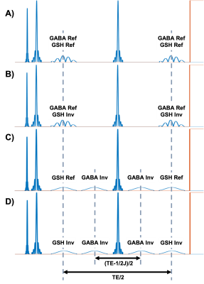 |
40 | Simultaneous J-difference editing of GABA and GSH with extended echo-times using ExTE-HERMES
Peter Truong1 and Jamie Near1,2
1Physical Sciences Research Platform, Hurvitz Brain Sciences Program, Sunnybrook Research Institute, Toronto, ON, Canada, 2Department of Medical Biophysics, University of Toronto, Toronto, ON, Canada
J-difference editing techniques, such as MEGA-PRESS, are often used to detect gamma-aminobutryic acid (GABA) and glutathione (GSH). However, two separate scans are needed to acquire both metabolites. The HERMES editing scheme does address this issue, allowing simultaneous acquisition of GABA and GSH. Even then, there are limitations as the optimal echo time (TE) can only be set for one metabolite at the expense of lower editing efficiency for the other. We propose the Extended TE (ExTE) HERMES editing scheme, which allows us to acquire edited spectra at TE=120ms, ensuring maximum GSH signal while still providing efficient GABA editing.
|
||
1034 |
41 | Cerebral Blood Flow Quantification using Pseudo-continuous Arterial Spin Labeling at 7T MRI
Salem Alkhateeb1, Tae Kim2, Howard J Aizenstein2, and tamer S. Ibrahim3
1University of Pittsburgh, PITTSBURGH, PA, United States, 2University of Pittsburgh, Pittsburgh, PA, United States, 3University of Pittsburgh, pittsburgh, PA, United States
The feasibility of implementing pCASL technique at 7T MRI as a routine protocol uncovers the difficulties and challenges that need to be overcome and taking into consideration the many variables that need to be addressed before running the sequence. We were able to successfully implement pCASL sequence with satisfactory results. CBF quantification for the 4 volunteers was within clinical normal ranges and CBF mapping agrees with published data. Future work will include the inclusion of neck coil module for improving labeling efficiency.[GU1] Where is figure 4 referenced [GU1] Please add the references
|
||
1035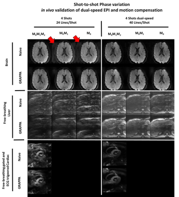 |
42 | Prospective correction of multi-shot diffusion imaging using motion compensation and dual-speed EPI
Kevin Moulin1,2, Tyler E Cork3,4, Thomas Troalen5, Daniel Ennis3,4, Pierre Croisille1,2, and Magalie Viallon1,2
1CREATIS, Lyon, France, 2Department of Radiology, University Hospital Saint-Etienne, Saint Etienne, France, 3Department of Radiology, Stanford University, Stanford, CA, United States, 4Division of Radiology, Veterans Administration Health Care System, Palo Alto, CA, United States, 5Siemens Healthcare SAS, Saint-Denis, France Multi-shot (MS) reduces image distortion in EPI caused by B0 field inhomogeneity and enables higher resolution diffusion imaging. However, interaction between physiologic motion and diffusion encoding gradients leads to strong image aliasing artifacts in MS-EPI due to phase discrepancies. We propose prospective strategies to reduce shot-to-shot phase variation for interleaved phase-segmented diffusion MS-EPI. First and second order motion compensated diffusion gradient waveforms and dual-speed EPI with oversampled k-space center were evaluated as a shot-to-shot phase mitigation strategy. Validations were performed in silico, in vitro, and in vivo in the brain, liver, and heart. |
||
1036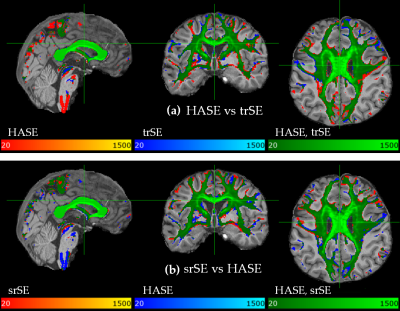 |
43 | Highly Asymmetric Acquisitions for Diffusion MRI Tractography
Manoj Shrestha1, Pavel Hok1,2, Ulrike Nöth1, and Ralf Deichmann1
1Brain Imaging Center (BIC), Goethe University Frankfurt, Frankfurt am Main, Germany, 2Department of Neurology, Palacky University Olomouc and University Hospital Olomouc, Olomouc, Czech Republic
Frequently, diffusion-weighting modules such as schemes based on the Stejskal-Tanner concept or the STEAM are combined with EPI readout for fast k-space sampling. In contrast to standard sequences, highly asymmetric spin-echo and highly asymmetric STEAM sequences (HASE, HASTEAM) offer shorter TE, maintaining constant diffusion preparation duration, independent of the matrix-size. In this study, HASE and HASTEAM sequences were implemented for neuronal fibre tracking, and results were compared with standard sequences. Both asymmetric sequences HASE and HASTEAM allow for more accurate estimations of the subsidiary fibre orientations, which may considerably improve the quality and quantity of the detected fibre tracts.
|
||
1037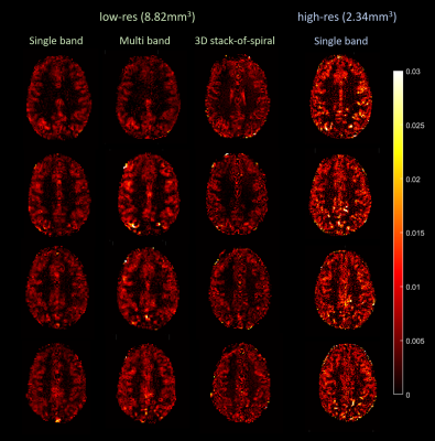 |
44 | Investigating Spiral Arterial Spin Labeling with Pulseq and Field Monitoring at 7T
Ruoyun Emily Ma1, Dimo Ivanov1, Renzo Huber1, Denizhan Kurban1, and Benedikt A Poser1
1Faculty of Psychology and Neuroscience, Maastricht University, Maastricht, Netherlands
We developed several ASL sequences at 7T with a FAIR-QUIPSS II labeling scheme and various spiral readout strategies using Pulseq. Iterative algebraic image reconstruction was performed with CG-SENSE, using the field evolution data measured with external NMR probes. Robust performance in detecting brain’s perfusion signal was observed in 2D single- and multi-band spiral acquisitions especially at relatively high spatial resolution, without the requirement for a longer scan time. 3D spiral acquisition showed reduced contrast level in perfusion maps and requires further investigation and optimization.
|
||
1038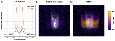 |
45 | 3D EPI sequence for indirect detection of hyperpolarized [2-13C]lactate with J-coupling artifact correction
Tyler Blazey1 and Cornelius von Morze1
1Washington University in St. Louis, St. Louis, MO, United States
Imaging of hyperpolarized (HP) 13C magnetization is typically done using direct detection of the 13C signal. However, indirect 1H-based detection via J-coupled protons has the advantage of reducing gradient requirements and enhancing sensitivity, as well as practical advantages. We have developed an INEPT-based EPI sequence, which we tested by acquiring 3D images of a 13C formate phantom after transfer of thermal polarization from 1H to 13C. We also tested a novel deconvolution method to correct ghosting artifacts introduced in EPI images by J-coupling. These methods will be translated into in vivo HP imaging experiments in the next phase of research.
|
||
1039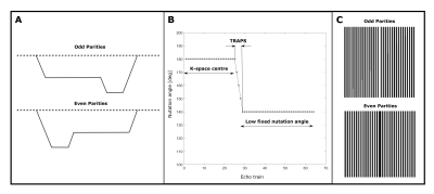 |
46 | Robust and Motion-Insensitive Approach to Diffusion-Weighted Half-Fourier Acquisition Single-Shot Turbo Spin-Echo Imaging
Aidin Arbabi1, Vitaliy Khlebnikov1, David. G. Norris1, and Jose P. Marques1
1Donders Institute, Radboud University, Nijmegen, Netherlands
Half-Fourier acquisition single-shot turbo spin-echo diffusion-weighted (DW-HASTE) is a clinically established magnetic resonance imaging tool for the detection of small lesions, particularly cholesteatoma. However, in the standard approach, half of the available signal is sacrificed through displacing one echo parity out of the acquisition window to fulfil the Carr-Purcell-Meiboom-Gill condition. We present a selective parity approach to tackle this problem. The proposed method features a near full sensitivity, a low specific absorption rate for long echo trains, and about two-fold increase in signal-to-noise ratio, compared to the manufacturer's method under the same conditions.
|
||
1040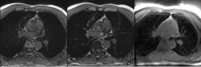 |
47 | Pulmonary imaging Using 3D Dual-Echo FID Ultra-short Echo Time MRI with Rosette k-space Pattern: Introduction and Feasibility.
Nathan Ooms1, Xin Shen2, Ali Caglar Ozen3, Serhat Ilbey3, Mark Chiew4, and Uzay Emir1
1School of Health Sciences, Purdue University, West Lafayette, IN, United States, 2Weldon School of Biomedical Engineering, Purdue University, West Lafayette, IN, United States, 3Radiology, Medical Center- University of Freiburg, Freiburg, Germany, 4Welcome Centre for Integrative Neuroimaging, University of Oxford, Oxford, United Kingdom
Pulmonary imaging traditionally uses techniques utilizing ionizing radiation such as x-rays and CT. Pediatrics, pregnant patients, and patients who will be exposed multiple times over long periods of time would benefit from a non-ionizing modality. MRI does not use ionizing radiation but suffers from low signal from the lungs and breathing motion artifact. We are proposing a novel MRI technique to combat these issues: a 3-dimensional Dual-Echo FID Ultra-short Echo Time (3D DE UTE) sequence utilizing Rosette k-space acquisition. Preliminary results demonstrated better pulmonary artery segmental branches and pleural wall definition compared to other commercially available MRI sequences.
|
||
1041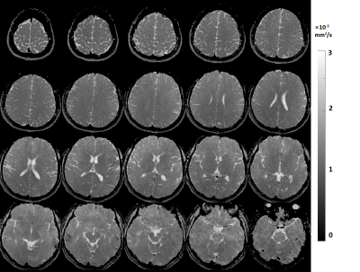 |
48 | Simultaneous ADC mapping and water-fat separation with B0 correction using a rosette acquisition
Kang Yan1,2, Huajun She2, and Yiping Du2
1Biomedical Engineering, University of Virginia, Charlottesville, VA, United States, 2Biomedical Engineering, Shanghai Jiao Tong University, Shanghai, China
In diffusion-weighted imaging (DWI) using echo-planar acquisition, fat signals are commonly suppressed by using chemical saturation or by spectral-spatial RF excitation. In this study, we present a technique using a rosette trajectory for simultaneous mapping of apparent diffusion coefficient (ADC) and water-fat separation with inherent B0 correction. The feasibility of using this technique is demonstrated in phantom, brain, and head-neck scans.
|
||
1042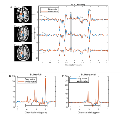 |
49 | SLOW-EPSI for the Whole Brain Detection of Phosphorylethanolamine (PE) at 7T
Guodong Weng1,2, Piotr Radojewski1,2, Philippe Schucht3, Roland Wiest1,2, and Johannes Slotboom1,2
1Institute for Diagnostic and Interventional Neuroradiology, Support Center for Advanced Neuroimaging (SCAN), INSEL Gruppe AG, University of Bern, Bern, Switzerland, 2Translational Imaging Center, sitem-insel AG, Bern, Switzerland, 3Department of Neurosurgery, Inselspital Bern and University Hospital, Bern, Switzerland Purpose: to detect phosphoethanolamine (PEtn or PE) in vivo for whole-brain MRSI, overcoming challenges of B1+-inhomogeneities and CSDE at 7T. Methods: An EPSI-based B1+ robust SLOW-editing was applied to three healthy subjects. TE = 90 ms, TR = 1500 ms, and TA = 9:04 mins. Results: the editing PE signal at 3.22 ppm was clearly present, and the B1+-homogenous result/map was obtained. Conclusions: whole-brain in vivo PE-editing can be performed in around 9 minutes measurement time and is therefore clinically applicable. |
||
1043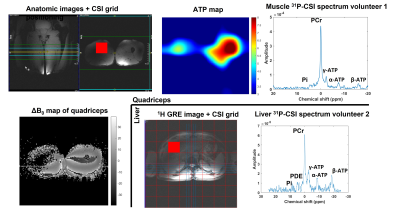 |
50 | A modular software “sequence building block” for interleaved MRS and MRI acquisition on 7T Terra
Jabrane Karkouri1, Christopher Wickens1, Daniel Atkinson1, and Christopher T. Rodgers1
1University of Cambridge, Cambridge, United Kingdom
In this work, we present a software “sequence building block” that enables interleaving MRS acquisitions (X-nuclear or 1H) inside a base imaging sequence (typically 1H) on the Siemens 7T Terra platform. We envisage using this approach for dynamic B0 shim and frequency offset correction to mitigate breathing artefacts in 31P-MRSI of heart and liver.
|
||
1044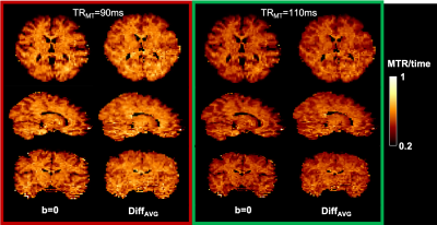 |
51 | Dual-encoding of magnetization transfer and diffusion for the characterization of tract-specific myelination
Ilana R Leppert1, Christopher D Rowley1,2, Jennifer SW Campbell1, Mark Nelson1,3, Bruce G Pike4, and Christine L Tardif1,2,3
1McConnell Brain Imaging Centre, Montreal Neurological Institute and Hospital, Montreal, QC, Canada, 2Department of Biomedical Engineering, McGill University, Montreal, QC, Canada, 3Department of Neurology and Neurosurgery, McGill University, Montreal, QC, Canada, 4Hotchkiss Brain Institute and Departments of Radiology and Clinical Neuroscience, University of Calgary, Calgary, Canada, Calgary, AB, Canada Most white matter voxels are comprised of a combination of microstructurally different tracts that kiss and cross. Conventional MRI contrasts that provide a single measurement per voxel fail to disentangle the microstructural properties of these tracts. Co-encoding diffusion with a complementary contrast offers additional insight into each tract’s individual contribution. Here we present a novel MT-prepared diffusion sequence, optimized for contrast in both MTR and orientation, acquired within a clinically acceptable scan time of under 7 min. The goal is to provide a data acquisition framework for the computation of tract-specific myelin indices and potentially more structurally specific connectomes. |
||
1045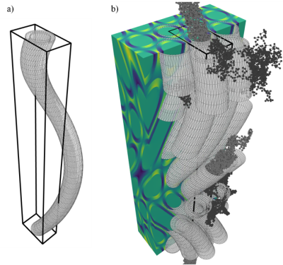 |
52 | Susceptibility-induced B-field inhomogeneity of undulating axons and effects on the DWI signal
Sidsel Winther1,2, Henrik Lundell2, and Tim B. Dyrby1,2
1Department of Applied Mathematics and Computer Science, Technical University of Denmark, Kongens Lyngby, Denmark, 2Danish Research Centre for Magnetic Resonance (DRCMR), Copenhagen University Hospital Hvidovre, Hvidovre, Denmark
Magnetic susceptibility induces morphology- and orientation-dependent perturbations of the $$$B_0$$$-field and hence the DWI signal. At the micro-structural scale of brain white matter, the main contribution to these perturbations comes from myelin. To understand how the perturbations are shaped by the intrinsically undulating morphology of axons, observed in 3D reconstructions of synchrotron imaging and electron microscopy, we have implemented a framework that enables us to systematically study how undulation affects the orientation-dependency of susceptibility-induced B-field inhomogeneity, and how this affects the DWI signal - both in the intra- and extra-axonal compartments.
|
||
1046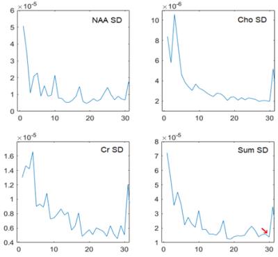 |
53 | Outlier Rejection for Fetal Brain MRS
Jack Highton1, Maria Yanez Lopez1, Anthony Price1, Vanessa Kyriakopoulou1, Lisa Story1, Mary A. Rutherford1, Jo Hajnal1, and Enrico De Vita1
1Biomedical Engineering and Imaging Sciences, King's College London, London, United Kingdom
Magnetic resonance spectroscopy (MRS) can measure the concentration of a range of metabolites to understand pathophysiological and normal development in the fetus, as well as provide diagnostic and prognostic markers [1]. However, MRS applications in the fetus are technically very challenging due to the inherently low SNR (fetal size, distance from the coil, the use of 1.5T scans) and signal disruption caused by unrestricted fetal motion during data collection. Given the limited available SNR, outlier rejection to remove motion corrupted averages and improve spectral quality is particularly important. Spectral phase shifts are associated with motion, both during signal acquisition and between MRS repeats [2]. We propose a threshold-free method for optimal outlier rejection of fetal MRS, “Ranked residual-water Phase Shift”: the phase shift in the residual water after water suppression is used as a metric linked to potential motion in individual transients/repeats.
|
||
1047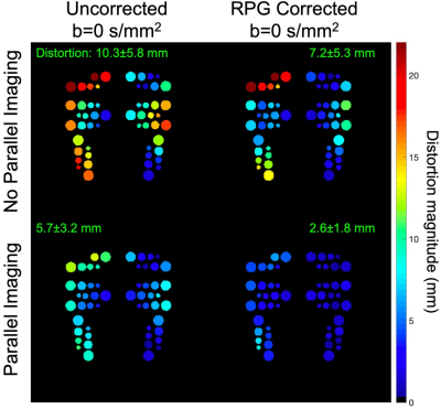 |
54 | Optimization of a breast EPI-DWI protocol with high b-values and anterior-posterior phase-encoding direction: initial results
Ana E Rodríguez-Soto1, Summer J Batasin1, Lauren K Fang1, Michael Carl2, Haydee Ojeda-Fournier1, Kathryn E Keenan3, and Rebecca A Rakow-Penner1,4
1Radiology, University of California, San Diego, La Jolla, CA, United States, 2GE Healthcare, University of California, San Diego, La Jolla, CA, United States, 3National Institute of Standards and Technology, Boulder, CO, United States, 4Bioengineering, University of California, San Diego, La Jolla, CA, United States
Diffusion-weighted imaging (DWI) acquired with echo-planar imaging (EPI) is utilized in breast MRI protocols to evaluate cancer lesions. While clinical implementation is limited by the distortion artifacts which affect EPI, methods such as reverse polarity gradient (RPG) and data collection with reduced field-of-view (FOV) have been used to reduce distortion. Evaluating these methods in minimizing distortion for images acquired with parallel imaging (PI) is a promising approach to improve EPI-DWI breast imaging. Here, we evaluated the distortion magnitude of EPI-DWI data collected with and without PI on a breast phantom and tested the feasibility of this protocol in vivo.
|
||
1048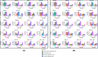 |
55 | Evaluating Within-Subject Test-Retest Replicability of Free-Water Corrected and Uncorrected Multi-Shell Diffusion MRI Measures.
Virendra R Mishra1, Ofer Pasternak2, Karthik R Sreenivasan1, and Dietmar Cordes1
1Imaging, Cleveland Clinic Lou Ruvo Center for Brain Health, Las Vegas, NV, United States, 2Harvard Medical School, Boston, MA, United States
Free-water (FW) estimation could be improved with higher spatial resolution diffusion MRI (dMRI) data acquired at multiple shells. However, the effect of multi-shell protocol features on the accuracy of estimating FW and FW-corrected fractional anisotropy (FA) across the brain structures is currently unknown. We evaluated test-retest reproducibility of FW and FW-corrected FA across 20 major white-matter tracts across four dMRI protocols, two protocols with ADNI-3 and two protocols with HCP sequences. Our analysis suggests higher spatial resolution HCP-style dMRI data acquisition with correction of FW-estimation could be reliably performed in routine clinical investigations.
|
||
1049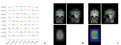 |
56 | MRSI Processing and Simulation Toolbox in the FID Appliance (FID-A)
Brenden Kadota1, Bernhard Strasser2,3, Wendy Oakden1, and Jamie Near1,4
1Physical Studies Research Platform, Sunnybrook Research Institute, Toronto, ON, Canada, 2Department of Biomedical Imaging and Image-Guided Therapy, Medical University of Vienna, Vienna, Austria, 3A. A. Martinos Center for Biomedical Imaging, Massachusetts General Hospital, Boston, MA, United States, 4Medical Biophysics, University of Toronto, Toronto, ON, Canada
Magnetic resonance spectroscopic imaging (MRSI) is a biomedical imaging tool that enables spatial mapping of metabolite signals in vivo. Dedicated software for MRSI data preprocessing, image reconstruction, visualization and simulation is vital to the development and routine application of MRSI but relatively few tools are available for this purpose. We present an open source toolbox in MATLAB for preprocessing, visualization and simulation of MRSI data. The processing toolbox includes functions for importing data, coil combination, B0 inhomogeneity correction, and non cartesian reconstruction, while the simulation toolbox can simulate MRSI acquisitions with arbitrary input k-space trajectories.
|
||
 Back to Meeting Home
 Back to the Program-at-a-Glance
The International Society for Magnetic Resonance in Medicine is accredited by the Accreditation Council for Continuing Medical Education to provide continuing medical education for physicians.
 Back to Meeting Home
 Back to the Program-at-a-Glance
 View the Presentation