Digital Poster
Data Acquisition for Diffusion, Perfusion & Spectroscopy II
Joint Annual Meeting ISMRM-ESMRMB & ISMRT 31st Annual Meeting • 07-12 May 2022 • London, UK

Digital Poster
Data Acquisition for Diffusion, Perfusion & Spectroscopy II
| Computer # | ||||
|---|---|---|---|---|
1142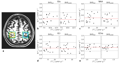 |
47 | Impact of the B0 and B1+-adjustments on the in vivo metabolite quantification accuracy of simultaneous 2voxel 1H brain MRS at 7T
Layla Tabea Riemann1, Christoph Stefan Aigner1, Ralf Mekle2, Sebastian Schmitter1,3,4, Bernd Ittermann1, and Ariane Fillmer1
1Physikalisch-Technische Bundesanstalt (PTB), Berlin, Germany, 2Center for Stroke Research Berlin, Charité Universitätsmedizin, Berlin, Germany, 3Center for Magnetic Resonance Research, University of Minnesota, Minneapolis, MN, United States, 4Medical Physics in Radiology, German Cancer Research Center (DKFZ), Heidelberg, Germany
For simultaneous multi-voxel spectroscopy (sMVS), it is necessary to optimize the B0 shimming and the B1+ adjustment simultaneously for two voxels. The impact of these adjustments on the smallest possible distance for simultaneous two-voxel MRS acquisitions is determined by Bloch simulations, and the influence on the spectral quality is assessed for three different brain regions. To this end, the previously introduced 2 spin-echo full-intensity acquired localization (2SPECIAL) sequence and the voxel-GeneRalized Autocalibrating Partial Parallel Acquisition (vGRAPPA) decomposition algorithm are utilized to simultaneously acquire and retrospectively decompose in vivo brain 1H-MRS data from two voxels at short echo times at 7T.
|
||
1143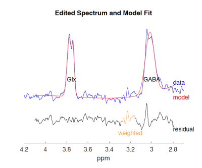 |
48 | Quantitative measurements of GABA in mouse brain using MR Spectroscopy at 7T Video Permission Withheld
Mohamed Tachrount1, Jason Lerch1,2, and Stuart Clare1
1Wellcome Centre for Integrative Neuroimaging (FMRIB), Nuffield Department of Clinical Neurosciences, University of Oxford, Oxford, United Kingdom, 2Mouse Imaging Centre (MICe), University of Toronto, Toronto, ON, Canada
The goals of this study were to implement a MEGA-sLASER sequence with prospective field drift correction on a preclinical scanner, develop the processing tools and validate them on a group of wild type mice.
|
||
1144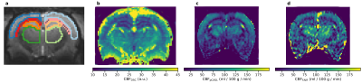 |
49 | Comparison of pCASL, DSC and FAIR for Cerebral Perfusion in Rats @9.4T
Lisa Y Hung1, Karl-Heinz Herrmann1, Renat Sibgatulin1, Lydiane Hirschler2,3, Jan Warnking3, Emmanuel L Barbier3, Otto W Witte4, Knut Holthoff4, Jürgen R Reichenbach1, and Alexander Joerk4
1Medical Physics Group, Institute of Diagnostic Radiology, Jena University Hospital, Jena, Germany, 2Department of Radiology, Leiden University Medical Center, Leiden, Netherlands, 3Grenoble Institut Neurosciences, Univ. Grenoble Alpes, Inserm, U1216, Grenoble, France, 4Hans-Berger Department of Neurology, Jena University Hospital, Jena, Germany
Attaining reproducible, time-efficient, high spatial resolution and quantitative MRI perfusion images in small animal MRI @9.4T is still challenging. To take a step toward this goal, perfusion data from arterial spin labeling (ASL), Flow-sensitive Alternating Inversion Recovery (FAIR), dynamic susceptibility contrast (DSC), and pseudo-continuous arterial spin labeling (pCASL) sequences are compared by assessing cerebral perfusion in 5 rats, each scanned 3 times. Differences in (cerebral blood flow) CBF in major brain regions were analyzed, and the repeatability of perfusion MRI was assessed.
|
||
1145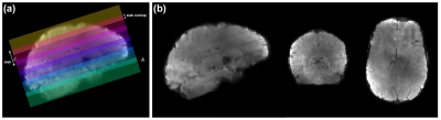 |
50 | The impact of optimal RF coil-combination on whole-brain sub-millimetre resolution perfusion imaging at 7T
Sriranga Kashyap1,2, Dimo Ivanov2, Roy A. M. Haast3, Francisco J. Fritz4, Robbert L. Harms2, Benedikt A. Poser2, and Kamil Uludag5,6
1Techna Institute, University Health Network, Toronto, ON, Canada, 2Department of Cognitive Neuroscience, Maastricht University, Maastricht, Netherlands, 3Aix-Marseille Universite, CNRS, CRMBM, Marseille, France, 4Institute for Systems Neuroscience, Universitätsklinikum Hamburg-Eppendorf, Hamburg, Germany, 5Techna Institute & Koerner Scientist in MR Imaging, University Health Network, Toronto, ON, Canada, 6Center for Neuroscience Imaging Research, Institute for Basic Science & Department of Biomedical Engineering, Sungkyunkwan University, Suwon, Korea, Republic of
In this work, we use 3D-EPI ASL to acquire perfusion maps of the human brain at an unprecedented spatial resolution of 0.7 mm isotropic at 7T. This multi-session, single-subject ASL dataset offers a unique opportunity to investigate the cortical distribution of baseline perfusion across and within brain areas, as well as studying the physiological basis for the interpretation of laminar and columnar fMRI. In this abstract, we use an open-source, memory-efficient, CPU/GPU accelerated coil-combine Python toolbox to probe the impact of using covariance-weighted sum-of-squares (CovSoS) and tSNR optimised RF coil-combination (STARC) on high-resolution perfusion imaging at 7T.
|
||
1146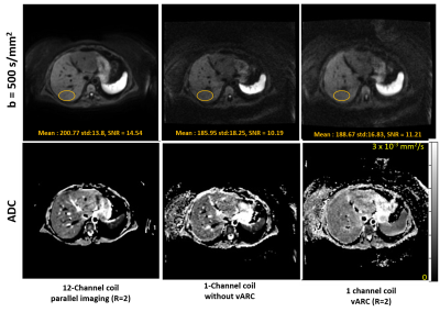 |
51 | Multi-Shot Liver Diffusion MRI Using Variable Auto-Calibrating (vARC) Sampling Across Averages
Ashok Kumar P Reddy1, Harsh Kumar Agarwal1, Rajdeep Das1, Rajagopalan Sundaresan1, Shaik Ahmed1, Sajith Rajamani1, Bhairav Mehta1, Gaohong Wu1, M Ramasubba Reddy2, and Ramesh Venkatesan1
1GE Healthcare, Bangalore, India, 2Indian Institute of Technology Madras, Chennai, India
Obesity is a key biomarker of liver pathology. Diffusion MRI in liver is key protocol in the absence on intravenous contrast agent and also provides additional insights on diffuse liver diseases and lesions However, its usage is limited by the inability to use multi-channel surface coil for obese liver patients in non-wide-bore MRI scanners. A variable k-space sampling scheme and auto-calibrating image reconstruction technique, vARC, is proposed in this abstract to reduce the amount of distortion and improve the image quality of Liver DWI acquired with single channel volume coil.
|
||
1147 |
52 | Intra-Subject Variability of Skeletal Muscle Glycogen Using 13C/1H MRS at 3T Using a Novel Standardization Method
Rajakumar Nagarajan1, Gwenael Layec2, and Jane A Kent2
1Human Magnetic Resonance Center, Institute for Applied Life Sciences, University of Massachusetts Amherst, Amherst, MA, United States, 2Kinesiology, University of Massachusetts Amherst, Amherst, MA, United States
Given its central role in substrate metabolism, the ability to quantify muscle glycogen (Gly) continuously and noninvasively using natural abundance 13C MR spectroscopy presents an important opportunity for understanding human metabolism. This study evaluated the reproducibility of Gly measured by 13C MRS in the quadriceps muscle using three methods for signal standardization at 3T: Gly/total creatine (TCr) from 13C, Gly/TCr using 13C and 1H, and Gly/water. The novel results from this study suggest that Gly/TCr measurements using the creatine peak from 1H MRS is highly reproducible and may be an advantageous approach to measuring muscle glycogen in the future.
|
||
1148 |
53 | Diffusion-weighted MEGA-PRESS spectroscopy
Emile Schjeldsøe Berg1 and John Georg Seland1
1Department of Chemistry, University of Bergen, Bergen, Norway
The editing capabilities of the MEGA-PRESS sequence are combined with diffusion-weighing to enable both quantification and diffusion measurements of less abundant brain metabolites. The technique, labelled DW-MEGA-PRESS, was tested for combined editing and diffusion-weighting of GABA. Obtained data give reliable results for GABA concentrations and corresponding diffusion coefficients from in vitro experiments. When applied to in vivo acquisitions, reliable diffusion coefficients for the dominating metabolites were obtained. GABA could be reliably quantified, but the signal-to-noise ratio was not sufficient for a reliable determination of its diffusion coefficient. This could be improved by further optimization of scanning and post-processing protocols.
|
||
1149 |
54 | Frequency sweep 31P MR spectroscopic imaging at 7T
Songi Lim1,2, Mark Stephan Widmaier1,2, and Lijing Xin1,3
1CIBM Center for Biomedical Imaging, Lausanne, Switzerland, 2Laboratory for Functional and Metabolic Imaging, EPFL, Lausanne, Switzerland, 3Animal Imaging and Technology, EPFL, Lausanne, Switzerland
Phase-cycled spectroscopic imaging (PCSI) method was implemented and validated for 31P PCSI imaging at 7T. The PCSI method uses a balanced steady-state free precession sequence with an ultra-low flip angle (<1°) to achieve sharp passband with 2.52-ms of TR, which enable to accelerate the acquisition. With prior knowledge of 31P spectra, it is feasible to acquire major 31P peaks by changing the frequency offset and non-uniform phase sweeping instead of acquiring full spectra uniformly. To investigate feasibility of the method, a multi-compartment KH2PO4 phantom with in vivo equivalent concentrations was prepared and 31P PCSI was compared with conventional FID-CSI.
|
||
1150 |
55 | A spectrally interleaved magnetic resonance spectroscopic imaging sequence incorporating semi-adiabatic pulses at ultrahigh field
Gaurav Verma1, Seena Dehkharghani2, Leeor Alon3, and Priti Balchandani1
1Biomedical Engineering and Imaging Institute, Icahn School of Medicine at Mount Sinai, New York, NY, United States, 2Radiology and Neurology, New York University, New York, NY, United States, 3Radiology, New York University, New York, NY, United States
A spectrally-interleaved semi-adiabatic magnetic resonance spectroscopic imaging sequence was developed to simultaneously acquire water and metabolite resonances within the same repetition time. A water interleave enables eddy current correction, absolute quantification through water reference and thermometry using empirically-derived formulae based on chemical shifts of temperature-sensitive water and temperature-insensitive metabolites. The sequence successfully acquired both 1-minute single voxel and 4-minute multi-voxel acquisitions in phantom and in vivo, producing temperature estimates of 21.2 °C, and 35.7 °C, respectively. LCModel fitting of metabolites provided reliable fitting of multiple metabolite peaks including separation of glutamate and glutamine.
|
||
1151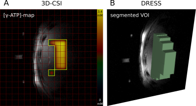 |
56 | Comparison of in vivo hepatic metabolic quantitation by localized 31P MRS at 7T
Lorenz Pfleger1,2, Wolfgang Bogner2, Albrecht Ingo Schmid3, Thomas Scherer1, Peter Wolf1, Michael Krebs1, and Martin Krššák1,4
1Division of Endocrinology and Metabolism, Department of Medicine III, Medical University of Vienna, Vienna, Austria, 2High Field MR Centre, Department of Biomedical Imaging and Image-guided Therapy, Medical University of Vienna, Vienna, Austria, 3High Field MR Centre, Center for Medical Physics and Biomedical Engineering, Medical University of Vienna, Vienna, Austria, 4Christian Doppler Laboratory for Clinical Molecular MR Imaging, Vienna, Austria This study focuses on the comparison of different in vivo data-acquisitions for quantifying hepatic 31P metabolite concentrations using a phantom replacement method. We have compared a 1D-slab localized DRESS, a single-voxel-spectroscopy (SVS) ISIS as well as a 3D-CSI 31P MRS acquisition at 7T. Each method has its own advantages and disadvantages and needs to be chosen according to the focus and design of the study. |
||
1152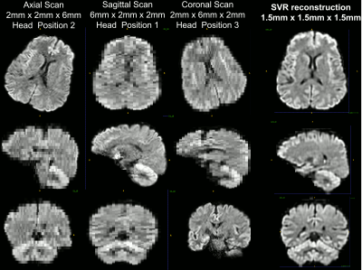 |
57 | Super-resolution mean diffusivity spectroscopic MRI in the human brain
Alexandru V Avram1,2, Magdoom Kulam1, Joelle E Sarlls3, Raisa Freidlin4, and Peter J Basser1
1Eunice Kennedy Shriver National Institute of Child Health and Human Development, National Institutes of Health, Bethesda, MD, United States, 2Center for Neuroscience and Regenerative Medicine, The Henry Jackson Foundation, Bethesda, MD, United States, 3National Institute of Neurological Disorders and Stroke, National Institutes of Health, Bethesda, MD, United States, 4Center for Information Technology, National Institutes of Health, Bethesda, MD, United States
We describe a comprehensive pipeline for super-resolution reconstruction of clinical diffusion-weighted MRIs acquired with isotropic diffusion encoding (IDE), or spherical tensor encoding. The pipeline integrates blip-up/down EPI distortion correction and slice-to-volume registration (SVR). Multiple low-resolution IDE-MRIs with different slice orientations relative to the brain are processed to reconstruct high-resolution IDE-MRIs. From high-resolution IDE-MRIs with a wide range of b-values we estimate spectra of subvoxel MD values to describe the distribution of water mobilities in microscopic brain tissue microenvironments. Integrating SVR-reconstruction with IDE is an important step in the clinical translation of MD spectroscopic MRI for fetal MRI applications.
|
||
1153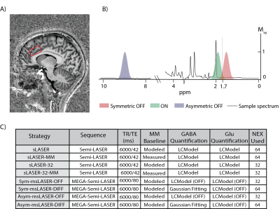 |
58 | Simultaneous Measurement of Glu and GABA at 7T
Tal Finkelman1, Edna Furman-Haran2, Rony Paz3, and Assaf Tal1
1Chemical and Biological Physics, Weizmann, Rehovot, Israel, 2Life Sciences Core Facilities, Weizmann, Rehovot, Israel, 3Brain Sciences, Weizmann, Rehovot, Israel
Glutamate (Glu) and γ‑aminobutyric-acid (GABA) are central neurotransmitters participating in many cognitive processes, and their combined measurement is of great relevance. Proton Magnetic Resonance Spectroscopy (1H-MRS) allows the no–invasive, in-vivo, measurement of GABA and Glu. At magnetic fields below 7T, GABA is detected by spectral editing strategies, while Glu is detected by non-editing strategies. We compared both edited and non‑edited approaches at 7T for simultaneously quantifying Glu and GABA from 21 volunteers, and found that non-edited sequences at TE=80ms provide better combined-reproducibility than either edited sequences at the same TE, or non-edited sequence at a shorter TE (42ms).
|
||
1154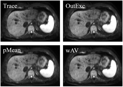 |
59 | Postprocessing Reduces Pulsation Artifacts and Increases Visibility of Liver Lesions in Flow-compensated Diffusion-weighted Imaging
Tobit Führes1, Marc Saake 1, Hannes Seuss1,2, Astrid Müller1, Sebastian Bickelhaupt1, Alto Stemmer3, Thomas Benkert3, Michael Uder1, Bernhard Hensel4, and Frederik Bernd Laun1
1University Hospital Erlangen, Erlangen, Germany, 2Klinikum Forchheim, Forchheim, Germany, 3Siemens Healthcare GmbH, Erlangen, Germany, 4Friedrich-Alexander-Universität Erlangen-Nürnberg (FAU), Erlangen, Germany
Diffusion-weighted imaging of the liver is prone to the cardiac pulsation artifact, which can lead to reduced lesion visibility. We addressed this problem with a two-fold approach. First, flow-compensated diffusion weightings were used, which are known to reduce this artifact. Using a dataset of 40 patients suffering from focal liver lesions, we addressed the remaining signal voids with different postprocessing techniques, namely weighted averaging, the p-mean approach, and an outlier exclusion algorithm. The algorithms substantially increased the lesion visibility and further reduced the pulsation artifact. An evaluation of CNR and calculation time showed that weighted averaging was suited best.
|
||
1155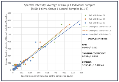 |
60 | Quality Assurance and Quality Control for HRMAS MRS Studies of Human Blood
Leo Cheng1, Matteo Sanchez-Dahl1, Isabella Muti1, JianXiang Weng1, and Anya Zhong1
1Massachusetts General Hospital, Charlestown, MA, United States
The lack of a standard universal protocol for sample preparation in MRS experiments results in instrumental and biological variability across reported studies. The purpose of this experiment is to evaluate a quality assurance protocol for performing HRMAS 1H MRS experiments with human biofluid samples, such as blood serum. The research objective is to determine if the spectral results measured from pooled samples can be used to assess results from individual samples as controls. These findings will help better develop efficient standard protocols for researchers and clinicians to use in the future.
|
||
 Back to Meeting Home
 Back to the Program-at-a-Glance
The International Society for Magnetic Resonance in Medicine is accredited by the Accreditation Council for Continuing Medical Education to provide continuing medical education for physicians.
 Back to Meeting Home
 Back to the Program-at-a-Glance
 View the Presentation