Digital Poster
MR Fingerprinting I
Joint Annual Meeting ISMRM-ESMRMB & ISMRT 31st Annual Meeting • 07-12 May 2022 • London, UK

| Computer # | ||||
|---|---|---|---|---|
2507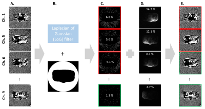 |
39 | Reducing streak artefacts in radial MR fingerprinting of the prostate through automated channel removal
Kaia Ingerdatter Sørland1, Cristopher George Trimble1, Elise Sandsmark2, Tone Frost Bathen1,2, Mattijs Elschot1,2, and Martijn A. Cloos3
1Department of Circulation and Medical Imaging, Norwegian University of Science and Technology, Trondheim, Norway, 2Department of Radiology and Nuclear Medicine, St. Olavs hospital, Trondheim University Hospital, Trondheim, Norway, 3Centre for Advanced Imaging, The University of Queensland, Brisbane, Australia
The extreme undersampling factors used in radial magnetic resonance fingerprinting (MRF) of the prostate lead to strong streak artefacts from the femoral arteries and veins. However, it turns out that only a subset of receiver channels is responsible for these streaks. In this work we automatically detected and removed these from the reconstruction pipeline. The method was applied to MRF acquired with various acceleration factors in seven asymptomatic volunteers, significantly reducing visible streaks in reference tissues surrounding the prostate without impairing the prostate T1 and T2 values in the MRF maps.
|
||
2508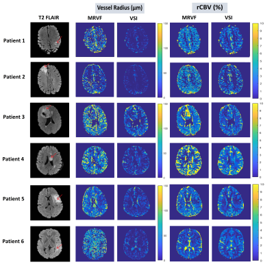 |
40 | Quantification of microvascular properties of gliomas using DSC – Hybrid EPI based MR Vascular Fingerprinting compared with Vessel Size Imaging
Krishnapriya Venugopal1, Fatemehsadat Arzanforoosh1, Daniëlle van Dorth2, Marion Smits1, Juan Antonio Antonio Hernandez-Tamames1,3, Esther A.H Warnert1, Matthias J.P van Osch2, and Dirk H.J Poot1
1Radiology and Nuclear Medicine, Erasmus MC, Rotterdam, Netherlands, 2Radiology, Leiden University Medical Center, Leiden, Netherlands, 3Medical Imaging, TU Delft, Delft, Netherlands
This study uses an MRVF approach to analyze the time evolution of a DSC hybrid -EPI (HEPI) sequence that simultaneously acquires gradient and spin echo images. HEPI properties are incorporated from the scanner into simulations including contrast agent extravasation, diffusion, and MR signal evolution and for varying the outcome parameters: CBV, mean vessel-size, and leakage. In vivo data of six glioma patients are used to compare MRVF output maps to those obtained from conventional VSI modelling. The results show reasonable agreement. Also, the noise sensitivity of both techniques was investigated.
|
||
2509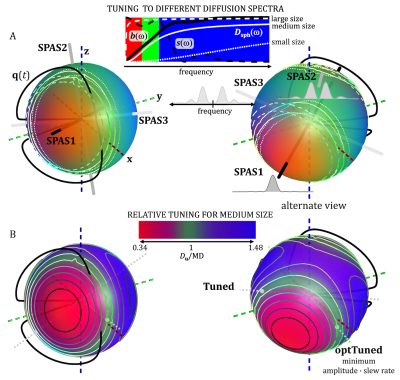 |
41 | Spectral principal axes system (SPAS) and tuning of tensor-valued encoding for time-dependent anisotropic diffusion
Samo Lasič1,2, Nathalie Just1, and Henrik Lundell1
1Centre for Functional and Diagnostic Imaging and Research, Copenhagen University Hospital Amager and Hvidovre, Danish Research Centre for Magnetic Resonance, Copenhagen, Denmark, 2Random Walk Imaging, Lund, Sweden
Tensor-valued diffusion encoding can be confounded by time-dependent diffusion (TDD). Matching sensitivity to TDD or tuning of b-tensors with different shapes is needed for unbiased microstructure assessment. We present a method for tuning linear tensor encoding (LTE) to spherical tensor encoding (STE), which could be optimized for different hardware constraints. Furthermore, we introduce the spectral principle axes system (SPAS), representing spectral anisotropy of STE. The SPAS LTEs could provide an alternative to tuning and enable disentangling effects of microscopic anisotropy and TDD, useful to correlate cell shape and size.
|
||
2510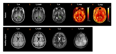 |
42 | Tailored MR fingerprinting of pediatric brain tumor patients
Pavan Poojar1,2, Enlin Qian1, Alexis B Maddocks3, and Sairam Geethanath1
1Columbia Magnetic Resonance Research Center, Columbia University, New York, NY, United States, 2Dayananda Sagar College of Engineering, Bangalore, India, 3Columbia University Irving Medical Center, New York, NY, United States
We demonstrated the utility of tailored MR fingerprinting (TMRF) of pediatric patients with brain tumor to 1) differentiate tumor from healthy tissue using quantitative maps; 2) tailor TMRF sequence to include T2 fluid-attenuated inversion recovery (FLAIR) contrast that could potentially substitute T2 FLAIR sequence used in routine pediatric tumor protocol. The mean土SD difference in T1 and T2 values between tumor and normal tissues for 3 patients were 1.05土0.09 and 0.05土0.02 seconds respectively. The TR and flip angle trains were tailored to include T2 FLAIR contrast for adult imaging. Current and future work includes optimizing T2 FLAIR for the pediatric population.
|
||
2511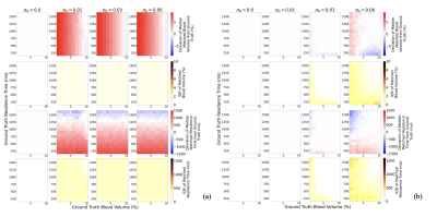 |
43 | Feasibility of Quantifying Cerebral Blood Volume and Blood-Brain Barrier Water Exchange using Non-Contrast MR Fingerprinting
Emma L Thomson1,2, Elizabeth Powell3, Claudia A M Gandini Wheeler-Kingshott2,4,5, and Geoff J M Parker1,2,6
1Centre for Medical Image Computing, Department of Medical Physics and Biomedical Engineering, University College London, London, United Kingdom, 2NMR Research Unit, Queen Square MS Centre, Department of Neuroinflammation, UCL Queen Square Institute of Neurology, Faculty of Brain Sciences, London, United Kingdom, 3Centre for Medical Image Computing, Department of Computer Science, University College London, London, United Kingdom, 4Department of Brain & Behavioural Sciences, University of Pavia, Pavia, Italy, 5Brain Connectivity Centre Research Department, IRCCS Mondino Foundation, Pavia, Italy, 6Bioxydyn Limited, Manchester, United Kingdom
We propose the use of magnetic resonance fingerprinting (MRF), applied using a spoiled gradient echo sequence, to quantify cerebral blood volume ($$$\nu_b$$$) and inter-vascular water exchange (1/$$$\tau_b$$$), without the need for contrast agents. Through a simulation study we optimise a simulated acquisition protocol and test the sensitivity of the measurement, and its accuracy in the presence of variations in blood T1, tissue T1, and B1. We demonstrate that voxel-wise simultaneous quantification of $$$\nu_b ,\tau_b , T_{1,b}, T_{1,e}$$$ and $$$B_1^+$$$ is likely feasible with an optimised acquisition.
|
||
2512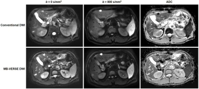 |
44 | Evaluation of multiband variable-rate selective excitation (MB-VERSE) diffusion-weighted imaging (DWI) of the liver MRI Video Permission Withheld
Ja Kyung Yoon1, Yong Eun Chung1, Jaeseung Shin1, Jin-Young Choi1, Mi-Suk Park1, and Myeong-Jin Kim1
1Radiology, Severance Hospital, Research Institute of Radiological Science, Yonsei University College of Medicine, Seoul, Korea, Republic of
In liver MRI, diffusion-weighted imaging (DWI) allows better detection and characterization of focal lesions, but requires a relatively long scan time. The multiband variable-rate selective excitation (MB-VERSE) echoplanar imaging for DWI provides accelerated acquisition time with some expected trade-off in image quality. Qualitative and quantitative image quality of MB-VERSE images as well as focal liver lesion detectability was evaluated by three readers. The MB-VERSE sequence as well as focal liver lesion detectability showed significant sacrifice in quantitative and qualitative overall image quality, but comparable focal lesion detectability.
|
||
2513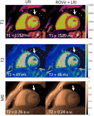 |
45 | Combining Region-optimized virtual coils with low rank reconstructions for accelerated cardiac MR Fingerprinting
Gastao Cruz1, Andrew Phair1, Carlos Velasco1, René M. Botnar1, and Claudia Prieto1
1King's College London, London, United Kingdom
Cardiac Magnetic Resonance Fingerprinting (MRF) produces co-registered, multi-parametric maps from highly accelerated acquisitions. Low rank methods leveraging temporal information have enabled highly undersampled MRF reconstructions. However, residual aliasing from these reconstructions can potentially propagate through the MRF framework into the parametric maps. Region-Optimized Virtual coils have recently been proposed for undersampled reconstructions in conventional imaging, by suppressing unwanted sources of aliasing signal. Here we combine both methods and investigate its performance in Cardiac Magnetic Resonance Fingerprinting.
|
||
 |
2514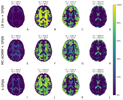 |
46 | Joint sparsity multi-component MRF reconstruction - directly from k-space to component maps
Martijn Nagtegaal1, Emiel Hartsema1, Kirsten Koolstra2, and Frans Vos1,3
1Imaging Physics, Delft University of Technology, Delft, Netherlands, 2Department of Radiology, Leiden University Medical Centre, Leiden, Netherlands, 3Department of Radiology and Nuclear Medicine, Erasmus MC, Rotterdam, Netherlands
The use of high undersampling factors and short flip angle trains leads to shorter acquisition times in MR Fingerprinting acquisitions. To obtain accurate multi-component estimates from this data advanced reconstructions are required. We study a low-rank ADMM based reconstruction method that adds a multi-component constraint to the inverse reconstruction problem (MC-ADMM). This method is combined with a joint-sparsity constraint yielding higher quality multi-component estimates with k-SPIJN than with previous methods. In simulations we observed increased stability to sequence truncation and in vivo multi-component estimates contained less noise-like effects.
|
|
2515 |
47 | Magnetic Resonance Fingerprinting with Total Nuclear Variation Regularisation
Imraj Ravi Devia Singh1, Olivier Jaubert1, Bangti Jin1, Kris Thielemans2, and Simon Arridge1
1Department of Computer Science, University College London, London, United Kingdom, 2Institute of Nuclear Medicine, University College London, London, United Kingdom
Magnetic Resonance Fingerprinting (MRF) accelerates quantitative magnetic resonance imaging. The reconstruction can be separated into two problems: reconstruction of a set of multi-contrast images from k-space signals, and estimation of parametric maps from the set of multi-contrast images. In this study we focus on the former problem, while leveraging dictionary matching for the estimation of parametric maps. Two different sparsity promoting regularisation strategies were investigated: contrast-wise Total Variation (TV) which encourages image sparsity separately; and Total Nuclear Variation (TNV) which promotes a measure of joint edge sparsity. We found improved results using joint sparsity.
|
||
2516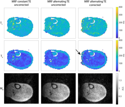 |
48 | 3D magnetic resonance fingerprinting at 50 mT with integrated estimation and correction of image distortions due to B0 inhomogeneities
Thomas O'Reilly1, Peter Börnert2, Andrew Webb1, and Kirsten Koolstra1
1Leiden University Medical Center, Leiden, Netherlands, 2Philips Research Hamburg, Hamburg, Germany
Strong B0 inhomogeneities in compact point-of-care low field permanent magnet systems can cause image distortions and reduced signal for large field-of-view images. MRF, as a flexible acquisition framework, can encode these effects into the acquisition process, to incorporate them into the reconstruction process. In this work, we use an alternating TE in the MRF sequence that supports ΔB0 estimation and image distortion correction of the MRF data. This method reduces distortions in the relaxation time maps without needing to acquire an additional ΔB0 map.
|
||
2517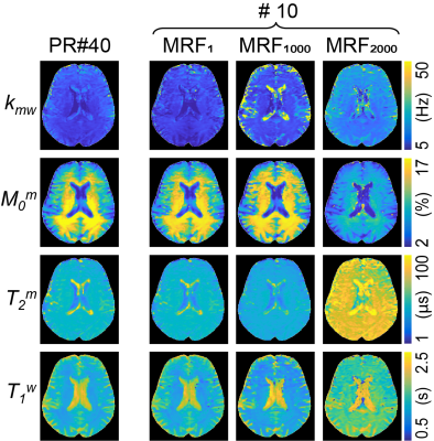 |
49 | Learning-based prediction of encoding capability for acquisition schedule of Magnetization Transfer Contrast MR fingerprinting
Beomgu Kang1, Hye-Young Heo2,3, and Hyunwook Park1
1School of Electrical Engineering, Korea Advanced Institute of Science and Technology, Daejeon, Korea, Republic of, 2Division of MR Research, Department of Radiology, Johns Hopkins University, Baltimore, MD, United States, 3F.M. Kirby Research Center for Functional Brain Imaging, Kennedy Krieger Institute, Baltimore, MD, United States
Magnetization transfer contrast MR fingerprinting (MTC-MRF) is used to quantify multiple tissue parameters of free bulk water and semisolid macromolecule using pseudo-randomized MRF schedules. An optimal design of the MRF schedule is important to improve the quantification accuracy and reduce the scan time. However, lack of the objective function that represents the encoding capability of MRF schedule hinders the reliable optimization. In this study, we propose a novel metric that represents the encoding capability of MRF schedule based on recurrent neural network. Unlike the conventional metrics based on indirect measurements, the proposed learning-based metric directly measures the tissue quantification errors.
|
||
2518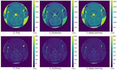 |
50 | Application of Deep Learning techniques to Magnetic Resonance Fingerprinting
Raffaella Fiamma Cabini1,2, Leonardo Barzaghi1,3, Davide Cicolari2,4, Anna Pichiecchio5,6, Silvia Figini2,7, Paolo Arosio8,9, Marta Filibian2,10, Alessandro Lascialfari2,4, and Stefano Carrazza8,9
1Department of Mathematics, University of Pavia, Pavia, Italy, 2INFN, Istituto Nazionale di Fisica Nucleare - Pavia Unit, Pavia, Italy, 3Department of Neuroradiology, Advanced Imaging and Radiomics, IRCCS Mondino Foundation, Pavia, Italy, 4Department of Physics, University of Pavia, Pavia, Italy, 5Department of Neuroradiology, IRCCS Mondino Foundation, Pavia, Italy, 6Department of Brain and Behavioural Sciences, University of Pavia, Pavia, Italy, 7Department of Social and Political Science, University of Pavia, Pavia, Italy, 8Department of Physics, University of Milano, Milano, Italy, 9INFN, Istituto Nazionale di Fisica Nucleare - Milano Unit, Milano, Italy, 10Centro Grandi Strumenti, University of Pavia, Pavia, Italy
We developed a Neural Network (NN) for the reconstruction of T1 and T2 parametric maps obtained with the Magnetic Resonance Fingerprinting (MRF) technique. The training phase was realized on experimental inputs, eliminating the use of simulated datasets and theoretical models. The set of optimal hyperparameters of the NN and the supervised training algorithm were established through an optimization procedure. The model achieved similar performances to the traditional reconstruction method, but the number of MRF images required was lower with respect to the dictionary-based method. If translated to the clinic, our results envisage a significant time shortening of MRI investigation.
|
||
2519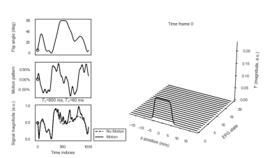 |
51 | Through-plane Motion in Magnetic Resonance Fingerprinting: Simulations and Phantom Experiments
Martijn Nagtegaal1, Charles McGrath2, Christian Günthner2, and Manuel Bauman3
1Imaging Physics, Delft University of Technology, Delft, Netherlands, 2Institute for Biomedical Enginering, ETH Zurich, Zurich, Switzerland, 3Philips Research Europe, Hamburg, Germany
-
|
||
2520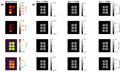 |
52 | A Scalable High-Performance Magnetic Resonance Fingerprinting Search Engine using GPUs
Gabriel Zihlmann1, Najat Salameh1, and Mathieu Sarracanie1
1Department of Biomedical Engineering, University of Basel, Allschwil, Switzerland
Dictionary search for the reconstruction of MR Fingerprinting-based sequences can become a bottleneck due to the exponential growth of dictionary size with the number of parameters. Several approaches have been proposed to accelerate this search, yet the availability of efficient and scalable software implementations remains limited. Here, we extended an existing, highly optimized similarity search library to be compatible with the requirements of MR Fingerprinting dictionary search. The evaluation of our GPU implementation shows an acceleration by two orders of magnitude over the brute-force search yielding identical results for 99.9 % of the voxels.
|
||
 Back to Meeting Home
 Back to the Program-at-a-Glance
The International Society for Magnetic Resonance in Medicine is accredited by the Accreditation Council for Continuing Medical Education to provide continuing medical education for physicians.
 Back to Meeting Home
 Back to the Program-at-a-Glance
 View the Presentation