Digital Poster
MR Fingerprinting II
Joint Annual Meeting ISMRM-ESMRMB & ISMRT 31st Annual Meeting • 07-12 May 2022 • London, UK

| Computer # | ||||
|---|---|---|---|---|
2589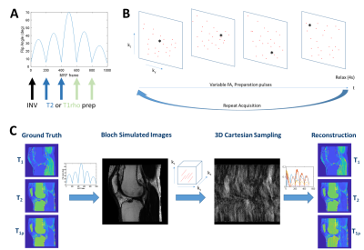 |
30 | 3D Cartesian T1rho Magnetic Resonance Fingerprinting Sequence Design for Evaluation of Cartilage and Skeletal Muscle in the Knee
Brendan L. Eck1, Jeehun Kim2, Mingrui Yang2, Dan Ma3, Mark A. Griswold3,4, and Xiaojuan Li2,3
1Imaging Institute, Cleveland Clinic, Cleveland, OH, United States, 2Biomedical Engineering, Cleveland Clinic, Cleveland, OH, United States, 3Department of Biomedical Engineering, Case Western Reserve University, Cleveland, OH, United States, 4Department of Radiology, Case Western Reserve University, Cleveland, OH, United States
Magnetic resonance fingerprinting offers a promising framework by which to rapidly quantify T1rho relaxation alongside other tissue properties such as T1 and T2. However, T1rho-MRF has only recently been reported and sequence optimization remains under-explored, particularly for 3D sequences. Using a digital phantom constructed from real knee tissue property maps, we investigate sequence parameters for a 3D Cartesian T1rho-MRF sequence including preparation pulse scheduling and timing, flip angles, number of readouts, and acceleration factor. T1, T2, and T1rho quantification errors in cartilage and skeletal muscle were evaluated.
|
||
2590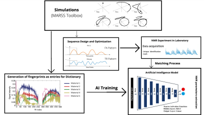 |
31 | Preliminary development of a Magnetic Resonance Fingerprinting framework for fast multiparametric low-field NMR relaxometry
Giovanni Vito Spinelli1, Leonardo Brizi1,2, Marco Barbieri3, Fabiana Zama4, Germana Landi4, Villiam Bortolotti5, Daniel Remondini1,2, and Claudia Testa1,2
1Department of Physics and Astronomy "Augusto Righi", University of Bologna, Bologna, Italy, 2INFN, Istituto Nazionale di Fisica Nucleare, Sezione di Bologna, Bologna, Italy, 3Department of Radiology, Stanford University, Stanford, CA, United States, 4Department of Mathematics, University of Bologna, Bologna, Italy, 5Department of Civil, Chemical, Environmental, and Materials Engineering, University of Bologna, Bologna, Italy A novel application of Magnetic Resonance Fingerprinting (MRF) on low-field NMR is presented. To successfully implement MRF, the correlation between the static and the radio-frequency fields has to be measured, because the evolution of the signal cannot be analyzed without accounting for magnetic fields inhomogeneities. Experimental results have been validated by simulations and compared using the RMSE. This preparatory evaluation allows the use of MRF for NMR parameters quantification. Then, an artificial intelligence approach for parameters reconstruction has been used to overcome the limitation of the standard dictionary approaches when several parameters have to be estimated. |
||
2591 |
32 | Searching for an MR Fingerprinting sequence to measure brain oxygenation without contrast agent
Thomas Coudert1, Aurélien Delphin1, Jan M Warnking1, Benjamin Lemasson1,2, Emmanuel Luc Barbier1, and Thomas Christen1
1Univ. Grenoble Alpes, Inserm, U1216, Grenoble Institut Neurosciences, GIN, Grenoble, France, 2MoGlimaging Network, HTE Program of the French Cancer Plan, Toulouse, France
We propose to use numerical simulations and Monte Carlo approaches to test 40 MR fingerprinting sequences for their ability to quantify the BOLD effect and provide maps of baseline blood oxygen saturation without the need for contrast agent injection. Our results suggest that several sequences can produce SO2 maps with less that 3% error on average even in the presence of B1 and B0 inhomogeneities. T2 and blood vessel radius could also be estimated with the same acquisitions.
|
||
2592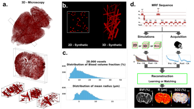 |
33 | Using 3D realistic blood vessel structures and machine learning for MR vascular Fingerprinting
Aurélien Delphin1, Fabien Boux1,2, Clément Brossard1,3, Jan M Warnking1, Benjamin Lemasson1,3, Emmanuel Luc Barbier1, and Thomas Christen1
1Univ. Grenoble Alpes, Inserm, U1216, Grenoble Institut Neurosciences, GIN, 38000, Grenoble, France, 2Univ. Grenoble Alpes, Inria, CNRS, G-INP, 38000, Grenoble, France, 3MoGlimaging Network, HTE Program of the French Cancer Plan, Toulouse, France
MR vascular fingerprinting aims at mapping cerebral vascular properties such as blood volume and blood oxygenation. We propose to improve the technique by generating dictionaries based on 3D vascular networks segmented from whole brain high-resolution (3 µm isotropic) microscopy datasets. In order to compensate for the limited number of available data and long computing times, we used a machine-learning reconstruction process to generalize our results and tested our approach in healthy, stroke and tumor animal models. Our results show high quality maps with expected contrast and baseline values in healthy animals as well as expected trends in pathological tissues.
|
||
2593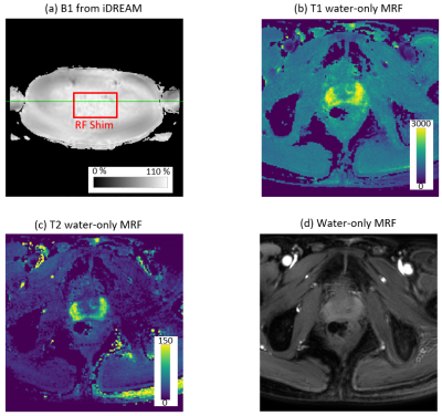 |
34 | Towards A Clinical Prostate MR Fingerprinting Protocol
Manuel Baumann1, Jochen Keupp1, Peter Mazurkewitz1, Peter Koken1, Kay Nehrke1, Jakob Meineke1, Thomas Amthor1, and Mariya Doneva1
1Philips Research Europe, Hamburg, Germany
We present an efficient MR Fingerprinting (MRF) protocol for prostate imaging at 3T. Full prostate coverage is achieved in 04:20 min with no considerable reconstruction latency. Dixon-MRF has been implemented in the scanner software and combined with a B1+ DREAM scan in a fully-integrated B1-corrected MRF reconstruction. Furthermore, flow compensation is included to suppress artifacts caused by blood flow in large vessels. The proposed prostate MRF protocol has been evaluated in six healthy volunteers.
|
||
2594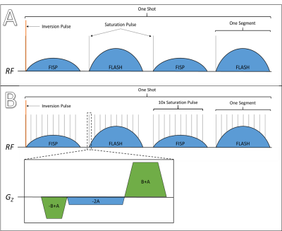 |
35 | Incorporating saturation bands into MR Fingerprinting reduces streaking artefacts
Christopher George Trimble1, Kaia Sørland1, Elise Sandsmark2, Mattijs Elschot1,2, Tone F. Bathen1,2, and Martijn A. Cloos3
1Department of Circulation and Medical Imaging, Norwegian University of Science and Technology, Trondheim, Norway, 2Department of Radiology and Nuclear Medicine, St. Olavs Hospital, Trondheim, Norway, 3Centre for Advanced Imaging, University of Queensland, Brisbane, Australia Magnetic Resonance Fingerprinting (MRF) enables fast quantitative MR imaging, which is appealing in prostate cancer diagnostics. In pelvic MR Fingerprinting, blood flow in the femoral vessels causes strong streak-like artefacts when a radial sampling strategy is used. Here we develop a strategy to incorporate saturation bands into MRF sequences. Our phantom study shows quantification of areas outside the saturation band are not significantly affected. Meanwhile, qualitative analysis of in vivo experiments indicates a marked reduction in streak intensity when saturation is applied. |
||
2595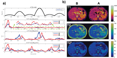 |
36 | Dictionary variance based optimization of MR Fingerprinting
Rasim Boyacioglu1, Sherry Huang2, Debra McGivney2, Yong Chen1, and Mark A Griswold1
1Radiology, Case Western Reserve University, Cleveland, OH, United States, 2Biomedical Engineering, Case Western Reserve University, Cleveland, OH, United States This study presents an optimization tool to investigate and validate the effects of MR sequence modules on MR Fingerprinting. Dictionary variance across the tissue property dimension can predict the outcome of a given change in an MRF sequence. An MRF sequence variant is optimized by varying three typical MRF sequence blocks of FA pattern, RF phase and inversion pulses. The observations from the dictionary variance align with in vivo data results. |
||
2596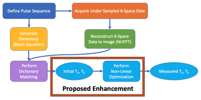 |
37 | Non-Linear Optimization for Enhanced Parameter Retrieval in MR Fingerprinting
John T. Lundstrom1,2, Megan E. Poorman3, Andrew Dienstfrey4, and Kathryn E. Keenan3
1Department of Physics, University of Colorado, Boulder, CO, United States, 2Associate of the National Institute of Standards and Technology, Boulder, CO, United States, 3Physical Measurement Laboratory, National Institute of Standards and Technology, Boulder, CO, United States, 4Information Technology Laboratory, National Institute of Standards and Technology, Boulder, CO, United States
We investigate non-linear optimization to produce quantitative parameter maps in MR Fingerprinting. Our Monte Carlo analysis shows non-linear optimization to be robust with respect to noise. We also find non-linear optimization to yield consistent results when initialized by dictionary matching using either sparse or densely populated dictionaries. Our research outcomes suggest non-linear optimization is an effective enhancement to the MR Fingerprinting pipeline, allowing for smaller dictionary sizes and more accurate parameter retrieval. Future work will include enhancements to, and analysis of, the computational efficiency of non-linear optimization.
|
||
2597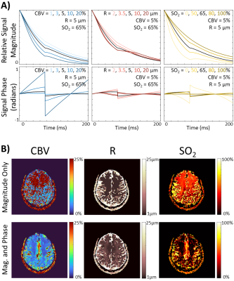 |
38 | Feasibility of Dynamic Contrast-free Vascular Magnetic Resonance Fingerprinting
Gregory J. Wheeler1, Quimby N. Lee2, Mary Kate Manhard3, Berkin Bilgic4, and Audrey P. Fan1,2
1Biomedical Engineering, University of California Davis, Davis, CA, United States, 2Neurology, University of California Davis, Davis, CA, United States, 3Radiology, Cincinnati Children's Hospital, Cincinnati, OH, United States, 4Radiology, Massachusetts General Hospital and Harvard Medical School, Boston, MA, United States
Vascular magnetic resonance fingerprinting (vMRF) using both magnitude and phase information can achieve reasonable vascular parameter maps of the brain without contrast agents. This approach combined with a rapid, multi-echo pulse sequence enables dynamic vascular function mapping of brain physiology. Here we begin to investigate the feasibility and tradeoffs of performing vMRF with only 5 echoes and a temporal resolution of 5 seconds. Initial findings indicate that reasonable fingerprint matching can be done with only 5 echoes and that the proposed sequence has adequate signal-to-noise ratio for reliable results.
|
||
2598 |
39 | Mitigation of Magnetisation Transfer Effects using Off-Resonance Pulses in MR Fingerprinting
Simran Kukran1,2, Iulius Dragonu3, Ben Statton4, Jack Allen5, Pete Lally6, Rebecca Quest7,8, Neal Bangerter7, Dow-Mu Koh9,10, Matthew Orton10,11, and Matthew Grech-Sollars1,12
1Surgery and Cancer, Imperial College London, London, United Kingdom, 2Radiotherapy and Imaging, Institute of Cancer Research, LONDON, United Kingdom, 3Research and Collaborations GB&I, Siemens Healthcare Ltd, Frimley, United Kingdom, 4London Institute of Medical Sciences,Medical Research Council, Imperial College London, London, United Kingdom, 5National Heart and Lung Institute, Imperial College London, London, United Kingdom, 6Brain Sciences, Imperial College London, London, United Kingdom, 7Department of Bioengineering, Imperial College London, London, United Kingdom, 8Department of Imaging, Imperial College Healthcare NHS Trust, London, United Kingdom, 9Department of Radiotherapy and Imaging, Institute of Cancer Research, London, United Kingdom, 10Department of Radiology, Royal Marsden Hospital, London, United Kingdom, 11Radiotherapy and Imaging, Institute of Cancer Research, London, United Kingdom, 12Department of Medical Physics, Royal Surrey NHS Trust, London, United Kingdom
Previous work has shown an inherent bias in quantitative T1 values for current standard MRF techniques as compared to gold standard methods. In our study we show how magnetisation transfer effects are a component of this bias and that the introduction of off-resonance pulses within an MRF sequence may mitigate these effects.
|
||
2599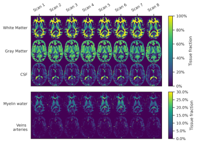 |
40 | Accuracy and repeatability of joint sparsity multi-component estimations in MR Fingerprinting
Martijn Nagtegaal1, Laura Nunez-Gonzalez2, Dirk Poot2, Jeroen de Bresser3, Thijs van Osch4, Juan Hernandez Tamames2, and Frans Vos1,2
1Imaging Physics, Delft University of Technology, Delft, Netherlands, 2Department of Radiology and Nuclear Medicine, Erasmus MC, Rotterdam, Netherlands, 3Department of Radiology, Leiden University Medical Centre, Leiden, Netherlands, 4C.J. Gorter Center for High Field MRI, Radiology Department, Leiden University Medical Centre, Leiden, Netherlands Multi-component MR Fingerprinting estimations with Sparsity Promoting Iterative Joint Non-negative least squares (SPIJN-MRF) provide the possibility to obtain partial volume tissue segmentations and myelin water maps. We evaluated the accuracy and repeatability of SPIJN-MRF estimations in simulations and 5 subjects, scanned 8 times. The obtained segmentations were compared to segmentations from SPM12 and FSL. In simulations, SPIJN-MRF showed the highest accuracy in total volume and voxel-wise measures. In vivo, higher variation was observed with SPIJN-MRF than with SPM12 and FSL, especially in WM and GM. In conclusion, SPIJN-MRF provides accurate and precise tissue relaxation parameter estimations with partial volume estimations. |
||
2600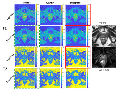 |
41 | Fast distortionless T1-T2 mapping in the prostate using MR fingerprinting with radial acquisition and subspace reconstruction
Victoria Y Yu1, Ouri Cohen1, Can Wu1, Ergys Subashi1, Manuel Baumann2, Peter Koken2, Mariya Doneva2, Michael Zelefsky3, Laura Cervino1, and Ricardo Otazo1
1Medical Physics, Memorial Sloan Kettering Cancer Center, New York, NY, United States, 2Philips Research, Hamburg, Germany, 3Radiation Oncology, Memorial Sloan Kettering Cancer Center, New York, NY, United States
The application of MR fingerprinting in the prostate is challenging due to the presence of image distortions produced by B0 inhomogeneities. This works presents the combination of radial sampling to minimize the effects of B0 inhomogeneity and temporal subspace reconstruction to accelerate the acquisition for fast distortionless T1 and T2 mapping in the prostate in less than 4 minutes.
|
||
 Back to Meeting Home
 Back to the Program-at-a-Glance
The International Society for Magnetic Resonance in Medicine is accredited by the Accreditation Council for Continuing Medical Education to provide continuing medical education for physicians.
 Back to Meeting Home
 Back to the Program-at-a-Glance
 View the Presentation