Digital Poster
Acquisition Strategies I
Joint Annual Meeting ISMRM-ESMRMB & ISMRT 31st Annual Meeting • 07-12 May 2022 • London, UK

| Computer # | ||||
|---|---|---|---|---|
1779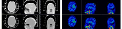 |
49 | Attenuation Correction in PET/MRI Pediatric Treated Brain Tumors: a Preliminary Comparison between the ZTE and Atlas Techniques
Paola Scifo1, Federico Fallanca1, Maurizio Barbera2, Annarita Savi1, Valentino Bettinardi1, Ilaria Neri1, Giovanna Gattuso3, Maura Massimino3, Paolo Verderio4, Sara Pizzamiglio4, Andrea Falini2,5, Luigi Gianolli1, Filippo Spreafico3, Maria Picchio1,5, and Cristina Baldoli2,5
1Nuclear Medicine, IRCCS San Raffaele Scientific Institute, Milan, Italy, 2Neuroradiology Unit, IRCCS San Raffaele Scientific Institute, Milan, Italy, 3Department of Medical Oncology and Hematology, Pediatric Oncology Unit, Fondazione IRCCS Istituto Nazionale dei Tumori, Milan, Italy, 4Unit of Bioinformatics and Biostatistics, Department of Applied Research and Technological Development, Fondazione IRCCS Istituto Nazionale dei Tumori, Milan, Italy, 5Vita-Salute San Raffaele University, Milan, Italy A preliminary comparison between the two Atlas and ZTE attenuation correction methodologies in a group of pediatric patients with brain tumors is presented. The differences of the two ACmaps and PET images were calculated and the PET parameters were compared. Our results show that using ZTE or Atlas to generate ACmaps modifies the SUVs, but the differences in values are very small. Moreover, when metallic artefacts occur, both ZTE and Atlas show loss of signals but ACmapsAtlas has smaller artefacts. |
||
1780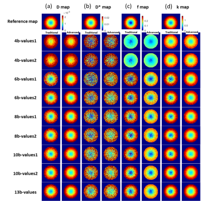 |
50 | Evaluating b-value combinations on IVIM-DKI analysis with parametric reconstruction method in pancreatic carcinoma: A digital phantom study
Archana Vadiraj Malagi1, Devasenathipathy Kandasamy2, Kedar Khare3, Fernando Calamante4, Raju Sharma2, and Amit Mehndiratta5,6
1Centre for Biomedical Engineering, Indian Institute of Technology Delhi, Navi Mumbai, India, 2Department of Radiodiagnosis, All India Institute of Medical Sciences Delhi, New Delhi, India, 3Department of Physics, Indian Institute of Technology Delhi, New Delhi, India, 4Sydney Imaging and School of Biomedical Engineering, University of Sydney, Sydney, Australia, 5Indian Institute of Technology Delhi, New Delhi, India, 6Department of Biomedical Engineering, All India Institute of Medical Sciences Delhi, New Delhi, India
Optimization of b-values in IVIM-DKI is required for parameter estimation accuracy. IVIM-DKI model with Total Variation correction approach was employed in this study, which avoids abrupt changes in parameter values and eliminates non-physiological heterogeneity in maps. In pancreatic cancer, different b-value combinations were used: 4,6,8,10, and 13b-values. In terms of accurate parameter estimations and comparable qualitative maps, 8-13 b-values exhibited a similar trend; in contrast, 4b-values indicated inaccurate and noisy maps. 8b-values can be employed in pancreatic cancer with a constrained technique for a shorter acquisition process and reduced noise in parameter maps at low SNR.
|
||
1781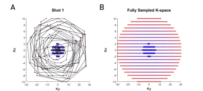 |
51 | A Novel Acquisition Strategy for Volumetric Diffusion-Weighted Imaging using Variable Density TSE-CASPR
Sebastian Eduardo Fonseca1, Sheng-Qing Lin1, Limin Zhou1, Yiming Wang1, and Ananth J Madhuranthakam1,2
1Department of Radiology, UT Southwestern Medical Center, Dallas, TX, United States, 2Advanced Imaging Research Center, UT Southwestern Medical Center, Dallas, TX, United States
Diffusion weighted imaging (DWI) is a highly valuable MR imaging technique in the clinic. Single-shot EPI is the reference standard sequence for DWI due to its speed, however it suffers from severe distortion artifacts that can obscure potential pathologies. In this abstract, we present a novel 3D acquisition strategy that uses a fast spin echo readout and variable density sampling for volumetric DWI. The oversampled center region serves as navigator to correct phase errors. We present preliminary results in brains of healthy volunteers compared to the standard sequences used in DWI.
|
||
1782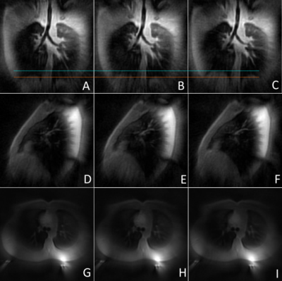 |
52 | Self-Gated Zero Echo Time Imaging of the Lung
Hanna Frantz1, Tobias Lobmeyer1, Patrick Metze1, Thomas Hüfken1, Kilian Stumpf1, Julien Rivoire2, Hizami Murad2, and Volker Rasche1
1Department of Internal Medicine II, Ulm University Medical Center, Ulm, Germany, 2RS2D, Mundolsheim, France
Zero echo time (ZTE) imaging is an established approach for three-dimensional imaging of tissues and materials with ultrashort T2* relaxation times. An intrinsic disadvantage of this approach are missing data points in the center region of k-space due to hardware limitations. These missing points are often retrospectively interpolated or subsequently acquired using single-point imaging approaches or additional ZTE acquisitions with decreased resolutions. The presented approach relies on an interleaved combination of ZTE read-outs with different resolutions in combination with Compressed Sensing, to acquire the k-space center, thus enabling k-space based self-gating.
|
||
1783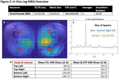 |
53 | Ultra-Short Echo Time 31P 3D MRSI at 3T with Novel Rosette k-space Trajectory
Brian Matthew Bozymski1, Xin Shen2, Ali Caglar Özen3, Serhat Ilbey3, Albert Thomas4, Mark Chiew5, Ulrike Dydak1,6, and Uzay Emir1,2
1School of Health Sciences, Purdue University, West Lafayette, IN, United States, 2Weldon School of Biomedical Engineering, Purdue University, West Lafayette, IN, United States, 3Department of Radiology, Medical Center, University of Freiburg, Freiburg, Germany, 4Department of Radiology, University of California, Los Angeles, CA, United States, 5Wellcome Centre for Integrative Neuroimaging, University of Oxford, Oxford, United Kingdom, 6Department of Radiology and Imaging Sciences, Indiana University School of Medicine, Indianapolis, IN, United States Feasibility of a novel, rosette trajectory UTE (70 μs) 3D 31P MRSI sequence is tested at 3T in phantoms and in upper leg scan with healthy subject. Braino phantom, proton resolution phantom, and highly concentrated 31P phantom test demonstrated proper reconstruction and metabolite localization. In vivo calculations of acquired PCr and β-ATP signals showed competitive SNR across 20 voxels of interest. With further acceleration, the sequence may serve as a superior alternative to conventional weighted 3D MRSI, allowing for greater SNR or resolution in briefer times. Notably, this unprecedentedly short UTE 31P MRSI intrinsically avoids baseline and phasing issues. |
||
1784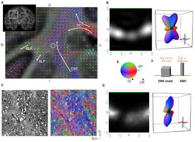 |
54 | Insights to the diffusion MRI ‘crossing fiber problem’: Characterizing the micro-structure in an MRI voxel using synchrotron radiation imaging
Hans Martin Kjer1,2, Mariam Andersson2, Alexandra Pacureanu3, Vedrana Andersen Dahl1, Anders Bjorholm Dahl1, and Tim Bjørn Dyrby1,2
1Technical University of Denmark, Kgs. Lyngby, Denmark, 2Danish Research Centre for Magnetic Resonance, Hvidovre, Denmark, 3European Synchrotron and Radiation Facility, Grenoble, France
Complex fiber regions where multiple tracts and bundles intersect are challenging to study with diffusion weighted MRI (DWI), due to the anatomical variation and 3D complexity within the volume covered by the measure voxel. Using x-ray nano-holotomography (XNH) we have been able to obtain and analyze a volume from a complex fiber region and make a comparison against DWI measurements from the same location of the same ex-vivo monkey brain. We derive comparable micro-structural features in the form of orientation distributions and anisotropy measures and show strong connections between them.
|
||
1785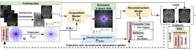 |
55 | Benchmarking learned non-Cartesian k-space trajectories and reconstruction networks
Chaithya G R1,2 and Philippe Ciuciu1,2
1NeuroSpin, Joliot, CEA, Université Paris-Saclay, F-91191, Gif-sur-Yvette, France, 2Inria, Parietal, Université Paris-Saclay, F-91120, Palaiseau, France We benchmark the current existing methods to jointly learn non-Cartesian k-space trajectory and reconstruction: PILOT1, BJORK2 and compare them with those obtained from recently developed generalized hybrid learning (HybLearn) framework3. We present the advantages of using projected gradient descent to enforce MR scanner hardware constraints as compared to using added penalties in the cost function. Further, we use the novel HybLearn scheme to jointly learn and compare our results through retrospective study on fastMRI validation dataset. |
||
1786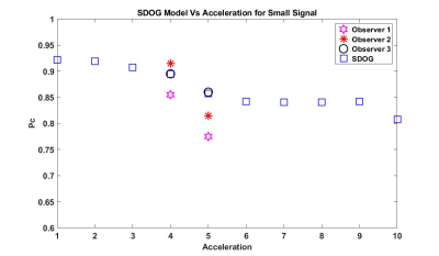 |
56 | Evaluation of Multicoil SENSE Reconstruction of Undersampled Data using a Human Observer Model of Signal Detection
Alexandra G O'Neill1, Tavianne M Kemp1, Sajan G Lingala2, and Angel R Pineda1
1Mathematics Department, Manhattan College, Riverdale, NY, United States, 2Roy J. Carver Department of Biomedical Engineering, University of Iowa, Iowa City, IA, United States
We evaluated undersampling in MRI using a multicoil SENSE reconstruction with no regularization based the detection of signals by humans. We used a sparse difference-of-Gaussians (S-DOG) model to predict human performance in the detection of a small and large signal in anatomical backgrounds. The prediction was then validated using human observer two-alternative forced choice (2-AFC) tasks. Our model predicted a decrease in performance for both the small and large signal from 4X to 5X acceleration. Our observer study validated that prediction. This approach may lead to a way of assessing image quality that predicts human performance with fewer observer studies.
|
||
1787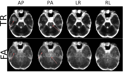 |
57 | Optimal Phase-Encoding Schemes for Regional Bias and Reproducibility of Diffusion MRI derived Measures
M. Okan Irfanoglu1, Grayson Clark1, and Carlo Pierpaoli1
1QMI, NIBIB/NIH, Bethesda, MD, United States
Our goal is to determine whether diffusion MRI acquisitions with different phase-encoding directions (PEDs) result in comparable quantitative measures in different regions of the human brain, or whether PEDs introduce a bias. Additionally, we aim to determine if for a given region, a set of PEDs has better reproducibility. For several regions, PA phase-encoding direction resulted in values that were significantly different than the other three. LR/RL phase-encoding was more reproducible compared to AP/PA in most brain regions, with frontal white matter being the exception. The main source of these reproducibility variations was identified as ghosts overlapping with brain regions.
|
||
 Back to Meeting Home
 Back to the Program-at-a-Glance
The International Society for Magnetic Resonance in Medicine is accredited by the Accreditation Council for Continuing Medical Education to provide continuing medical education for physicians.
 Back to Meeting Home
 Back to the Program-at-a-Glance
 View the Presentation