Digital Poster
Acquisition Strategies II
Joint Annual Meeting ISMRM-ESMRMB & ISMRT 31st Annual Meeting • 07-12 May 2022 • London, UK

| Computer # | ||||
|---|---|---|---|---|
1858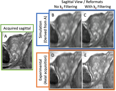 |
38 | Development of Non-Rectangular Excitation Pulses for Improved kZ Fidelity in Super-Resolution T2-Weighted Spin-Echo MRI
Eric Allen Borisch1, Alexandru C. Florea1,2, Roger C. Grimm1, Akira Kawashima3, and Stephen J. Riederer1
1Radiology, Mayo Clinic, Rochester, MN, United States, 2Gustavus Adolphus College, Saint Peter, MN, United States, 3Radiology, Mayo Clinic, Phoenix, AZ, United States
Results from continuing investigation of non-rectangular slice excitation profiles with the goal of providing improved results when used to reconstruct overlapping-slice 2D acquisitions leveraging knowledge of the slice profile are presented. A simulation of the excitation/acquisition process and accompanying reconstruction has been developed to aid the exploration of non-traditional slice profiles and their impact on the final reconstruction quality.
|
||
1859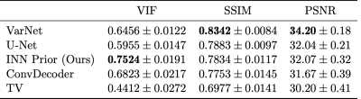 |
39 | Accelerated Magnetic Resonance Imaging with Flow-Based Priors Video Permission Withheld
Frederik Fraaz1 and Reinhard Heckel1,2
1Department of Electrical and Computer Engineering, Technical University of Munich, Munich, Germany, 2Department of Electrical and Computer Engineering, Rice University, Houston, TX, United States
Convolutional neural networks trained end-to-end achieve state-of-the-art image quality for accelerated MRI. But end-to-end networks are trained for a specific undersampling operator. A more flexible approach that can work with any undersampling operator at inference is to train a generative image prior and impose it during reconstruction. In this work, we train a flow-based generator on image patches and then impose it as a prior in the reconstruction. We find that this method achieves slightly better reconstruction quality than state-of-the-art un-trained methods and slightly worse quality than neural networks trained end-to-end on the 4x accelerated multi-coil fastMRI dataset.
|
||
1860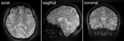 |
40 | Radial Echo-Planar Imaging with Subspace Reconstruction for Brain MRI
Zhengguo Tan1 and Martin Uecker1,2
1Institute for Diagnostic and Interventional Radiology, University Medical Center Goettingen, Goettingen, Germany, 2Institute of Medical Engineering, Graz University of Technology, Graz, Austria
While echo planar imaging (EPI) is successful in many MRI applications, this study demonstrates that radial EPI is applicable to brain MRI as well. Initial volumetric brain experiments at 1 mm isotropic resolution show that radial EPI is capable of rendering high-quality T1- and T2*-weighted images at 2.5 and 1.3 minutes scan time, respectively. Further, radial EPI requires neither magnetization preparation nor fat saturation.
|
||
1861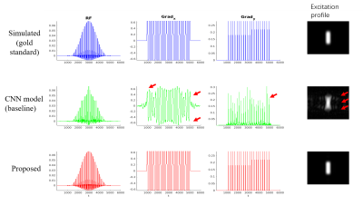 |
41 | Robust and fast RF pulse design of rectangle excitations using a hybrid CNN framework with signal modeling
Yajing Zhang1, Guohui Ruan1, Yan Zhao2, Xianrui Huang2, and Marc Van Cauteren3
1BU-MR Clinical Science, Philips Healthcare, Suzhou, China, 2BU-MR R&D, Philips Healthcare, Suzhou, China, 3BU-MR Clinical Science, Philips Healthcare, Tokyo, Japan
Radio frequency (RF) pulses play a key role in magnetic resonance imaging. However, current MRI systems mostly use pre-defined RF waveforms with minimal adaptions. In this work, we propose a novel hybrid framework to combine convolutional neural network (CNN) with signal modeling by feature extractions. The result demonstrates that the proposed method provides more robust pulse design estimation result than using a generalized CNN framework for any desired rectangle excitations.
|
||
1862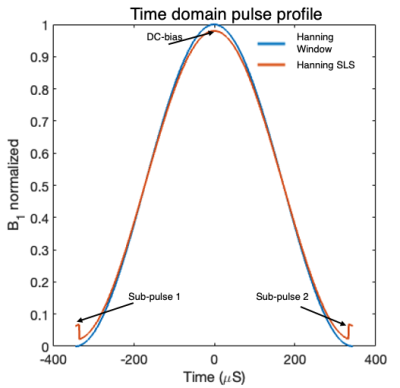 |
42 | Practical RF-pulse shape designs to minimize off-resonance artifacts in dissolved-phase hyperpolarized 129Xe MRI
Junlan Lu1, Suphachart Leewiwatwong2, Bastiaan Driehuys2, and John Mugler3
1Medical Physics, Duke University, Durham, NC, United States, 2Biomedical Engineering, Duke University, Durham, NC, United States, 3Radiology and Medical Imaging, University of Virginia, Charlottesville, VA, United States
Minimizing artifacts in 129Xe gas exchange MRI requires selectively exciting the broad dissolved phase resonances, without exciting the much larger gas phase pool. This requires not only a short, selective pulse, but also overcoming pulse distortions imposed by the RF amplifier. Here, we demonstrate a novel, practical RF pulse based on augmenting the traditional Hanning window pulse with tunable DC bias and sub-lobes in the time-domain. We characterized the performance through simulations, phantom, and in vivo testing and determined that the tunable DC bias parameter creates a robust dissolved-phase excitation while minimizing gas-phase contamination.
|
||
1863 |
43 | Reduction of Acquisition time using RAPID-SI technique: Preliminary in-vitro results and comparison with CSI at Ultra-High-Field.
Hacene Serrai1
1Carle Foundation Hospital, Center for Clinical Imaging Research, Urbana, IL, United States
It has been shown that RAPID-SI, implemented at 3 Tesla magnet, allows for reduction of acquisition time while providing accurate metabolite information as compared to CSI. To benefit from the higher sensitivity of the Ultra-High-Field (UHF), RAPID-SI was implemented on a Siemens Terra 7T scanner, and phantom MRSI data were acquired, with less averaging, and compared in terms of acquisition time, signal-to-noise ratio (SNR), and data analysis to those collected using CSI method. As expected, and in addition to the acquisition time reduction, proportional to the speed factor R, RAPID-SI provides similar quantification results with similar SNR values, afterward data reconstruction, as compared to CSI.
|
||
1864 |
44 | Characterization of SNR for radial spin echo imaging
Neta Stern1, Kai T Block2, Chen Solomon1, Tamar Blumenfeld-Katzir1, and Noam Ben-Eliezer1,2,3
1Biomedical Engineering, Tel Aviv University, Tel Aviv, Israel, 2New-York University Langone Medical Center, New York, NY, United States, 3Sagol School of Neuroscience, Tel Aviv University, Tel Aviv, Israel
To optimize the use of radial spin-echo sequences, SNR characterization for relevant T1 and T2 ranges is required. In this work, we studied the interplay between the number of excitations (Nexc) and the turbo factor (NTF - number of echoes within each TR) on the SNR of images acquired using the RAISE (RAdIal Spin Echo) sequence. SNR was evaluated for phantom and in-vivo images. Phantom analysis focused on SNR per T1 and T2 ranges appropriate for known gray and white matter relaxation times. Selected parameter sets were later used for in vivo scans for qualitative evaluation.
|
||
1865 |
45 | Single-Shot Multi-b-value (SSMb) Diffusion-weighted MRI Using Spin Echo and Stimulated Echoes with Variable Flip Angles
Guangyu Dan1,2, Kaibao Sun1, Qingfei Luo1, and Xiaohong Joe Zhou1,2,3
1Center for Magnetic Resonance Research, University of Illinois at Chicago, Chicago, IL, United States, 2Department of Biomedical Engineering, University of Illinois at Chicago, Chicago, IL, United States, 3Departments of Radiology and Neurosurgery, University of Illinois at Chicago, Chicago, IL, United States
In diffusion-weighted imaging with multiple b-values, the acquisition times using a spin-echo EPI sequence can be long because each b-value requires a new shot. The lengthy acquisition time imposes issues with patient compliance and increases venerability to motion-induced errors in quantitative analysis. We herein report a novel pulse sequence that acquires multiple diffusion-weighted images with difference b-values in a single shot by utilizing the combination of a spin echo and a train of stimulated echoes with variable flip angles. This sequence has been demonstrated in a quantitative diffusion phantom and the human brain to realize a 4-fold scan time reduction.
|
||
1866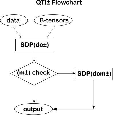 |
46 | Diffusivity-limited q-space trajectory imaging
Deneb Boito1,2, Magnus Herberthson3, and Evren Özarslan1,2
1Department of Biomedical Engineering, Linköping University, Linköping, Sweden, 2Center for Medical Image Science and Visualization (CMIV), Linköping University, Linköping, Sweden, 3Department of Mathematics, Linköping University, Linköping, Sweden
Q-space trajectory imaging (QTI) characterizes microstructures through the statistical moments of the diffusion tensor distribution. A constrained estimation framework named QTI+ was recently proposed to achieve mathematically and physically acceptable estimates of these moments. Here we consider expanding QTI+ with a new set of conditions based on the theoretical maximum value of water diffusivity. We show where these conditions are violated for different bulk diffusivity values, and how the new constraints affect the QTI scalar maps. The results show violations occurring almost exclusively in voxels containing gray matter and CSF. Imposing the constraints produces metrics closer to the expected values.
|
||
1867 |
47 | Quantitative Reduced Field-of-view Imaging using 3D Tailored Inner Volume Excitation and Pattern Recognition
Yun Jiang1, Jon-Fredrik Nielsen2, Vikas Gulani1, and Nicole Seiberlich1
1Department of Radiology, University of Michigan, Ann Arbor, MI, United States, 22Department of Biomedical Engineering, University of Michigan, Ann Arbor, MI, United States
The goal of this study is to achieve small field-of-view (FOV) T1 and T2 mapping using 3D tailored inner volume (IV) excitation and pattern recognition based on MR Fingerprinting.
|
||
1868 |
48 | Pressure Pain Activation Revealed in Simultaneous Brain, High-Res Brainstem, and High-Res Spinal Cord fMRI.
Christine S W Law1, Ken A Weber1, Sean Mackey1, and Gary H Glover1
1Stanford University, Stanford, CA, United States
We present a novel fMRI acquisition that mitigates susceptibility-induced off-resonance and simultaneously captures brainstem, spinal cord, and brain. Brainstem and spinal cord are imaged in high spatial resolution whereas brain resolution is standard. This fMRI acquisition is applied in a noxious pressure-pain experiment from which we observe activation in brain, spinal cord, and brainstem pons & medulla.
|
||
1869 |
49 | Comparison of Imaging Methods for Fast Whole-Body MRI Screening
Marc Rea1, Alex Barrett1, and Christopher Romaniuk2
1Physics, Clatterbridge Cancer Centre, Liverpool, United Kingdom, 2Radiology, Clatterbridge Cancer Centre, Liverpool, United Kingdom
Improvements in image quality and scan time coupled with AI reading of images, is enabling the possibility of quickly screening all patients who attend MRI for malignant tumours. This research explores the imaging and clinical requirements for such a programme, and reports on the ability of various sequences and patient configurations to meet those requirements.
|
||
 Back to Meeting Home
 Back to the Program-at-a-Glance
The International Society for Magnetic Resonance in Medicine is accredited by the Accreditation Council for Continuing Medical Education to provide continuing medical education for physicians.
 Back to Meeting Home
 Back to the Program-at-a-Glance
 View the Presentation