Digital Poster
Machine Learning/Artificial Intelligence: Clinical Application I
Joint Annual Meeting ISMRM-ESMRMB & ISMRT 31st Annual Meeting • 07-12 May 2022 • London, UK

Digital Poster
Machine Learning/Artificial Intelligence: Clinical Application I
| Computer # | ||||
|---|---|---|---|---|
1428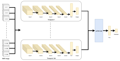 |
52 | Understanding Alzheimer's disease through fMRI and deep learning
Sarah Wacher1, Jinglei Lv1,2, and Mariano Cabezas1
1Brain and Mind Centre, The University of Sydney, Sydney, Australia, 2School of Biomedical Engineering, The University of Sydney, Sydney, Australia Deep learning approaches for Alzheimer's disease (AD) classification have primarily focused on modelling the structural changes associated with the condition, neglecting the changes in functional brain dynamics. These functional changes may be detectable earlier than structural atrophy providing an avenue for earlier diagnosis and treatment. We therefore proposed a convolutional neural network combined with a long short-term memory unit to decode fMRI signals. The model was able to classify AD from healthy control with a balanced accuracy of 0.69. Whilst there is room to improve network performance, the study already provides promising insights into the possibilities of resting-state fMRI classification. |
||
1429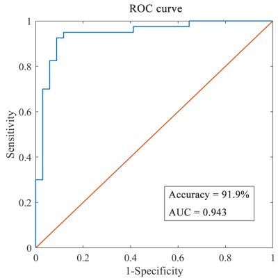 |
53 | Classification of Chemotherapy-Related Subjective Cognitive Complaints in Breast Cancer Using Multi-Level Features of Functional MRI
Lei Wang1 and Fuqing Zhou1
1Department of Radiology, The First Affiliated Hospital of Nanchang University, Nanchang, China
Chemotherapy-related cognitive impairment (CRCI), especially subjective cognitive complaints (SCC), had been often reported in breast cancer survivors, which affects their life and work. The identification of biomarkers for early diagnosis and prognosis prediction of SCC remains a crucial challenge of important clinical implications. The resting-state functional magnetic resonance imaging (rs-fMRI) has been widely used to detect abnormalities of brain activity in CRCI. The machine learning method combined with rs-fMRI features could effectively identify breast cancer survivors with chemotherapy-related SCC from healthy controls (HC).
|
||
1430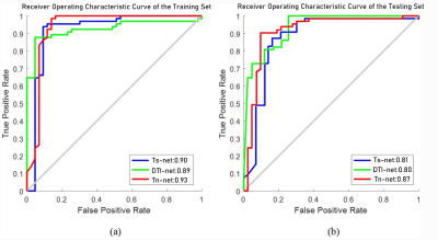 |
54 | Multi-Channel Deep Learning for IDH Mutation Status Prediction in Gliomas:A Multimodal Approach Video Not Available
Jin Wang1, Jiayang Deng2, Rui Wang1, and Jing Zhang1
1Department of Magnetic Resonance, LanZhou University Second Hospital, Lanzhou, China, 2Vipshop (China) Co. LTD, Guangzhou, China
Isocitrate dehydrogenase (IDH) mutation status has emerged as an important prognostic marker in gliomas.Currently, reliable IDH mutation determination requires invasive surgical procedures. Various studies have demonstrated the efficacy of deep learning in classify IDH status using Magnetic resonance images(MRI)data.In this study we propose a multi-channel architecture of 3D convolutional neural networks (CNNs) based on deep learning to predict the IDH status.We utilize traditional structures imaging and various diffusivity metric maps derived from diffusion tensor imaging (DTI) as input to the network.The final model achieved the AUC value of 0.93.
|
||
1431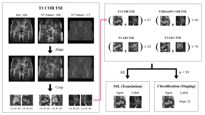 |
55 | MR-only staging of osteonecrosis of the femoral head (ONFH) using self-supervised learning for multiple MR protocols
Bomin Kim1, Geun Young Lee2, and Sung-Hong Park1
1Department of Bio and Brain Engineering, Korea Advanced Institute of Science and Technology, Daejeon, Korea, Republic of, 2Deparment of Radiology, Chung-Ang University Hospital, Seoul, Korea, Republic of
Diagnosing stages of osteonecrosis of the femoral head (ONFH) based on MR images can reduce additional cost and radiation exposure caused by CT scan. We propose a deep learning network that enables 4-way classification of ONFH stages, utilizing the information from MR images with different contrasts and planes. Given the limited number of available data, we enhanced the network performance by using self-supervised learning based on MR-to-CT translation task, which increased AUC significantly. We also investigated the diagnostic results from different MR protocols, and obtained more precise and robust results by combining them.
|
||
1432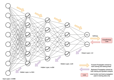 |
56 | Towards a personalized MRgFUS treatment for tremor disorders: A study on the number of ablations using deep learning and structural connectivity
Zihao Tang1,2, Mariano Cabezas2, Kain Kyle2,3, Arkiev D'Souza2, Stephen Tisch4, Ben Jonker4, Yael Barnett4, Joel Maamary4, Jerome Maller5, Michael Barnett2,3, Weidong Cai1, and Chenyu Wang2,3
1School of Computer Science, University of Sydney, Sydney, Australia, 2Brain and Mind Centre, University of Sydney, Sydney, Australia, 3Sydney Neuroimaging Analysis Centre, Sydney, Australia, 4St Vincent's Hospital Sydney, Sydney, Australia, 5GE Healthcare Australia, Melbourne, Australia Disabling tremor is the most common symptom of tremor-dominated Parkinson’s disease (PD) patients. Recently, MR guided focused ultrasound (MRgFUS) has been applied in the clinical environment to treat tremor. However, patients can have different treatment responses to the ablation. Tremor suppression can be observed on some patients after ablating the ventral intermediate nucleus (Vim), while others require further ablations. To provide a tailored treatment, a deep learning technique is introduced in this work to predict whether a subject undergoing MRgFUS will respond to a single ablation in the VIM or require additional lesioning in other regions. |
||
1433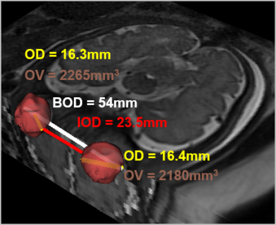 |
57 | Improved assessment of fetal ocular pathologies in MRI using ocular ratios and machine-learning multi-parametric classification
Netanell Avisdris1,2, Daphna Link Sourani1, Liat Ben Sira3,4,5, Leo Joskowicz2, Gustavo Malinger4,6, Simcha Yagel7, Elka Miller8, and Dafna Ben Bashat1,4,5
1Sagol Brain Institute, Tel Aviv Sourasky Medical Center, Tel Aviv, Israel, 2School of Computer Science and Engineering, The Hebrew University of Jerusalem, Jerusalem, Israel, 3Division of Pediatric Radiology, Tel Aviv Sourasky Medical Center, Tel Aviv, Israel, 4Sackler Faculty of Medicine, Tel Aviv University, Tel Aviv, Israel, 5Sagol School of Neuroscience, Tel Aviv University, Tel Aviv, Israel, 6Division of Ultrasound in Obstetrics & Gynecology, Lis Maternity Hospital, Tel Aviv, Israel, 7Obstetrics and Gynecology Division, Hadassah Hebrew University Medical Center, Jerusalem, Israel, 8Medical Imaging, CHEO, University of Ottawa, Ottawa, ON, Canada
Brain MRI of 296 fetuses (22-40 weeks’ gestational-age, normal n=244, hypo/hyper-telorism n=52 were included. Binocular, interocular, and ocular diameters, and ocular volume were measured using automatic methods. Two new parameters, binocular-ratio and interocular-ratio, were defined. In normal fetuses, all four measurements increased with gestational-age. However, constant values were detected across all gestational-ages of binocular-ratio=1.56±0.05 and interocular-ratio=0.60±0.05. A random-forest classifier achieved the best results (out of eight classifiers) with AUC-ROC=0.90±0.03 for classification between normal and fetuses with hypo/hyper-telorism. mainly based on the two new ratios. Machine-learning multi-parametric classification and the new ratios are suggested to be used in routine clinical practice.
|
||
1434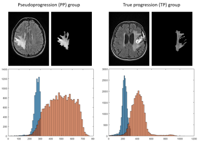 |
58 | Radiomics signature from multiparametric MRI as early in-vivo biomarkers for pseudoprogression in recurrent glioblastoma patients. Video Permission Withheld
Lucie Piram1, Acquitter Clément2, Julia Gilhodes3, Umberto Sabatini4, Elizabeth Cohen-Jonathan Moyal1,5, Benjamin Lemasson2, and Soleakhena Ken5,6,7
1Department of Radiotherapy, Institut Universitaire du Cancer de Toulouse - Oncopole, Toulouse, France, 2Grenoble Institut des Neurosciences, Grenoble, France, 3Department of Clinical Trials, Institut Universitaire du Cancer de Toulouse - Oncopole, Toulouse, France, 4Dipartimento di Scienze Mediche e Chirurgiche, Università Magna Graecia, Catanzaro, Italy, 5U1037, RADOPT Team, Cancer Research Center of Toulouse, Toulouse, France, 6Department of Engineering and Medical Physics, Institut Universitaire du Cancer de Toulouse - Oncopole, Toulouse, France, 7MINDS Team UMR 5505, Institut de Recherche en Informatique de Toulouse, Toulouse, France Radiomic features computed from multiparametric MRI were found to be relevant as early in-vivo biomarkers for pseudoprogression evaluation in recurrent glioblastoma patients. At baseline, predictive biomarker for pseudoprogression outcome was related to kurtosis parameter of FLAIR histogram plotted from abnormal hyper-intense signal area. When considering variation between baseline and first event (either pseudoprogression or true progression), four early biomarkers were found for entropy of T1-weighted, T1-weighted-post-contrast morphological MRI and Apparent Diffusion Coefficient maps derived from diffusion-weighted MRI. Such early in-vivo biomarkers easily computed from automatic segmentation and first order radiomics analysis could be useful for the assessment of treatment response. |
||
1435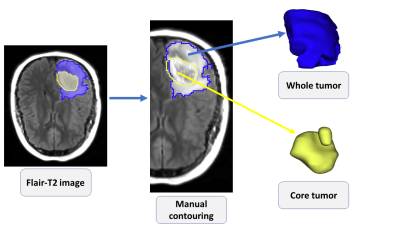 |
59 | MRI based Radiomics for Distinguishing IDH-mutant from IDH wild-type Grade-4 Astrocytomas
Seyyed Ali Hosseini1, Isaac Shiri2, Ghasem Hajianfar3, Stephen Bagley4, MacLean Nasrallah5, Donald M O’Rourke 6, Suyash Mohan7, and SANJEEV CHAWLA7
1Medical physics and biomedical engineering, Tehran university of medical science, Karaj, Iran (Islamic Republic of), 2Medical Imaging, University of Geneva, Geneva, Switzerland, 3Cardiovascular Medical and Research Center, Iran University of Medical Science, Tehran, Iran (Islamic Republic of), 4Hematology-Oncology, Perelman School of Medicine at the University of Pennsylvania, Philadelphia, PA, United States, 5Pathology and Lab Medicine, Perelman School of Medicine at the University of Pennsylvania, Philadelphia, PA, United States, 6Neurosurgery, Perelman School of Medicine at the University of Pennsylvania, Philadelphia, PA, United States, 7Radiology, Perelman School of Medicine at the University of Pennsylvania, Philadelphia, PA, United States
To distinguish IDH-mutant from IDH wild-type grade-4 astrocytomas, conventional MR imaging (post-contrast T1-weighted and T2-Flair images) was acquired from 57 patients [IDH-mutant (n=23) and IDH-wild-type (n=34) grade-4 astrocytomas]. Post-contrast T1-weighted and T2-FLAIR images were resliced, resampled and co-registered. Neoplasms were segmented into whole tumor, enhancing region, central necrotic region, edema region, and core tumor (contrast enhancing + necrotic region). A total of 105 first-, second-, higher-order and shape based radiomics features were extracted from each ROI. Readily interpretable and quantitative features from different sub-regions of neoplasms were observed with high diagnostic performances in distinguishing IDH-mutant from IDH wild-type grade-4 astrocytomas.
|
||
1436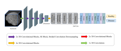 |
60 | Exploring Hybrid CNN-Transformer for Schizophrenia Classification using Structural MRI
Vishwanatha Mitnala Rao1, Junhao Zhang1, and Jia Guo2
1Department of Biomedical Engineering, Columbia University, New York, NY, United States, 2Department of Psychiatry, Columbia University, New York, NY, United States Schizophrenia diagnosis is clinically difficult due to the lack of biomarkers associated with the disease. While machine learning algorithms and convolutional neural networks (CNNs) have found success using neuroimaging inputs to diagnose the disease, they have historically not performed or generalized well enough for clinical use. We propose 3D-MIC-Transformer, the first transformer-based deep learning architecture applied to neurological disease classification that demonstrates state-of-the-art schizophrenia classification performance and generalization using structural MRI inputs. 3D-MIC-Transformer outperforms prior CNN implementations (AUROC: 0.985, accuracy: 0.933), and we believe 3D-MIC-Transformer can serve as a backbone for other disease classification tasks in the future. |
||
1437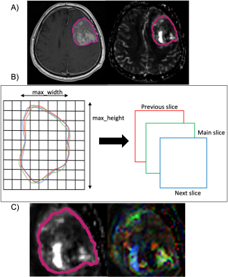 |
61 | Differentiation of NF2 Loss and S100 Positivity in Meningioma Using Dynamic Susceptibility Contrast MRI with Machine Learning at 3T
Buse Buz-Yalug1, Ayca Ersen Danyeli2,3, Kubra Tan4, Ozge Can5, Necmettin Pamir3,6, Alp Dincer3,7, Koray Ozduman3,8, and Esin Ozturk-Isik1
1Institute of Biomedical Engineering, Bogazici University, Istanbul, Turkey, 2Department of Medical Pathology, Acibadem Mehmet Ali Aydinlar University, Istanbul, Turkey, 3Center for Neuroradiological Applications and Reseach, Acibadem Mehmet Ali Aydinlar University, Istanbul, Turkey, 4Health Institutes of Turkey, Istanbul, Turkey, 5Department of Medical Engineering, Acibadem Mehmet Ali Aydinlar University, Istanbul, Turkey, 6Department of Neurosurgery, Acibadem Mehmet Ali Aydinlar University, Istanbul, Turkey, 7Department of Radiology, Acıbadem Mehmet Ali Aydinlar University, Istanbul, Turkey, 8Department of Neurosurgery, Acıbadem Mehmet Ali Aydinlar University, Istanbul, Turkey
Meningiomas are the most frequent primary intracranial and spinal tumors. In this work, we studied the relative cerebral blood volume (rCBV) changes in the meningiomas with NF2 loss and S100 positivity. Meningiomas with NF2-L had lower rCBV than NF2-NL, and S100 positive group had also lower rCBV than S100 negative group. The highest classification accuracies obtained using machine learning applications were 75.6% for NF2 molecular subsets and 75.2% for S100 molecular subsets.
|
||
1438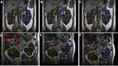 |
62 | Deep Learning based Total Kidney Volume Segmentation in Autosomal Dominant Polycystic Kidney Disease
Anish Raj1, Fabian Tollens2, Anika Strittmatter1, Laura Hansen1, Dominik Noerenberg2, and Frank G Zöllner1
1Computer Assisted Clinical Medicine, Mannheim Institute for Intelligent Systems in Medicine, Medical Faculty Mannheim, Heidelberg University, Mannheim, Germany, 2Department of Clinical Radiology and Nuclear Medicine, Medical University Center Mannheim, Medical Faculty Mannheim, Heidelberg University, Mannheim, Germany
The total kidney volume (TKV) increases with ADPKD progression and hence, can be used to quantify disease progression. The TKV calculation requires accurate delineation of kidney volumes which is usually performed manually by an expert physician. However, this is time consuming and automated segmentation is warranted, e.g., using deep learning. The implementation of the latter is usually hindered due to a lack of large, annotated datasets.In this work, we address this problem by implementing the cosine loss function and a technique called Sharpness Aware Minimization (SAM) into the U-Net to improve TKV estimation in small sized datasets.
|
||
 Back to Meeting Home
 Back to the Program-at-a-Glance
The International Society for Magnetic Resonance in Medicine is accredited by the Accreditation Council for Continuing Medical Education to provide continuing medical education for physicians.
 Back to Meeting Home
 Back to the Program-at-a-Glance
 View the Presentation