Digital Poster
Relaxation & Multiparameter Mapping I
Joint Annual Meeting ISMRM-ESMRMB & ISMRT 31st Annual Meeting • 07-12 May 2022 • London, UK

Digital Poster
Relaxation & Multiparameter Mapping I
| Computer # | ||||
|---|---|---|---|---|
1259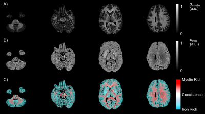 |
55 | Determinants of MR Relaxation in the Extremely Preterm Brain at Adolescence: Myelin and Iron
Ryan McNaughton1,2, Ning Hua2, Lei Zhang3, David Kennedy4, T. Michael O'Shea3, Karl Kuban2, and Hernan Jara2
1Mechanical Engineering, Boston University, Boston, MA, United States, 2Boston University Medical Center, Boston, MA, United States, 3University of North Carolina at Chapel Hill, Chapel Hill, NC, United States, 4University of Massachusetts, Worcester, MA, United States
Purpose: To describe the dependencies of multiparametric quantitative MRI (MP-qMRI) on myelin and iron content in the extremely preterm born brain at adolescence. Methods: Algorithms for a fast exchange relaxation (FER) model and MP-qMRI create maps of R1, R2, myelin, and iron in 30 participants using MR images obtained with the triple TSE pulse sequence at age 15 years. Results: R1 and R2 have linear dependencies with myelin and iron content, respectively. Conclusion: Application of a FER model produces coregistered maps of myelin and iron content which exhibit unique influences on the relaxation of white matter and gray matter regions.
|
||
1260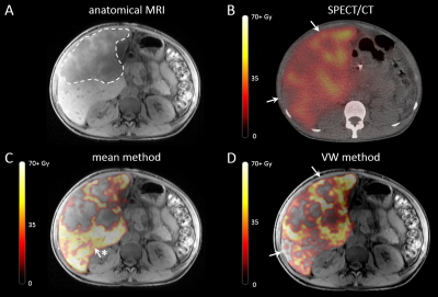 |
56 | Improved ΔR2* calculation through voxelwise subtraction for MRI-based dosimetry of holmium-166 transarterial radioembolization
Meike W.M. van Wijk1, Joey Roosen1, Lovisa E.L. Westlund Gotby1, Mark J. Arntz1, Marcel J.R. Janssen1, Daphne Lobeek1, Gerrit H. van de Maat2, Christiaan G. Overduin1, and J. Frank W. Nijsen1
1Department of Medical Imaging, Radboud University Medical Center, Nijmegen, Netherlands, 2Imaging & Software Solutions, Quirem Medical B.V., Deventer, Netherlands Transarterial radioembolization (TARE) is a treatment for liver cancer, during which radioactive microspheres are administered through the hepatic artery. Microspheres containing holmium-166 enable MRI-based dosimetry, based on subtraction of pre- and post-treatment $$$R_2^*$$$ values. This subtraction is performed using a mean pre-treatment $$$R_2^*$$$ value. This does however not take pre-existing differences of $$$R_2^*$$$ values into account, introducing an error in the dosimetry. In this work a voxelwise subtraction method is presented, using deformable registration to transform the pre-treatment $$$R_2^*$$$ map to the post-treatment $$$R_2^*$$$ map, enabling voxel-by-voxel subtraction. This method does take $$$R_2^*$$$ differences into account and improves MRI-based dosimetry. |
||
1261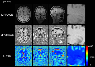 |
57 | The Compressed Sensing MP2RAGE as a surrogate to the MPRAGE for neuro-imaging at 3T
Aurélien J Trotier1, Bixente Dilharreguy2, Serge Anandra3, Nadège Corbin1, William Lefrançois1, Valery Ozenne1, Sylvain Miraux1, and Emeline Julie Ribot1
1Centre de Résonance Magnétique des Systèmes Biologiques, UMR 5536, CNRS, Bordeaux, France, 2UMS3767, CNRS, Bordeaux, France, 3UMS3767, Université de Bordeaux, Bordeaux, France
A Compressed Sensing Magnetization prepared Two Rapid Gradient Echo (CS-MP2RAGE) sequence was developed to provide images in <10min including reconstruction time. It was applied on 13 participants each scanned 3 to 4 times with repositioning. Compared to the standard MPRAGE sequence, it provides higher contrast morphological images. Similar intra-volunteer variabilities in volume segmentations of the brain structures were obtained. Additionally, high resolution T1 maps provided T1 values of white and gray matters and several deep grey nuclei consistent with the literature, and show very low variability (<1%). The CS-MP2RAGE can be used in future clinical research protocols.
|
||
1262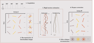 |
58 | Whole brain 3D radial T2 mapping with retrospective motion correction capabilities.
Nadège Corbin1,2, Aurélien J Trotier1, Sylvain Miraux1, and Emeline J Ribot1
1Centre de Résonance Magnétique des Systèmes Biologiques, UMR 5536, Bordeaux, France, 2Wellcome Centre for Human Neuroimaging, UCL Queen Square Institute of Neurology, University College London, London, United Kingdom
T2 mapping is usually very time consuming and long acquisition time often result in high sensitivity to intra-scan motion. Here we propose a T2 mapping method relying on 3D radial sampling and advanced iterative reconstruction to retrospectively correct for motion and/or reduce the acquisition. First, in-vivo experiments at 3T showed the capability of the technique to correct for intra-scan motion and therefore improve the quality of resulting T2 maps. Second, acquisitions varying from 7 to 33 min of whole-brain single and multi-compartment T2 maps were obtained, showing the potential of the method to accelerate and therefore reduce sensitivity to motion.
|
||
1263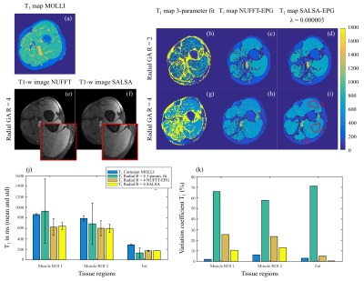 |
59 | Improving T1 mapping with a golden-angle radial MOLLI and a model-based regularized reconstruction SALSA-EPG
Andreia C Freitas1, Andreia S Gaspar1, Nuno A Silva2, José M Bioucas-Dias3, and Rita G Nunes1
1ISR Lisboa/LARSyS and Department of Bioengineering, Instituto Superior Técnico - Universidade de Lisboa, Lisbon, Portugal, 2Hospital da Luz Learning Health, Luz Saúde, Lisbon, Portugal, 3Instituto de Telecomunicações, Instituto Superior Técnico - Universidade de Lisboa, Lisbon, Portugal
T1 mapping provides valuable information regarding cardiovascular pathologies. Clinical approach consists of using a Cartesian MOLLI sequence and fitting a 3-parameter model. Although easy and straightforward, this method can require long breath-holds and does not explicitly include other factors affecting T1 estimation. We propose coupling an accelerated golden-angle radial MOLLI with a model-based regularized reconstruction (SALSA). The proposed method was tested in phantom and in vivo data at 1.5T. Improved T1 precision and good accuracy was found for the phantom data. Lower T1 was estimated with SALSA compared to the commercial sequence in vivo, future work will address this.
|
||
1264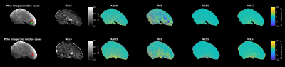 |
60 | Robust and efficient R2* estimation in human brain using log-linear weighted least squares
Luke J. Edwards1, Siawoosh Mohammadi1,2, Kerrin J. Pine1, Martina F. Callaghan3, and Nikolaus Weiskopf1,4
1Department of Neurophysics, Max Planck Institute for Human Cognitive and Brain Sciences, Leipzig, Germany, 2Department of Systems Neuroscience, University Medical Center Hamburg-Eppendorf, Hamburg, Germany, 3Wellcome Centre for Human Neuroimaging, UCL Queen Square Institute of Neurology, University College London, London, United Kingdom, 4Felix Bloch Institute for Solid State Physics, Faculty of Physics and Earth Sciences, Leipzig University, Leipzig, Germany
In vivo maps of R2* in human brain hold promise for neuroscientific investigations. We tested several R2* fitting routines (nonlinear least squares [NLLS; silver standard], auto-regression on linear operations [ARLO], log-linear weighted least square [WLS], and log-linear ordinary least squares [OLS]) for dual flip angle multi-echo FLASH data in simulations and challenging 400 µm resolution in vivo 7T data. Log-linear WLS was found to give a good trade-off between accuracy, precision, and computational time. The method will be available in a future version of the open source hMRI toolbox (hmri.info).
|
||
1265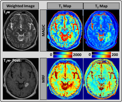 |
61 | Quantitative Relaxometry Metrics for Normal and Brain Tumor Tissues using MR Fingerprinting and Magnetic Resonance Image Compilation Methods
Amaresha Shridhar Konar1, Akash Deelip Shah2, Abhay Dave3, Suchandrima Banerjee4, Maggie Fung5, Vaios Hatzoglou2, and Amita Shukla-Dave1,2
1Medical Physics, Memorial Sloan Kettering Cancer Center, New York City, NY, United States, 2Radiology, Memorial Sloan Kettering Cancer Center, New York, NY, United States, 3Touro College of Osteopathic Medicine, New York, NY, United States, 4GE Healthcare, New York, NY, United States, 5GE Healthcare, New York City, NY, United States
This work aims to assess the relaxometry maps generated using MRF and MAGiC in brain tumor (n=27). A total of 14 brain tumor tissue regions (Pre-Tx n=5, Post-Tx n=9) were used for comparison. The T1 and T2 values measured at tumor and normal appearing (contralateral) region showed significant difference in both MAGiC and MRF generated maps. The results show that the relaxometry values estimated using MRF and MAGiC methods can differentiate Tumor and normal appearing tissues. The utility of this technique needs to be further explored in larger sample studies and it could be further extendable to classify tumor types.
|
||
1266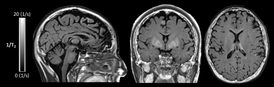 |
62 | Feasibility of fast whole brain T2 mapping using CAIPIRINHA accelerated SPACE sequences
Christoph Birkl1, Christian Kremser2, Alexander Rauscher3, and Elke Ruth Gizewski1
1Department of Neuroradiology, Medical University of Innsbruck, Innsbruck, Austria, 2Department of Radiology, Medical University of Innsbruck, Innsbruck, Austria, 3UBC MRI Research Centre, University of British Columbia, Vancouver, BC, Canada We investigated whether multiple SPACE sequences accelerated using CAIPIRINHA and acquired with different echo times can be used to obtain high resolution isotropic whole brain T2 maps within a clinical feasible scan time. In a phantom study, we could show that the SPACE based T2 values are comparable with T2 values derived using multiple single spin echo sequences. By reducing the number of echoes we could acquire 1 mm isotropic in vivo whole brain T2 maps in under 3 minutes. |
||
1267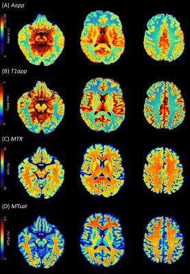 |
63 | Magnetisation transfer saturation (MTsat) and MTR: relationship with T1 recovery in multiple sclerosis and healthy brain
Elizabeth N. York1, Rozanna Meijboom1, Agniete Kampaite1, Maria Valdes Hernandez1, Michael J. Thrippleton1, and Adam D. Waldman1
1Centre for Clinical Brain Sciences, University of Edinburgh, Edinburgh, United Kingdom
Magnetisation transfer saturation (MTsat) shows improved tissue contrast compared with magnetisation transfer ratio (MTR), due to T1 correction, and is more sensitive to subtle myelin loss in multiple sclerosis (MS). It is not clear how MTsat performs in severely demyelinated tissue with markedly increased T1. We examine the relationship between T1app, MTsat and MTR in healthy and MS brains. We show a negative linear relationship between MTsat and T1app in white matter, which breaks down in grey matter and white matter lesions. MTsat is sensitive to modest myelin disruption, while T1app may better reflect severe damage in MS lesions.
|
||
1268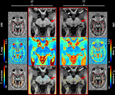 |
64 | High-resolution T1 atlas for subject-specific abnormality detection at 7T
Gian Franco Piredda1,2,3, Piotr Radojewski4,5, Arun Joseph5,6,7, Gabriele Bonanno5,6,7, Karl Egger8, Shan Yang8, Punith B. Venkategowda9, Ricardo A. Corredor-Jerez1,2,3, Bénédicte Maréchal1,2,3, Roland Wiest4,5, Jean-Philippe Thiran2,3, Tom Hilbert1,2,3, and Tobias Kober1,2,3
1Advanced Clinical Imaging Technology, Siemens Healthcare AG, Lausanne, Switzerland, 2Department of Radiology, Lausanne University Hospital and University of Lausanne, Lausanne, Switzerland, 3LTS5, École Polytechnique Fédérale de Lausanne (EPFL), Lausanne, Switzerland, 4Support Center for Advanced Neuroimaging, Institute for Diagnostic and Interventional Neuroradiology, Inselspital, Bern University, Bern, Switzerland, 5Translational Imaging Center, sitem-insel AG, Bern, Switzerland, 6Advanced Clinical Imaging Technology, Siemens Healthcare AG, Bern, Switzerland, 7Magnetic Resonance Methodology, Institute of Diagnostic and Interventional Neuroradiology, University of Bern, Bern, Switzerland, 8Department of Neuroradiology, Medical Center – University of Freiburg, Faculty of Medicine, University of Freiburg, Freiburg, Germany, 9Siemens Healthcare Pvt. Ltd., Bangalore, India
Previous studies at 3T have shown that T1 relaxometry enables personalized characterization of brain tissues by comparing physical properties of a single patient to a normative atlas. Ultra-high field imaging allows exploiting this concept at even higher resolutions, which can be crucial to detect certain diseases. To this end, here we established an atlas of normative T1 values at 7T from acquisitions with 0.6$$$\times$$$0.6$$$\times$$$0.6 mm3 isotropic resolution. Additionally, the clinical potential and improvement of 7T vs. 3T imaging is shown in two case reports from patients scanned at both field strengths.
|
||
1269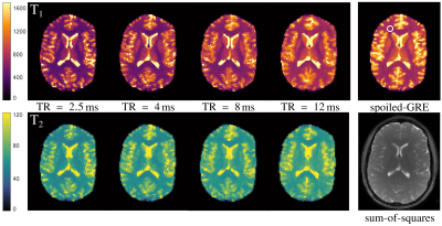 |
65 | TR dependence of phase-cycled bSSFP relaxometry in brain tissue
Jessica Schäper1,2 and Oliver Bieri1,2
1Department of Biomedical Engineering, University of Basel, Basel, Switzerland, 2Division of Radioligical Physics, Department of Radiology, University of Basel Hospital, Basel, Switzerland
Brain relaxometry with phase-cycled bSSFP shows systematically lower T1 values, if compared to spoiled-GRE or inversion-recovery spin echo methods. One explanation can be the pronounced asymmetry in the bSSFP's frequency profile, observed for tissues. It was recently shown that this asymmetry decreases towards shorter TR, possibly leading to an adjustment of T1 estimates from bSSFP to spoiled-GRE. Here, it was investigated how T1 and T2 quantification is influenced by TR. Contrary to expectation, a stronger mismatch between bSSFP and spoiled-GRE was observed towards shorter TR. The origin of this mismatch can thus not be attributed to the bSSFP profile asymmetry.
|
||
1270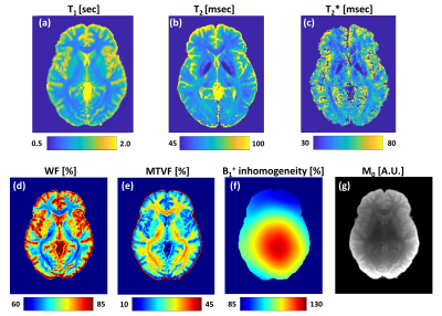 |
66 | A comprehensive protocol for multiparametric brain MRI
Dvir Radunsky1, Chen Solomon1, Tamar Blumenfeld-Katzir1, Neta Stern1, Shir Filo2, Aviv Mezer2, Anita Karsa3, Karin Shmueli3, Lucas Soustelle 4, Guillaume Duhamel4, Olivier M. Girard4, and Noam Ben-Eliezer1,5,6
1Biomedical Engineering, Tel Aviv University, Tel Aviv, Israel, 2The Edmond and Lily Safra Center for Brain Sciences, The Hebrew University of Jerusalem, Jerusalem, Israel, 3Department of Medical Physics and Biomedical Engineering, University College London, London, United Kingdom, 4Aix Marseille University, CNRS, CRMBM, Marseille, France, 5Sagol School of Neuroscience, Tel Aviv University, Tel Aviv, Israel, 6Center for Advanced Imaging Innovation and Research (CAI2R), University Langone Medical Center, New York, Israel
The clinical utility of quantitative MRI (qMRI) techniques was demonstrated in numerous pathologies. This work investigated a range of qMRI pulse-sequences and processing methods with proven clinical applicability, aiming to establish a comprehensive and standard qMRI scan protocol for the brain. This multiparametric protocol provides a wide range of numeric maps (e.g., T1, T2, T2*, PD, M0, B1+, water and macromolecular fractions, susceptibility, mean diffusivity, and more) with whole-brain coverage, diverse set of clinical biomarkers, and the ability for stable longitudinal and multi-center investigations. Limitations and practical tips are provided for users interested in quantitative brain imaging.
|
||
1271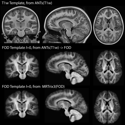 |
67 | Improved spatial normalization of white matter fiber orientation distributions using T1-weighted contrast
Jose M Guerrero-Gonzalez1,2, Olivia Surgent2,3, Nagesh Adluru2,4, Steven R Kecskemeti2, Gregory R Kirk2, Douglas C Dean III1,2,5, Brittany G Travers2,6, and Andrew L Alexander1,2,7
1Medical Physics, University of Wisconsin - Madison, Madison, WI, United States, 2Waisman Center, University of Wisconsin - Madison, Madison, WI, United States, 3Neuroscience Training Program, University of Wisconsin - Madison, Madison, WI, United States, 4Radiology, University of Wisconsin - Madison, Madison, WI, United States, 5Pediatrics, University of Wisconsin - Madison, Madison, WI, United States, 6Kinesiology Occupational Therapy Program, University of Wisconsin - Madison, Madison, WI, United States, 7Psychiatry, University of Wisconsin - Madison, Madison, WI, United States Fiber orientation distributions (FOD) derived from diffusion magnetic resonance imaging (dMRI) enable resolution of multiple fiber populations within a voxel. FOD-based white matter studies include voxel-based analysis, atlas-based labeling, and group average fiber tracking. These methods require spatial normalization of the FODs. This work describes an alternative approach for FOD spatial normalization based on co-registering individual dMRI to the T1-weighted (T1w) images, non-linear spatial normalization of the T1w images to a template, and applying the transformations to the FOD maps. This approach is compared to the conventional approach of directly aligning FOD maps. |
||
1272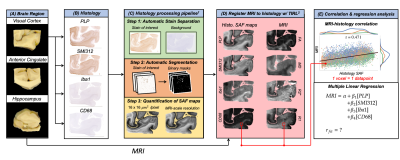 |
68 | Disentangling the contributions of myelin, neurofilament & microglia to MR contrast: an automated pipeline for voxelwise MR-histology analysis
Daniel Z.L. Kor1, Saad Jbabdi1, Jeroen Mollink1, Istvan N. Huszar1, Sean Foxley2, Menuka Pallebage-Gamarallage3, Connor Scott3, Adele Smart3, Olaf Ansorge3, Karla L. Miller1, and Amy F.D. Howard1
1Wellcome Centre for Integrative Neuroimaging, FMRIB, Nuffield Department of Clinical Neurosciences, University of Oxford, Oxford, United Kingdom, 2Department of Radiology, University of Chicago, Chicago, IL, United States, 3Neuropathology, Nuffield Department of Clinical Neurosciences, University of Oxford, Oxford, United Kingdom
Acquisition of MRI and histology in the same ex-vivo tissue sample enables direct correlation between MR and histologically-derived metrics. Here, we analysed immunohistochemistry images of human visual cortex, anterior cingulate and hippocampus to produce stained area fraction maps for myelin, neurofilaments and microglia. We performed voxelwise correlations between MR parameters (FA, MD, R2*, R1) and histology maps to generally characterise the strength of relationships. We then used partial correlation to identify the unique variance in MR parameters explained by each histological feature, and multiple linear regression to explore how well multiple microstructural properties can together explain MR parameters.
|
||
1273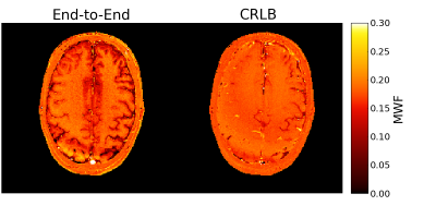 |
69 | End-to-End Scan Parameter Optimization for Improved Myelin Water Imaging
Steven T. Whitaker1, Jon-Fredrik Nielsen2, and Jeffrey A. Fessler1
1Electrical Engineering and Computer Science, University of Michigan, Ann Arbor, MI, United States, 2Biomedical Engineering, University of Michigan, Ann Arbor, MI, United States
Optimization techniques can be used to design scan parameters for quantitative imaging. The Cramér-Rao Lower Bound (CRLB) is often used for such designs, but it only characterizes unbiased estimators. We propose an end-to-end approach to scan design that optimizes scan parameters with a particular estimator in mind. We compare CRLB-based and end-to-end scan designs in the context of myelin water imaging. The end-to-end scan design results in lower estimation error in simulation and an in vivo myelin water fraction (MWF) map with improved contrast. The proposed end-to-end scan design approach is thus a promising alternative to using the CRLB.
|
||
 Back to Meeting Home
 Back to the Program-at-a-Glance
The International Society for Magnetic Resonance in Medicine is accredited by the Accreditation Council for Continuing Medical Education to provide continuing medical education for physicians.
 Back to Meeting Home
 Back to the Program-at-a-Glance
 View the Presentation