Digital Poster
Diffusion, Perfusion, Susceptibility & More I
Joint Annual Meeting ISMRM-ESMRMB & ISMRT 31st Annual Meeting • 07-12 May 2022 • London, UK

Digital Poster
Diffusion, Perfusion, Susceptibility & More I
| Computer # | ||||
|---|---|---|---|---|
1976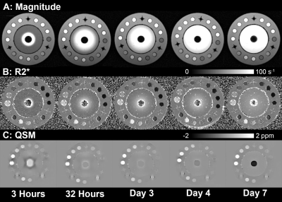 |
71 | Quantitative MRI to characterize diffusion-controlled release of gadolinium from a calcium sulphate matrix
Greg Hong1,2,3, Tina Khazaee1,2,3, Santiago F. Cobos1,2,3, Spencer D. Christiansen1,2, Junmin Liu2, Maria Drangova1,2,3, and David W. Holdsworth1,2,3
1Medical Biophysics, Western University, London, ON, Canada, 2Robarts Research Institute, London, ON, Canada, 3Bone & Joint Institute, London, ON, Canada
Calcium sulfate (CS) is commonly used to deliver antibiotics to treat periprosthetic joint infection, which is a leading cause of early revision. There is an unmet need for non-invasive measurement of antibiotic release from CS, which would improve understanding of antibiotic delivery in-vivo. Through the use of a gadolinium-based contrast agent as a surrogate, this study shows that quantitative MRI (R2* and QSM) can be used to track diffusion-controlled release. We demonstrate this in a phantom study consisting of a cylindrical CS core surrounded by agar, where gadolinium diffuses out of the core and through the agar sample.
|
||
1977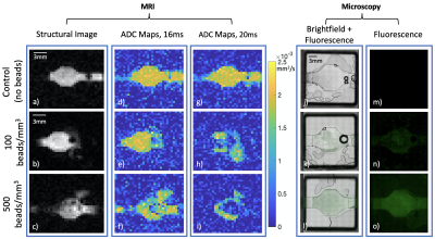 |
72 | Use of MRI- and microscopy-compatible bioreactor for validation of qMRI diffusion measurements via fluorescence microscopy
Megan F LaMonica1, Megan E Poorman2, David A Hormuth, II1, Thomas E Yankeelov1, and Kathryn E Keenan2
1Biomedical Engineering, UT Austin, Austin, TX, United States, 2NIST, Boulder, CO, United States
Quantitative magnetic resonance imaging (qMRI) could complement qualitative MRI analysis by providing quantitative diagnostic information standardized across patients. However, it is difficult to determine how microscopic tissue properties affect MR signal. A microscopy- and MRI-compatible bioreactor was filled with different concentrations of fluorescent beads in gel to explore this relationship. Higher bead count was associated with lower ADC. A 59% ADC decrease was observed at 100 beads/mm3 when varying gradient pulse separation from 16 to 20 ms, illustrating sensitivity to different length scales per the Einstein-Smoluchowski relationship. This work is a step towards validating qMRI with fluorescence microscopy.
|
||
1978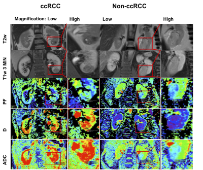 |
73 | Advanced diffusion-weighted MRI and proteomic analysis as potential predictors of renal tumor histopathology
Octavia Bane1,2, Jorge Daza3, Berengere Salome4, John Sfakianos3, Andrew Charap4, Kirolos Meilika3, Kennedy Okhawere3, Amir Horowitz4, Bachir Taouli1,2, Ketan Badani3, and Sara Lewis1,2
1Department of Radiology, Icahn School of Medicine at Mount Sinai, New York, NY, United States, 2BioMedical Engineering and Imaging Institute, Icahn School of Medicine at Mount Sinai, New York, NY, United States, 3Department of Urology, Icahn School of Medicine at Mount Sinai, New York, NY, United States, 4Division of Oncological Sciences, Icahn School of Medicine at Mount Sinai, New York, NY, United States
In this prospective, single-center study, we evaluated the use of intravoxel incoherent motion model of diffusion (IVIM-DWI) and high-throughput urine proteomics associated with inflammation for diagnosis and characterization of small renal masses. IVIM-DWI parameters had good to excellent diagnostic performance in identifying clear-cell renal cell carcinomas (ccRCC), as well as in distinguishing between stages of ccRCC. In a subset of patients with IVIM-DWI and proteomics, we observed significant correlations between IVIM and proteomic analytes which had differential expression between ccRCC and non-ccRCC, and are involved in promoting the recruitment, expansion and activation of innate and adaptive immune cells.
|
||
1979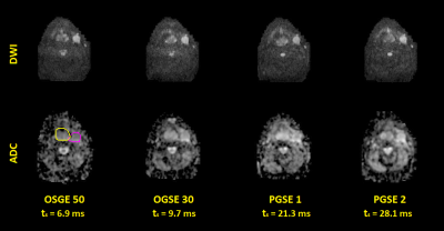 |
74 | Microstructure Imaging of Head and Neck Cancers using an Oscillating Diffusion Gradient Sequence and a Random Walk with Barriers Model
Siamak Nejad-Davarani1, Michelle Mierzwa1, Thorsten Feiweier2, Keith Casper3, and Yue Cao1,4
1Department of Radiation Oncology, University of Michigan, Ann Arbor, MI, United States, 2Siemens Healthcare GmbH, Erlangen, Germany, 3Department of Otolaryngology, University of Michigan, Ann Arbor, MI, United States, 4Department of Radiology, University of Michigan, Ann Arbor, MI, United States In vivo microstructure imaging based on diffusion weighted imaging can provide valuable information on tumor characteristics and response to cancer therapy. An oscillating diffusion gradient spin-echo sequence was used to acquire DW images with short diffusion times, along with those acquired by a pulsed diffusion gradient spin-echo sequence. Using a random walk with barriers model, microstructure/diffusion parameters in primary and nodal head and neck tumors were estimated prior to, and after two weeks of cancer treatment. Results show feasibility of this method to detect significant changes in these parameters, across the two imaging time points. |
||
1980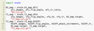 |
75 | Erwin - a quantitative MRI toolbox
Julien Lamy1, Mary Mondino1, and Paulo Loureiro de Sousa1
1ICube, Université de Strasbourg-CNRS, Strasbourg, France
Erwin is an open-source Python toolbox dedicated to the computation of quantitative maps from MRI data. It provides a unified interface, through either its Python API or its command-line tool, to well-known qMRI methods (e.g. B1 mapping using Actual Flip Angle (AFI) or T1 mapping using Variable Flip Angle (VFA)) and external toolboxes (e.g. MRtrix or MEDI). Erwin also provides tools to create complex pipelines where the dependencies between steps are tracked: a change in a source image or a method parameter will only trigger the recomputation of a minimal set of subsequent steps.
|
||
1981 |
76 | dtiRIM: A recurrent inference machine for diffusion tensor estimation
Emanoel R. Sabidussi1, Stefan Klein1, Ben Jeurissen2, and Dirk H. J. Poot1
1Radiology, Erasmus Medical Center, Rotterdam, Netherlands, 2Department of Physics, Imec-Vision Lab, University of Antwerp, Antwerp, Belgium In Diffusion MRI, the large variability of acquisition schemes limits broader use of Deep Learning for parameter estimation, with data-specific models needed for high-quality predictions. To reduce dependency on training data, we present dtiRIM, a recurrent neural network that learns a regularized solution to a model-based inverse problem. Using the diffusion model allows independent parameters (e.g. gradient directions) to also influence the estimation. We show that a single dtiRIM model predicts diffusion parameters for multiple datasets with lower error than the current state-of-the-art. Our study suggests that dtiRIM has the potential to be the first general learning method for DTI. |
||
1982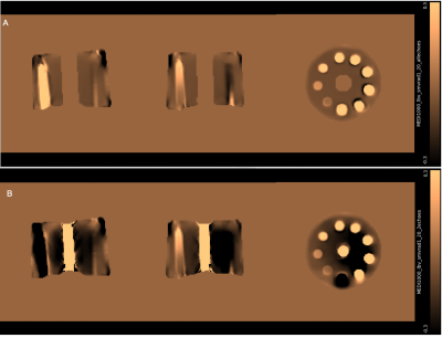 |
77 | Quantitative Susceptibility Mapping (QSM) Using High-resolution Ultra-Short Echo Time (UTE) MRI with Rosette k-space Pattern
Aparna Karnik1, Xin Shen2, Humberto Monsivais3, Antonia Sunjar2, Ali Caglar Özen4, Serhat Ilbey4, Mark Chiew5, Mukerrem Cakmak6, Joseph Rispoli1,2, and Uzay Emir3
1Elmore Family School of Electrical and Computer Engineering, Purdue University, West Lafayette, IN, United States, 2Weldon School of Biomedical Engineering, Purdue University, West Lafayette, IN, United States, 3School of Health Sciences, Purdue University, West Lafayette, IN, United States, 4Department of Radiology, Medical Physics, Medical Center – University of Freiburg, Faculty of Medicine, University of Freiburg, Freiburg, Germany, 5Wellcome Centre for Integrative Neuroimaging, University of Oxford, Oxford, United Kingdom, 6School of Materials Engineering, Purdue University, West Lafayette, IN, United States
We developed a method of acquiring high-resolution MRI for the generation of Quantitative Susceptibility Mapping (QSM) using Ultra-short echo time (UTE) MRI with a novel 3D rosette k-space trajectory. This method was used to generate high-resolution (0.94 mm3 isotropic) magnetic susceptibility maps in an Iron-Chloride phantom and the human brain. Generated maps were then compared with the susceptibility maps reconstructed from low-resolution data acquired using multi-echo UTE acquisition. Susceptibility values were comparable with current literature demonstrating the promise of this novel method in the generation of QSM.
|
||
1983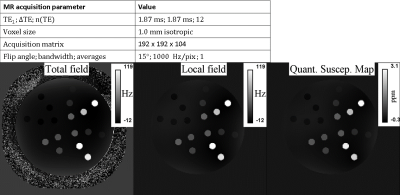 |
78 | Development of a Quantitative Susceptibility Mapping (QSM) phantom for validation of acquisition strategies and post-processing tools at 3 T
Padriac Hooper1,2, Monique Tourell1,2, Kieran O'Brien2,3, Jin Jin2,3, Simon Daniel Robinson1,4,5,6, and Markus Barth1,2,7
1Centre for Advanced Imaging, The University of Queensland, Brisbane, Australia, 2ARC Training Centre for Innovation in Biomedical Imaging Technology, Brisbane, Australia, 3Siemens Healthcare Pty Ltd, Brisbane, Australia, 4High Field MR Center, Department of Biomedical Imaging and Image-Guided Therapy, Medical University of Vienna, Vienna, Austria, 5Karl Landsteiner Institute for Clinical Molecular MR in Musculoskeletal Imaging, Vienna, Austria, 6Department of Neurology, Medical University of Graz, Graz, Austria, 7School of Information Technology and Electrical Engineering, The University of Queensland, Brisbane, Australia
The phantom was developed to cover the full range of physiological and pathological magnetic susceptibility seen in iron- and calcium-containing tissue, using 4 materials within the one phantom. The phantom facilitates quality assurance for acquisition strategies and post-processing tools.
|
||
1984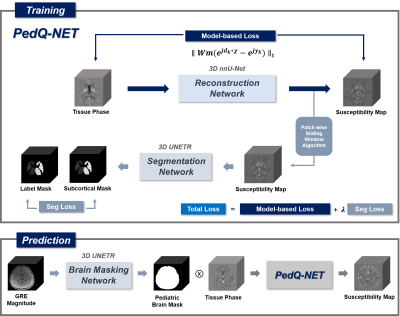 |
79 | PedQ-NET : Unsupervised Model-Based Joint-Loss Deep learning for Pediatric QSM
Siyun Jung1, Soohyun Jeon1, Yoonho Nam2, Hyun Gi Kim3, and Dong-Hyun Kim1
1Department of Electrical and Electronic Engineering, Yonsei University, Seoul, Korea, Republic of, 2Division of Biomedical Engineering, Hankuk University of Foreign Studies, Yongin, Korea, Republic of, 3Department of Radiology, Catholic University of Korea, Eunpyeong St. Mary's Hospital, Seoul, Korea, Republic of
In conventional QSM methods, it is difficult to analyze gray matter subcortical in pediatrics, because of small contrast difference between gray matter and white matter. In addition, there are no deep learning approaches that reflect the characteristics of the pediatric brain. In this study, we propose an unsupervised model-based joint-loss deep learning network for pediatric QSM, PedQ-NET. The proposed method achieved better edge preservation on the deep gray matter subcortical areas in pediatric QSM. According to the evaluation of in-vivo pediatric data, QSM generated by PedQ-NET shows enhanced edges in the subcortical areas and clear details with reduced artifacts.
|
||
1985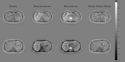 |
80 | Realistic in silico abdominal QSM phantom
Javier Silva1,2,3, Carlos Milovic4, Mathias Lambert1,2,3, Cristian Montalba2,3, Cristobal Arrieta1,2,3, Sergio Uribe2,3,5, and Cristian Tejos1,2,3
1Department of Electrical Engineering, Pontificia Universidad Catolica de Chile, Santiago, Chile, 2Biomedical Imaging Center, Pontificia Universidad Catolica de Chile, Santiago, Chile, 3Millennium Nucleus for Cardiovascular Magnetic Resonance, Santiago, Chile, 4Department of Medical Physics and Biomedical Engineering, University College London, London, United Kingdom, 5Department of Radiology, School of Medicine, Pontificia Universidad Catolica de Chile, Santiago, Chile
Compared to Quantitative Susceptibility Mapping (QSM) in the brain, abdominal QSM faces additional issues due to the presence of gas and fatty tissue. Recent works in abdominal QSM are more focused on its feasibility as a biomarker for disease diagnosis than improving or assessing the robustness and quality of the reconstructions. In this work, we present an abdominal QSM phantom with realistic tissue textures. Our flexible simulation pipeline allows emulating different stages of diseases and MRI signal contributions. Our reconstruction experiments show the potential of our phantom to compare QSM algorithms in different scenarios.
|
||
1986 |
81 | A CNN for Oxygen Extraction Fraction Mapping with combined QSM and qBOLD
Patrick Kinz1 and Lothar Schad1
1Computer Assisted Clinical Medicine, Medical Faculty Mannheim, Heidelberg University, Mannheim, Germany
MRI-based mapping of oxygen extraction fraction with QSM and qBOLD is a non-invasive diagnostic tool with many possible applications. But current reconstruction methods based on quasi-Newton (QN) methods are very dependent on accurate parameter initialization. Artificial Neural Networks showed a lot of potential in our previous works. Using a Convolutional Neural Network improves the reconstruction, since neighboring voxels can provide additional information. Using a GESFIDE sequence to sample the qBOLD signal instead of a standard mGRE that samples only the FID, improves the reconstruction accuracy of R2, Y and χnb a lot.
|
||
1987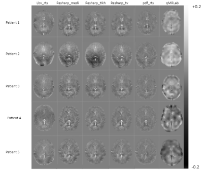 |
82 | Development of an image reconstruction algorithm to assess brain oxygenation in neonatal asphyxia using quantitative susceptibility mapping
Daniel Ridani1,2, Noée Ducros-Chabot1,2, Mathieu Dehaes2,3, and Benjamin De-Leener1,2,4
1NeuroPoly Lab, Institute of Biomedical Engineering, Polytechnique Montréal, Montreal, QC, Canada, 2Research Center, Ste-Justine Hospital University Centre, Montreal, QC, Canada, 3Department of Radiology, Radio-oncology, and Nuclear Medicine, Université de Montréal, Montreal, QC, Canada, 4Department of Computer and Software Engineering, Polytechnique Montréal, Montreal, QC, Canada
Neonatal asphyxia is a condition that causes lack of oxygen in the infant brain which lead to adverse neurodevelopmental outcomes. MRI provides a very clear image of the brain which lead to a detection of a variety of brain abnormalities. Quantitative susceptibility mapping (QSM) is a MRI tool to quantify brain oxygenation. In this study, we developed QSM reconstruction algorithms and tested them in data acquired in patients following neonatal asphyxia. Results include the comparison of five methods to generate QSM images in the brain specific segmented regions. Our preliminary data suggest that image intensity depends on the reconstruction method.
|
||
 Back to Meeting Home
 Back to the Program-at-a-Glance
The International Society for Magnetic Resonance in Medicine is accredited by the Accreditation Council for Continuing Medical Education to provide continuing medical education for physicians.
 Back to Meeting Home
 Back to the Program-at-a-Glance
 View the Presentation