Digital Poster
Diffusion, Perfusion, Susceptibility & More II
Joint Annual Meeting ISMRM-ESMRMB & ISMRT 31st Annual Meeting • 07-12 May 2022 • London, UK

Digital Poster
Diffusion, Perfusion, Susceptibility & More II
| Computer # | ||||
|---|---|---|---|---|
 |
2064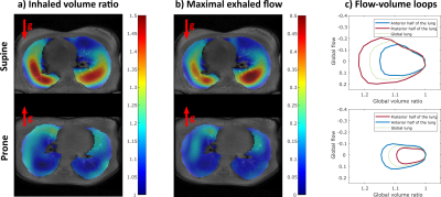 |
54 | 3D MR spirometry sensitivity to gravity lung dependence in healthy volunteers
Nathalie Barrau1, Claire Pellot Barakat1, Zhongzheng He1, Tolga Emre1, Angéline Nemeth1, Anass Afkir1, Wenpei Cai1, Vincent Brulon1, Tanguy Boucneau2, Brice Fernandez2, Florent Besson1, Vincent Lebon1, and Xavier Maître1
1Université Paris-Saclay, CEA, CNRS, Inserm, BioMaps, Orsay, France, 2General Electric Healthcare, Buc, France
Spirometry is a standard exam in pulmonary function testing for assessing the efficiency of global ventilation from flow-volume loops measured at the mouth during forced respiratory cycles. A newly developed technique, 3D MR spirometry, makes use of dynamic magnetic resonance imaging during spontaneous breathing to produce regional characterization of the lung function. The sensitivity of the technique was challenged with respect to gravity by extracting the associated ventilation gradients in prone and supine healthy volunteers.
|
|
2065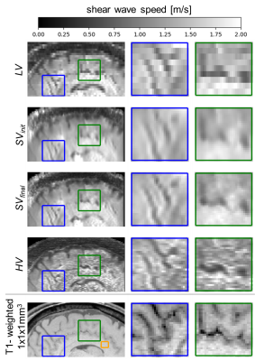 |
55 | 3D super-resolution MR elastography of the brain
Simone Hufnagel1, Matthias Anders2, Christoph Stefan Aigner1, Helge Herthum2, Heiko Tzschaetzsch2, Tobias Schaeffter1,3,4, Ingolf Sack2, and Christoph Kolbitsch1
1Physikalisch-Technische Bundesanstalt (PTB), Braunschweig and Berlin, Germany, 2Radiology, Charité - Universitätsmedizin Berlin, Berlin, Germany, 3School of Imaging Sciences and Biomedical Engineering, King's College London, London, United Kingdom, 4Department of Biomedical Engineering, Technical University of Berlin, Berlin, Germany MR elastography (MRE) provides valuable quantitative information about the mechanical properties of brain tissues. However, due to SNR limitations, often only low through-plane resolution is possible. We present super-resolution MRE based on multiple stacks of complex 3D wavefields of the brain resulting in elastograms with isotropic (1×1×1) mm3 resolution. The approach was evaluated for the in-vivo brain and showed improved visibility of fine structures while presenting consistent shear wave speed values. |
||
2066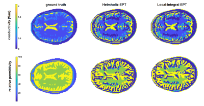 |
56 | Local Integral EPT - the alter ego of Helmholtz EPT
Luca Zilberti1, Alessandro Arduino1, Umberto Zanovello1, and Oriano Bottauscio1
1INRIM, Torino, Italy
A local-integral solution of the Electric Properties Tomography (EPT) problem is proposed. The method exploits a trick to consider the Helmholtz equation that regulates EPT as a Poisson equation. Then, it inverts the equation under the local homogeneity assumption, using Green's theorem. The approach turns out to be equivalent to the traditional Helmholtz-EPT.
|
||
2067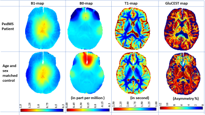 |
57 | GluCEST in Deep Gray Matter and Adjoining White Matter of pediatric-onset Multiple Sclerosis Brains using a Robust B1-calibration Approach
Dushyant Kumar1, Ritobrato Datta2, Micky K Bacchus2, Narayan Datt Soni1, Ravi Prakash Reddy Nanga1, Brenda Banwell 2, and Ravinder Reddy1
1Radiology, Center for Advanced Metabolic Imaging in Precision Medicine (CAMIPM), University of Pennsylvania, Philadelphia, PA, United States, 2Neurology, Children's Hospital of Philadelphia, Philadelphia, PA, United States
Several proton magnetic resonance spectroscopy (1H-MRS) based studies have reported regional alterations of glutamate metabolism in both acute and chronic multiple sclerosis (MS) pathologies, including in normal appearing white matter and mixed tissues. Despite its well-established capability for detecting altered glutamate metabolism, 1H-MRS is hampered by the low spatial resolution that precludes the measurements from small MS lesions. We demonstrate the feasibility of performing glutamate weighted imaging using chemical exchange saturation transfer with B0- and B1- corrections in pediatric-onset MS subjects. The proposed model for B1-calibration is a major improvement over the phenomenological method previously proposed by our group.
|
||
2068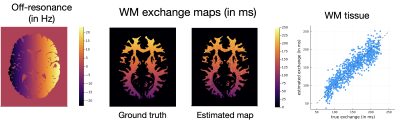 |
58 | Quantifying myelin water exchange using optimized bSSFP sequences
Naveen Murthy1, Jon-Fredrik Nielsen2, Steven T. Whitaker1, Melissa W. Haskell1,2, Scott D. Swanson3, Nicole Seiberlich3, and Jeffrey A. Fessler1
1Electrical Engineering and Computer Science, University of Michigan, Ann Arbor, MI, United States, 2fMRI Lab, University of Michigan, Ann Arbor, MI, United States, 3Radiology, University of Michigan, Ann Arbor, MI, United States
Mapping myelin water exchange rates could potentially be useful in helping us understand and characterize different diseases in the brain. It is a challenging quantitative parameter mapping problem to estimate multi-compartment exchange. This work focuses on scan design, with an aim of estimating exchange maps with good precision. In particular, a set of balanced steady-state free precession (bSSFP) sequences are optimized to estimate exchange rates in white matter (WM), for a two-pool model. Quantitative exchange maps are shown for a simulated phantom, using this optimized scan design.
|
||
2069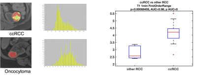 |
59 | MRI-based radiomics quantification for characterization of clear cell renal cell carcinoma.
Octavia Bane1,2, Christopher Kyriakakos1, Nicolas Gillingham1, Jordan Cuevas1,2, Kirolos Meilika3, Jorge Daza3, Amir Horowitz4, Bachir Taouli1,2, Ketan Badani3, and Sara Lewis1,2
1Department of Radiology, Icahn School of Medicine at Mount Sinai, New York, NY, United States, 2BioMedical Engineering and Imaging Institute, Icahn School of Medicine at Mount Sinai, New York, NY, United States, 3Department of Urology, Icahn School of Medicine at Mount Sinai, New York, NY, United States, 4Division of Oncological Sciences, Icahn School of Medicine at Mount Sinai, New York, NY, United States
In this prospective, single-center study, we evaluated the use of qualitative features and quantitative radiomics features from MRI for diagnosis and characterization of clear cell renal cell carcinoma (ccRCC). Qualitative features in isolation were of no value for diagnosing ccRCC, while the ccRCC likelihood score, which takes into account qualitative T2w signal and contrast enhancement pattern, had comparable diagnostic performance to the quantitative radiomics features. Quantitative radiomics features had good to excellent diagnostic performance in identifying ccRCC, as well as correlated to grade of ccRCC.
|
||
2070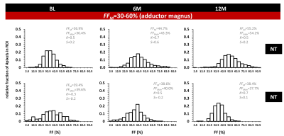 |
60 | Using MRI-based fat fraction histograms in dystrophic muscle to assess individual patient disease evolution
Harmen Reyngoudt1,2, Pierre-Yves Baudin1,2, Ericky Caldas de Almeida Araujo1,2, Brenda L Wong3,4, Pierre G Carlier5, and Benjamin Marty1,2
1NMR Laboratory, Neuromuscular Investigation Center, Institute of Myology, Paris, France, 2NMR Laboratory, CEA/DRF/IBFJ/MIRCen, Paris, France, 3Cincinnati Children’s Hospital Medical Center, Cincinnati, OH, United States, 4UMass Memorial Medical Center, Worcester, MA, United States, 5University Paris-Saclay, CEA, EA, DRF, Service Hospitalier Frédéric Joliot, Orsay, France
In most clinical quantitative MRI studies, using fat-water imaging, a mean fat fraction value per region of interest is generally used. The aim of this study was to look into the longitudinal changes in FF distribution on an individual patient basis, in dystrophic muscle, and at the same time, investigate whether differences were observed between treated and non-treated patients. As shown here, even if the mean fat fraction is the same between two patients or across time, there might be significant differences in the respective FF distributions, and might reveal, in retrospect, differential clinical or functional changes.
|
||
2071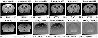 |
61 | Data-driven B1 non-uniformity correction in ultra-high-field small-animal macromolecular proton fraction and R1 mapping
Vasily L. Yarnykh1, Alena A. Kisel1, Andrey E. Akulov2, and Alexander Drobyshevsky3
1Radiology, University of Washington, Seattle, WA, United States, 2Institute of Cytology and Genetics, Siberian Branch of the Russian Academy of Sciences, Novosibirsk, Russian Federation, 3NorthShore University HealthSystem, Evanston, IL, United States
The data-driven surrogate B1 field correction algorithm was recently developed to eliminate B1-related spatial errors in the macromolecular proton fraction (MPF) and R1 maps obtained using the fast single-point method. The algorithm obviates the need for acquisition of actual B1 field maps, thus extending the routine use of fast MPF mapping. Initially, the surrogate B1 correction was applied to the human brain imaging at 3T. In this study, the method has been adopted and validated for small-animal imaging utilizing dedicated ultra-high-field MRI scanners. Particularly, we report guidelines on using surrogate B1 field correction in 9.4T and 11.7T magnetic fields.
|
||
2072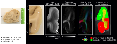 |
62 | Towards validation of in vivo U-fibre mapping using post-mortem DWI tractography of V1-V2 connections Video Permission Withheld
Fakhereh Movahedian Attar1, Evgeniya Kirilina 1,2, Christian Schneider1,3, Daniel Haenelt1, Luke J. Edwards1, Kerrin J. Pine1, Carsten Jäger1,4, Katja Reimann1, Andreas Pohlmann5, Joao Periquito5,6, Tobias Streubel7, Siawoosh Mohammadi1,7, Thoralf Niendorf5,8, Markus Morawski4, and Nikolaus Weiskopf1,9
1Department of Neurophysics, Max Planck Institute for Human Cognitive and Brain Sciences, Leipzig, Germany, 2Department of Education and Psychology, Center for Cognitive Neuroscience Berlin, Free University Berlin, Berlin, Germany, 3Faculty of Physics and Earth Sciences, Leipzig University, Leipzig, Germany, 4Paul Flechsig Institute of Brain Research, University of Leipzig, Leipzig, Germany, 5Berlin Ultrahigh Field Facility, Max Delbrück Center for Molecular Medicine in the Helmholtz Association, Berlin, Germany, 6Institute of Physiology, Charité- Universitätsmedizin Berlin, Berlin, Germany, 7Department of Systems Neuroscience, University Medical Center Hamburg-Eppendorf, Hamburg, Germany, 8Experimental and Clinical Research Center, a joint cooperation between the Charité Medical Faculty and the Max Delbrück Center for Molecular Medicine in the Helmholtz Association, Berlin, Germany, 9Felix Bloch Institute for Solid State Physics, Faculty of Physics and Earth Sciences, Leipzig University, Leipzig, Germany
U-fibres are the most abundant white matter fibres and yet are highly underrepresented in the MRI-derived human brain connectome. Development of validated in vivo pipelines for comprehensive mapping of these short association fibres is therefore essential. Here, we show the correspondence of the geometries of U-fibres connecting early visual cortices mapped in vivo using sub-millimetre resolution diffusion weighted imaging (DWI) tractography to those obtained in a post-mortem brain tissue sample using ultra-high resolution DWI tractography.
|
||
 Back to Meeting Home
 Back to the Program-at-a-Glance
The International Society for Magnetic Resonance in Medicine is accredited by the Accreditation Council for Continuing Medical Education to provide continuing medical education for physicians.
 Back to Meeting Home
 Back to the Program-at-a-Glance
 View the Presentation