Digital Poster
Modeling, Analysis & Evaluation I
Joint Annual Meeting ISMRM-ESMRMB & ISMRT 31st Annual Meeting • 07-12 May 2022 • London, UK

Digital Poster
Modeling, Analysis & Evaluation I
| Computer # | ||||
|---|---|---|---|---|
2693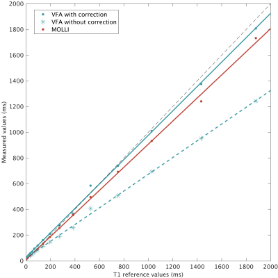 |
57 | A model for fat-suppressed variable flip angle T1-mapping and dynamic contrast enhanced MRI
Myrte Wennen1,2, Manon Moll3, Tim Marcus2, Joost Kuijer2, Leo Heunks1, Christina Lavini2, Gustav Strijkers2, Aart Nederveen2, and Oliver Gurney-Champion2
1Intensive Care, Amsterdam UMC, Amsterdam, Netherlands, 2Radiology and Nuclear Medicine, Amsterdam UMC, Amsterdam, Netherlands, 3University of Twente, Enschede, Netherlands
Although fat suppression can substantially improve image quality, the pulses corrupt the steady-state in a spoiled GRE sequence used for DCE and T1-relaxometry. Consequently, the conventional signal equations for quantifying tissue properties (T1, Ktrans, etc) do not hold. We have now included the effect of fat-suppression pulses in the signal equation that forms the basis for T1-relaxometry and DCE modelling. We have validated our equation with T1-mapping in a phantom and show its performance in healthy subjects and patients. Our equation enables accurate T1-mapping and pharmakinetic modelling for sequences that use fat suppression, including GRASP.
|
||
2694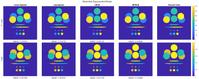 |
58 | Spin-Lock Times selection for Optimized T1ρ-Mapping of Knee Cartilage on Bi-Exponential and Stretched-Exponential Models
Hector Lise de Moura1, Marcelo Victor Wust Zibetti1, and Ravinder Regatte1
1Radiology, NYU Langone Health, New York, NY, United States T1ρ-Mapping requires acquisition with multiple Spin-Lock Times (TSLs) in order to fit the free parameters of the model. Reducing the #TSLs poses a trade-off between acquisition time and estimation error. Also, the choice of TSLs has a significant influence on the fitting results. A previous study analyzed the Cramér-Rao Lower Bound (CRLB) as an optimization criterium for the TSL choices using different #TSLs for Mono-Exponential models. This study extends the use of CRLB for Stretched- and Bi-exponential models using complex-valued fitting via Non-linear Least Squares. Experiments show that optimized TSL sequences improve parameter estimation when compared to non-optimized TSL sequences. |
||
2695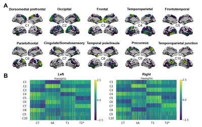 |
59 | Multivariate analysis of morphometric and quantitative magnetic resonance imaging metrics in aging and Alzheimer’s disease
Aurelie Bussy1,2, Raihaan Patel1,3, Alyssa Salaciak1, Sarah Farzin1, Stephanie Tullo1,2, Sandra Pelleieux4, Sylvia Villeneuve4,5, Judes Poirier4,5, John CS Breitner4,5, Gabriel A. Devenyi1,5, Christine L. Tardif3,6, and M. Mallar Chakravarty1,2,3,5
1Computational Brain Anatomy (CoBrA) Laboratory, Cerebral Imaging Centre, Douglas Mental Health University Institute, Montreal, QC, Canada, 2Integrated Program in Neuroscience, McGill University, Montreal, QC, Canada, 3Department of Biomedical Engineering, McGill University, Montreal, QC, Canada, 4Centre for the Studies on the Prevention of AD, Douglas Mental Health University Institute, Montreal, QC, Canada, 5Department of Psychiatry, McGill University, Montreal, QC, Canada, 6McConnell Brain Imaging Centre, Montreal Neurological Institute, McGill University, Montreal, QC, Canada
Morphometric and quantitative magnetic resonance imaging techniques have rarely been used simultaneously to characterise healthy aging and Alzheimer’s disease (AD) progression. Here, we are extracting four vertex-wise cortical metrics : cortical thickness, surface area, T1 value (myelin) and T2* values (iron). All these metrics were analysed using non-negative matrix factorization and linear models. Overall, cortical thinning seemed to be linked to both aging and AD progression, while decrease in myelin seemed to be a phenomenon mostly related to aging. No significant patterns of changes were seen in the prodromal phase of AD.
|
||
2696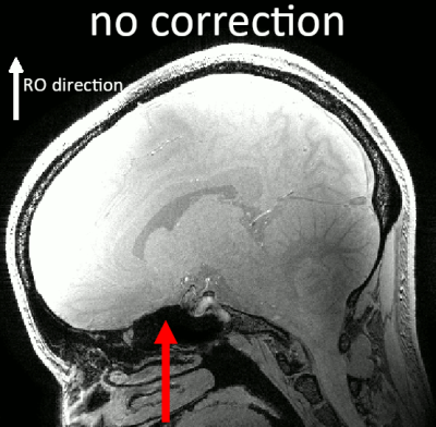 |
60 | Characterising the impact of distortion correction in high-resolution 3D GRE R2* mapping at 7T.
Barbara Dymerska1, Oliver Josephs1, and Martina Callaghan1
1Wellcome Centre for Human Neuroimaging, UCL Queen Square Institute of Neurology, University College London, London, United Kingdom
R2*-mapping delivers quantitative information about tissue microstructure. We investigate the scale of image distortions in high-resolution, bipolar, multi-echo GRE acquisitions at 7T and their effects on R2*-mapping. We study reduction in variance by applying field map-based distortion correction while also considering the confounding effect of local smoothing. We show that for regions with signal displacement > 0.5 voxels the reduction in variance comes largely, not from smoothing, but from the appropriate repositioning of the tissue signal. Therefore, correcting distortions, also in sub-voxel regime, improves data consistency in bipolar GRE acquisitions, facilitating more robust R2*-mapping in high-resolution laminar studies for instance.
|
||
2697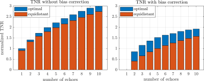 |
61 | Analysis on Echo Placement for Multi-echo Proton Resonance Frequency Shift Thermometry
Sven Nouwens1, Kemal Sumser2, Maarten Paulides2,3, Bram de Jager1, and Maurice Heemels1
1Mechanical Engineering, Control Systems Technology, Technical University Eindhoven, Eindhoven, Netherlands, 2Radiotherapy, Erasmus MC, Rotterdam, Netherlands, 3Electrical Engineering, Electromagnetics for Care & Cure, Technical University Eindhoven, Eindhoven, Netherlands
Proton resonance frequency shift based MR thermometry is widely used to non-invasively monitor thermal therapies in vivo. Multi-echo gradient-echo sequences are being studied to increase the effective temperature to noise ratio, however, choosing the optimal echo-times is non-trivial. We developed an optimization framework based on measurement noise models to compute the optimal echo placement for schemes that either correct or do not correct for the conductivity bias. In a 4-echo phantom experiment, we demonstrated a reduction in the noise standard deviation of 22% when comparing optimal to equidistant echo placements.
|
||
2698 |
62 | MAGiC in glioma: Are pre-contrast quantitative MRI parameters different in tumors with versus without enhancement?
Laura Nunez-Gonzalez1, Karin A. van Garderen1,2, Marion Smits1,2, Jaap Jaspers2, Alejandra Méndez Romero2, Dirk H. J. Poot1, and Juan A. Hernandez-Tamames1,3
1Erasmus MC, Rotterdam, Netherlands, 2Erasmus MC Cancer Institute, Rotterdam, Netherlands, 3TU Delft, Delft, Netherlands
We analyze quantitative values in glioma obtained with MAGiC, which allows obtaining quantitative T1, T2 and PD maps in one single acquisition of less than 6 minutes for a whole brain. The maps were obtained before contrast-agent injection in 14 patients with glioma. We investigated the possibility of characterizing tumor regions based on quantitative maps acquired without contrast-agent. The results showed significant differences among tumoral tissue, tissue with T1w-enhancement, and normal white matter. Voxel-wise, this allowed to distinguish tumoral tissue but did not allow to accurately predicting T1w-enhancement. However, promising results were found predicting T1w-enhancement inside the tumor.
|
||
2699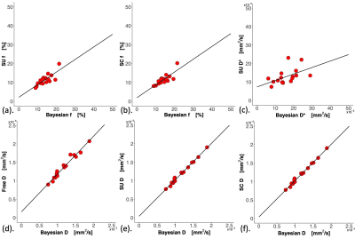 |
63 | Comparisons of Bayesian against non-Bayesian algorithms in intravoxel incoherent motion (IVIM) diffusion-weighted imaging in breast cancer
Sai Man Cheung1, Wing Shan Wu1, Nicholas Senn1, Ravi Sharma2, Trevor McGoldrick2, Tanja Gagliardi1,3, Ehab Husain4, Yazan Masannat5, and Jiabao He1
1Institute of Medical Sciences, University of Aberdeen, Aberdeen, United Kingdom, 2Oncology Department, Aberdeen Royal Infirmary, Aberdeen, United Kingdom, 3Radiology Department, Royal Marsden Hospital, London, United Kingdom, 4Pathology Department, Aberdeen Royal Infirmary, Aberdeen, United Kingdom, 5Breast Unit, Aberdeen Royal Infirmary, Aberdeen, United Kingdom
Intravoxel incoherent motion (IVIM) model approximates the tissue perfusion as a form of pseudo-diffusion, extracted as fast diffusion component in diffusion weighted imaging (DWI). Despite the central role of tissue perfusion in the angiogenesis in cancer, the clinical application of IVIM is hampered due to the susceptibility to noise and tendency of overfitting bi-exponential decay function. Recent introduction of Bayesian algorithm significantly enhanced the robustness and accuracy of IVIM method, renewing its potential as a clinical tool. We therefore set out to examine current IVIM algorithms in the context of neoadjuvant chemotherapy on patients with breast cancer preceding the treatment.
|
||
2700 |
64 | Performance Analysis of Undersampling Strategies for Multi-Shell Diffusion MRI
Elif Aygun1,2 and Emine Ulku Saritas1,2,3
1Department of Electrical and Electronics Engineering, Bilkent University, Ankara, Turkey, 2National Magnetic Resonance Research Center (UMRAM), Bilkent University, Ankara, Turkey, 3Neuroscience Graduate Program, Bilkent University, Ankara, Turkey
Multi-shell dMRI metrics quantify information on micro-properties of neural tissue and can be used as markers for neurological diseases. The protocols used to acquire dMRI data often have prolonged acquisition times. In this work, we propose different undersampling strategies that reduce the acquisition time to half, and evaluate how the performances of multi-shell dMRI metrics change under these strategies. The results show that, while the best performing strategy changes for each metric, 3-shell gradient schemes with small variance of b-vector density on consecutive shells demonstrate improved performance. Additionally, more complex dMRI metrics exhibit relatively increased sensitivity to undersampling.
|
||
2701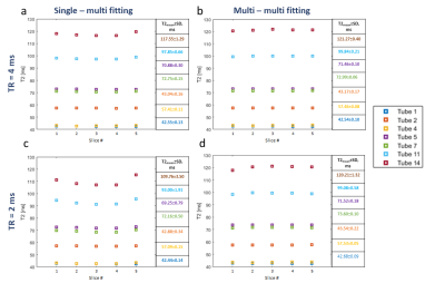 |
65 | T2 mapping for contiguous multi-slice 3D volume: A new EMC-based technique taking into account slice cross-talk effects Video Permission Withheld
Ekaterina A Brui1, Charles A de Mayenne 2, Stanislas Rapacchi 3, Thomas Troalen 4, Christophe Vilmen3, and David Bendahan1,3
1School of Physics and Engineering, ITMO University, Saint-Petersburg, Russian Federation, 2National Higher French Institute of Aeronautics and Space, Toulouse, France, 3Aix-Marseille Universite, CNRS, Centre de Résonance Magnétique Biologique et Médicale, Marseille, France, 4Siemens Healthcare SAS, Saint-Denis, France
Previously, an elegant method based on the generation of echo modulation curves (EMC) demonstrated a very high accuracy of T2 measurements. However, the original approach considered EMC dictionary modelling only for a single slice mode of multi-echo spin echo (MESE) sequence. For some applications, such as T2 mapping of cartilage in small joints, contiguous MRI is required. As a result, slice cross-talk effect may affect the accuracy of T2 estimation. In this work, we report a modified version of the EMC technique, which can be used to quantify T2 values for zero-gap multi-slice MESE acquisitions in tissues with long T1.
|
||
2702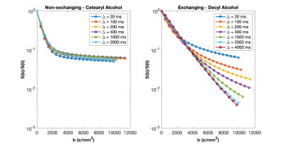 |
66 | Nanoscopic materials for quantitative water exchange phantoms
Scott D. Swanson1, Dariya I. Malyarenko1, and Thomas L. Chenevert1
1Radiology, University of Michigan, Ann Arbor, MI, United States
Nanoparticle lipid vesicles are developed with controllable water exchange to provide a ground-truth benchmark for MR methods which measure exchange. Diffusion data are collected as a function of diffusion delay Δ. An exchange time, τ, is measured to be 164 ms in a phantom with a highly fluid membrane. These materials should help to clarify water dynamics in complex systems.
|
||
2703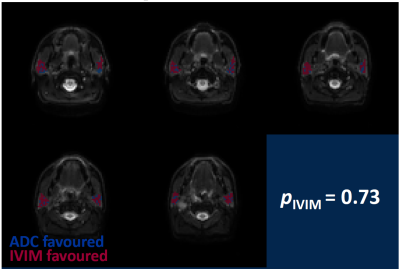 |
67 | Identifying perfused tissue regions through IVIM and ADC model comparison: technical validation
Damien J McHugh1,2, Michael J Dubec1,2, James Price2,3, Chris Moore1, David L Buckley1,4, James P. B. O'Connor2,5,6, David J Thomson3, and Andrew McPartlin3
1Christie Medical Physics and Engineering, The Christie NHS Foundation Trust, Manchester, United Kingdom, 2Division of Cancer Sciences, The University of Manchester, Manchester, United Kingdom, 3Department of Clinical Oncology, The Christie NHS Foundation Trust, Manchester, United Kingdom, 4Biomedical Imaging, University of Leeds, Leeds, United Kingdom, 5Department of Radiology, The Christie NHS Foundation Trust, Manchester, United Kingdom, 6Division of Radiotherapy and Imaging, The Institute of Cancer Research, London, United Kingdom Intravoxel incoherent motion (IVIM) can provide information about tissue perfusion in tumours and organs at risk, but the model may not be applicable in all voxels in a region of interest. Here, a model comparison framework is developed, based on comparing IVIM and apparent diffusion coefficient model fits, aiming to identify perfused voxels and quantify the proportion of tissue containing a perfusion component, pIVIM. Technical validation of the framework is performed, with simulations and phantom data showing that SNR and diffusion properties can bias pIVIM, while in vivo parotid gland data show that pIVIM exhibits good repeatability. |
||
2704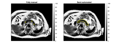 |
68 | Validation of semi-automated analysis of pancreas MRI-PDFF using a scan re-scan cohort from the Long COVID-19 study (COVERSCAN)
Alexandre Triay Bagur1, Michael Brady2, Arun Jandor2, Paul Aljabar2, and Daniel Bulte1
1Department of Engineering Science, University of Oxford, Oxford, United Kingdom, 2Perspectum Ltd, Oxford, United Kingdom
Impairment of the pancreas has been shown in initial findings of the Long COVID-19 study (COVERSCAN), that uses quantitative MRI and expert manual analysis of the pancreas. Quantitative MRI analysis in such large-scale studies benefits from the decision support made possible by advanced image analysis methods. In this work, we validate a semi-automated pancreas processing pipeline that involves manual slice selection and automated segmentation and MRI-PDFF quantification. We show good agreement of the semi-automatic method with the reference manual processing by experts, as well as repeatable quantification in a scan re-scan subset of healthy subjects within COVERSCAN (n=35).
|
||
 Back to Meeting Home
 Back to the Program-at-a-Glance
The International Society for Magnetic Resonance in Medicine is accredited by the Accreditation Council for Continuing Medical Education to provide continuing medical education for physicians.
 Back to Meeting Home
 Back to the Program-at-a-Glance
 View the Presentation