Digital Poster
Emerging Techniques in Data Acquisition & Analysis II
Joint Annual Meeting ISMRM-ESMRMB & ISMRT 31st Annual Meeting • 07-12 May 2022 • London, UK

Digital Poster
Emerging Techniques in Data Acquisition & Analysis II
| Computer # | ||||
|---|---|---|---|---|
0967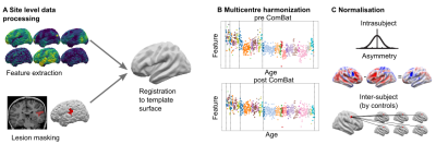 |
87 | Creating a multi-centre harmonised surface-based MRI dataset for the Multi-centre Epilepsy Lesion Detection Project
Mathilde Ripart1, Hannah Spitzer2, Russell Shinohara3, MELD Consortium4, Torsten Baldeweg1, Sophie Adler1, and Konrad Wagstyl5
1Great Ormond Street Institute for Child Health, UCL, London, United Kingdom, 2Institute of Computational Biology, Helmholtz Zentrum München, Munich, Germany, 3Penn Statistics in Imaging and Visualization Center, University of Pennsylvania, Philadelphia, PA, United States, 4UCL, London, United Kingdom, 5Wellcome Centre for Human Neuroimaging, UCL, London, United Kingdom
The Multi-centre Epilepsy Lesion Detection (MELD) project presents a methodology to harmonise a large heterogenous cohort of surface-based MRI data. Structural features were extracted from T1w and FLAIR images and pre-processed to reduce systematic site, scanner, and age-specific biases. The harmonised dataset enabled the characterisation of subtle radiological markers of focal cortical dysplasia (FCD), a cortical abnormality causing drug-resistant epilepsy. Machine-learning algorithms trained on the harmonised dataset improved the classification of FCD histopathologies. With open-source protocols and code, the MELD preprocessing pipeline offers a reproducible method to prepare large heterogeneous datasets for statistical analysis and machine-learning tasks.
|
||
0968 |
88 | Female patients with atopic dermatitis have reduced functional connectivity in the precuneus and inferior parietal lobe Video Permission Withheld
Tie-Qiang Li1, Klas Nordlind2, Elvar Theodorsson3, and Tomas Jonsson4
1Karolinska Institutet, Stockholm, Sweden, 2Division of Dermatology and Venereology, Karolinska Institutet, Stockholm, Sweden, 3Department of Biomedical and Clinical Science, Linköping University, Linköping, Sweden, 4CLINTEC, Karolinska Institutet, Stockholm, Sweden
A cohort of 32 atopic dermatitis (AD) patients and 32 matched healthy controls (HC) were recruited to study the neurological abnormalities which might be associated with this pruritic skin condition. The main findings are 1) reduced general functional connectivity in the precuneus, inferior parietal lobe and visual cortex was detected in females AD patients; 2) Severity of the symptoms was negatively correlated with functional connectivity in inferior parietal lobe, while salivary cortisol was positively associated. Resting-state fMRI can provide useful insight into the altered neurophysiology and clinically relevant assessment for the AD disorder.
|
||
0969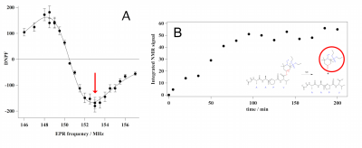 |
89 | Enzymatic activity monitoring through Dynamic Nuclear Polarization in Earth magnetic field
Dahmane Boudries1, Jean-Michel Franconi1, Sylvain R.A Marque2, Philippe Massot1, Philippe Mellet3, Elodie Parzy1, and Eric Thiaudiere1
1CNRS, Bordeaux, France, 2CNRS, Marseille, France, 3INSERM, Bordeaux, France
Earth-field MRI can provide new contrasts leading to the observation of pathologies at the biochemical level. However detection sensitivity is poor at low-field. In a preliminary spectroscopic approach, it is proposed here to detect protease-driven hydrolysis of a nitroxide probe thanks to electron-nucleus Overhauser enhancement in a homemade double resonance system. The nitroxide probe is a six-line nitroxide whose lines are shifted according to its substrate/product state. The Overhauser enhancement frequency dependence was in agreement with theoretical calculations. Enzymatic conversion of the nitroxide substrate was observed which opens the way for the design of new low-field DNP-MRI systems.
|
||
0970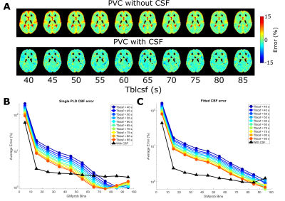 |
90 | The Effect of Label Crossing the Blood-CSF barrier on Partial Volume Correction: source of error or opportunity for quantification?
Leonie Petitclerc1,2,3, Lydiane Hirschler1,3, Iris Asllani4,5, and Matthias J.P. van Osch1,2,3
1C.J. Gorter Center for High Field MRI, Department of Radiology, Leiden University Medical Center, Leiden, Netherlands, 2Leiden Institute for Brain and Cognition (LIBC), Leiden, Netherlands, 3Department of Radiology, Leiden University Medical Center, Leiden, Netherlands, 4Clinical Imaging Science Center, University of Sussex, Sussex, United Kingdom, 5Biomedical Engineering, Rochester Institute of Technology, Rochester, NY, United States
Partial volume correction in ASL typically neglects CSF signal, however we have recently shown that some labeled signal crosses the blood-CSF barrier at a measurable level. Here we show the effect of including CSF in PVC, in simulated and in-vivo data. CSF-PVC reduced error on GM perfusion by ~10% in simulated data, with higher error when more CSF is present (including at longer LD and PLD). The difference between CSF-PVC and non-CSF-PVC GM signal in vivo was also ~10%. Comparing PVC signal in CSF to long-TE ASL signal suggests that BCSFB characterization may be possible without the acquisition of ultra-long-TE.
|
||
0971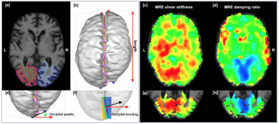 |
91 | The relationship between structural asymmetry and shear stiffness asymmetry in the human brain
Lily Xiang1, Lucy Victoria Hiscox2, Curtis L. Johnson3, and Neil Roberts1
1University of Edinburgh, Edinburgh, United Kingdom, 2University of Bath, Bath, United Kingdom, 3University of Delaware, Newark, DE, United States
On average the human brain has a global structural asymmetry comprising a counter-clockwise twist in the transverse plane and which is referred to as the cerebral torque. We have investigated whether there is a relationship between the magnitude of the torque and measures of brain shear stiffness obtained using Magnetic Resonance Elastography (MRE) in a group of Older Adults (AD) and a group of patients with Alzheimers DIsease (AD).
|
||
0972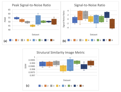 |
92 | Quantifiable study of magnetic resonance super resolution reconstruction in Placenta Accreta Spectrum using Image Quality Metrics
Joanna Chappell1, Nada Mufti1,2, Patrick O'Brien3, George Attilakos3, Magda Sokolska4, Priya Narayanan2, Rosalind Aughwane2,3, Giles Kendall2,3, David Atkinson5, Sebastien Ourselin 1, Anna L David2,3,6, and Andrew Melbourne1,4
1School of Biomedical Engineering and Imaging Sciences (BMEIS), Kings College London, London, United Kingdom, 2Elizabeth Garrett Anderson Institute for Women's Health, University College London, London, United Kingdom, 3Women's Health Division, University College London, London, United Kingdom, 4Depart. Medical Physics and Biomedical Engineering, University College London, London, United Kingdom, 5Centre for Medical Imaging, University College London, London, United Kingdom, 6WEISS (Wellcome /EPSRC Centre for Medical Imaging, University College London, London, United Kingdom
Magnetic Resonance Imaging is increasingly used for assessment of placental complications1. Here we apply MRI to examine Placenta Accreta Spectrum (PAS) a condition where the placenta is abnormally adherent or invasive. Super-Resolution Reconstruction (SRR) provides a high-resolution 3D reconstruction from 2D MRI slices to improve the visibility of structures for future clinical use2. Image Quality metrics allows quantitative evaluation of the SRR images to compare with the original 2D images. These metrics are found to be statistical significant, providing an objective assessment of the SRR images.
|
||
0973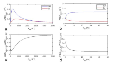 |
93 | NOE imaging in tumors using asymmetric analysis of 2pi-CEST signals
Jing Cui1,2 and Zhongliang Zu1,2
1Vanderbilt University Institute of Imaging Science, Nashville, TN, United States, 2Department of Radiology and Radiological Sciences, Vanderbilt University Medical Center, Nashville, TN, United States Nuclear overhauser enhancement (NOE) mediated chemical exchange saturation transfer (CEST) effect at -3.5 ppm has shown clinical interests in diagnosing tumors. Asymmetric analysis is a fast and simple method to measure and quantify the NOE effect, but has contamination from the downfield amide proton transfer (APT) effect at 3.5 ppm. In this abstract, we provide a new NOE quantification method using asymmetric analysis of CEST signals acquired with 2pi saturation pulses (2pi-CEST) which is also fast but has reduced contamination from the APT effect. |
||
0974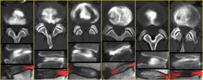 |
94 | Refined feature selection method for the depiction of pathology by texture analysis – Applicated on MR images of intervertebral discs fissures
Christian Waldenberg1,2, Stefanie Eriksson1,2, Hanna Hebelka1,2, Helena Brisby1,2, and Kerstin Lagerstrand1,2
1University of Gothenburg, Gothenburg, Sweden, 2Institute of Clinical Sciences, Sahlgrenska Academy, University of Gothenburg, Gothenburg, Sweden
Texture analysis combined with attention mapping has the potential to identify the position of pathology normally not visible in MR images by exploiting inter-pixel relationships in magnetic resonance (MR) images. However, as pathology can influence adjacent tissue, the features used to identify the position of the pathology have to be selected with care. Here we present an easily implemented method that efficiently selects texture features that are only sensitive to the pathology and can improve the localization of pathology even when not visible in MR images.
|
||
 Back to Meeting Home
 Back to the Program-at-a-Glance
The International Society for Magnetic Resonance in Medicine is accredited by the Accreditation Council for Continuing Medical Education to provide continuing medical education for physicians.
 Back to Meeting Home
 Back to the Program-at-a-Glance
 View the Presentation