Digital Poster
Spectroscopy I
Joint Annual Meeting ISMRM-ESMRMB & ISMRT 31st Annual Meeting • 07-12 May 2022 • London, UK

| Computer # | ||||
|---|---|---|---|---|
2533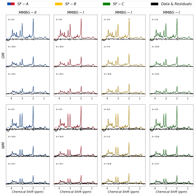 |
65 | Macromolecular background signals for gray and white matter regions in the human brain as defined from strong diffusion-weighted MR spectroscopy
Kadir Simsek1,2, André Döring3, André Pampel4, Harald E. Möller4, and Roland Kreis1,2
1Magnetic Resonance Methodology, Institute of Diagnostic and Interventional Neuroradiology, University of Bern, Bern, Switzerland, 2Translational Imaging Center, sitem-insel, Bern, Switzerland, 3Cardiff University Brain Research Imaging Centre (CUBRIC), School of Psychology, Cardiff University, Cardiff, United Kingdom, 4Max-Planck Institution for Human Cognitive and Brain Sciences, Leipzig, Germany
Diffusion-weighted MRS was successfully implemented at short TE, reaching ultra-high b values >20 ms/μm2 on a 3T Siemens Connectom system. With simultaneous fitting for different b-value spectra, macromolecular background patterns are estimated using different approaches to model metabolite diffusion and different macromolecular signal parameterization in gray matter and white matter.
|
||
2534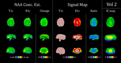 |
66 | B1+ Correction for 7T FID-CRT-MRSI
Philipp Lazen1, Bernhard Strasser1, Cornelius Cadrien1,2, Sukrit Sharma1, Lukas Hingerl1, Eva Niess1, Stanislav Motyka1, Alexandra Lipka1, Benjamin Spurny-Dworak3, Christoph Brandner4, Stephan Gruber1, Wolfgang Bogner1, Rupert Lanzenberger3, Karl Rössler2, Siegfried Trattnig1,5, and Gilbert Hangel1,2
1Department of Biomedical Imaging and Image-guided Therapy, Medical University of Vienna, Vienna, Austria, 2Department of Neurosurgery, Medical University of Vienna, Vienna, Austria, 3Department of Psychiatry and Psychotherapy, Medical University of Vienna, Vienna, Austria, 4Center for Medical Physics and Biomedical Engineering, Medical University of Vienna, Vienna, Austria, 5Institute for Clinical Molecular MRI, Karl Landsteiner Society, St. Poelten, Austria We implemented B1+ correction for our metabolite concentration estimation and analyzed the influence of B1+ inhomogeneities. Since the B1+ field is relatively homogeneous in the center of the brain anyways, the biggest effects were observed closer to the fringes of the brain. The concentration estimates increased between 1.0% and 5.6% for different metabolites when comparing B1+ correction to simple T1 correction, remaining within the spectrum of values established by previous literature. |
||
2535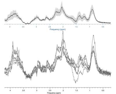 |
67 | Measuring the macromolecular baseline in the neonatal brain
Maria Yanez Lopez1, Georg Oeltzschner2, Anthony N Price1, Nicolaas AJ Puts3, Emer J Hughes1, Grainne M McAlonan3, Tomoki Arichi1, Richard AE Edden2, and Enrico De Vita4
1Centre for the Developing Brain, School of Biomedical Engineering and Imaging Sciences, King’s College London, London, United Kingdom, 2Russell H. Morgan Department of Radiology and Radiological Science, The Johns Hopkins University School of Medicine, Baltimore, MD, United States, 3Department of Forensic and Neurodevelopmental Sciences, Institute of Psychiatry, Psychology & Neuroscience, King's College London, London, United Kingdom, 4Biomedical Engineering Department, School of Biomedical Engineering and Imaging Sciences, King’s College London, London, UK, London, United Kingdom The doublet nature of the 3 ppm peak previously reported in neonates indicates that a lower macromolecular contribution to the GABA+ 3 ppm signal is likely to be present in this population. Detailed characterisation of age‐related MM contribution rates is required to improve the MRS fitting process, and therefore, to further increase the accuracy of metabolic measurements in neonates. As a first step, we here measure the macromolecular baseline using metabolite-nulling MRS in healthy term neonatal participants. |
||
2536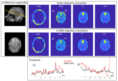 |
68 | Reducing Artifacts in Readout-Segmented Spectroscopic Imaging at 7T
Amir Seginer1, Graeme A. Keith2, David A. Porter2, and Rita Schmidt3
1Siemens Healthcare Ltd, Rosh Ha'ayin, Israel, 2Institute of Neuroscience and Psychology, University of Glasgow, Glasgow, Scotland, 3Department of Brain Sciences, Weizmann Institute of Science, Rehovot, Israel
Readout-Segmented COKE (RS-COKE) is an Echo Planar Spectroscopic Imaging (EPSI) variant supporting larger spectral-widths and is therefore useful for spectral imaging at 7T. However, mismatches between the segments at the overlaps lead to artifacts, especially if the lipids signal is not suppressed (to avoid adversely affecting the metabolites signal). We developed a procedure to measure and reduce the readout segment mismatches – through signal and trajectory corrections – leading to improved spectral images and spectra. The procedure was tested by scanning both a special 3D head-shaped phantom – which includes a lipid layer – and human volunteers.
|
||
2537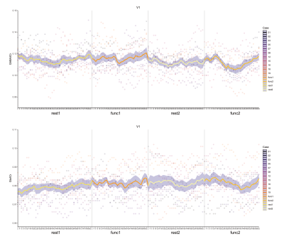 |
69 | GABA and Glutamate response to social processing; a functional MRS study
Duanghathai Pasanta1,2, David J. White3, Jason L. He1, Nicolaas A. Puts1,4, and Talitha Ford3,5
1Department of Forensic and Neurodevelopmental Sciences, Sackler Institute for Translational Neurodevelopment, Institute of Psychiatry, Psychology, and Neuroscience, King's College London, London, United Kingdom, 2Department of Radiologic Technology, Faculty of Associated Medical Sciences, Chiang Mai University, Chiang Mai, Thailand, 3Centre for Human Psychopharmacology & Swinburne Neuroimaging, School of Health Sciences, Swinburne University of Technology, Melbourne, Australia, 4MRC Centre for Neurodevelopmental Disorders, King’s College London, London, United Kingdom, 5Cognitive Neuroscience Unit, Faculty of Health, Deakin University, Geelong, Australia
We performed functional magnetic resonance spectroscopy (fMRS) to measure the dynamic response of GABA and Glutamate in the superior temporal sulcus (STS) and visual cortex (V1) while viewing social stimuli. MEGA-PRESS fMRS spectra were analyzed in both block and event-related design. Sliding window analyses were used to investigate GABA and Glutamate dynamics at higher temporal resolution. A small decrease in GABA level was observed during stimulus presentation in V1, but no change was observed in STS. We highlight the feasibility of using fMRS to assess changes in metabolite response during social processing in health and disease.
|
||
2538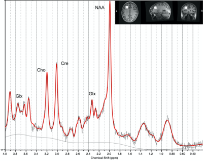 |
70 | Does Magnetic Resonance Spectroscopy Show Changes in Brain Metabolites At 3-Months Post Concussion in Pediatric Patients?
Robyn Walker1, Parker La2, Tiffany Bell 2, Julie M Joyce 2, Miriam Beauchamp3,4, William Craig5,6, Quynh Doan 7,8, Roger Zemek9,10, Keith Yeates 11, and Ashley Harris2
1University of Calgary, Calgary, AB, Canada, 2Radiology, University of Calgary, Calgary, AB, Canada, 3Psychology, University of Montreal, Montreal, QC, Canada, 4Psychology, Ste Justine Hospital, Montreal, QC, Canada, 5Pediatrics, University of Alberta, Edmonton, AB, Canada, 6Pediatrics, Stollery Children's Hospital, Edmonton, AB, Canada, 7Pediatrics, University of British Columbia, Vancouver, BC, Canada, 8Pediatrics, BC Children's Hospital, Vancouver, AB, Canada, 9Pediatrics and Emergency Medicine, University of Ottawa, Ottawa, ON, Canada, 10Pediatrics and Emergency Medicine, Children's Hospital of Eastern Ontario, Ottawa, ON, Canada, 11Psychology, University of Calgary, Calgary, AB, Canada
This study uses 1H magnetic resonance spectroscopy (MRS) to determine metabolite differences in the chronic phase (~3-month) of pediatric concussion compared to orthopedic injury (OI) controls. In the first analysis, significant differences in N-acetylaspartate (NAA), choline (Cho) and inositol (Ins) were seen between concussion and OI controls. Secondly, in a series of 3-way ANCOVAs across OI, symptomatic and asymptomatic concussion groups showed group differences in NAA, Cho and Ins depending on the symptom scale. When metabolites were different in this analysis by symptoms, it was generally driven by the asymptomatic group.
|
||
2539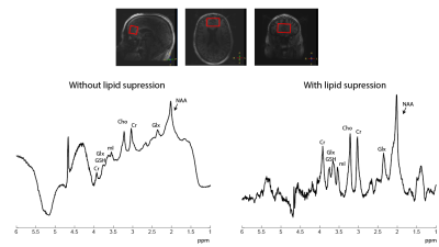 |
71 | Suppression of extracranial lipids in short TE STEAM of the human brain at 7T with an external crusher coil
Evita Wiegers1, Alex Bhogal1, Sarah Jacobs1, Mark Gosselink1, Jeanine Prompers1, and Dennis Klomp1
1Radiology, University Medical Center Utrecht, Utrecht, Netherlands
Short TE STEAM is an appealing MRS sequence especially for the quantification of low-concentration J-coupled spin systems. However, in comparison to sLASER, STEAM is associated with larger chemical shift displacement errors in all three directions. With an inhomogeneous B1+-field, slice profiles degrade, resulting in artefacts from extracranial lipids. Here we demonstrate that lipid artefacts can be removed through the use of an external crusher coil. Without use of the crusher coil, large extracranial lipid artefacts are present when selecting a large voxel. Upon activation of the crusher coil, these signals are eliminated, yielding 1H spectra free of large baseline distortions.
|
||
2540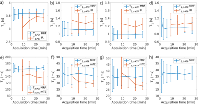 |
72 | Time-efficient and reproducible relaxation measurements by 31P-MR fingerprinting in human brain at 7T
Mark Stephan Widmaier1,2, Song-I Lim1,2, and Lijing Xin1,3
1CIBM Center for Biomedical Imaging, Lausanne, Switzerland, 2Laboratory for Functional and Metabolic Imaging, EPFL, Lausanne, Switzerland, 3Animal Imaging and Technology, EPFL, Lausanne, Switzerland In this abstract, we report the reproducibility of the new 31P-MRF technique on healthy volunteers. We show that relaxation and concentration rates can be estimated fast and accurate in good agreement with state-of-the-art methods in the human brain and in phantoms at 7T. This novel efficient technique is able to achieve a 5-fold acquisition time reduction of 31P relaxation parameter measurements, using only 6 min scan time. |
||
2541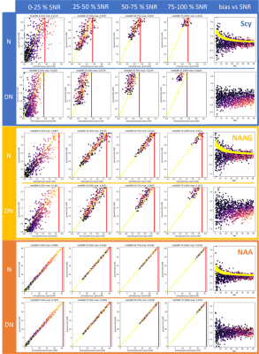 |
73 | Denoising MR spectra by deep learning: miracle or mirage?
Martyna Dziadosz1,2, Rudy Rizzo1,2, Sreenath P Kyathanahally3, and Roland Kreis1,2
1Magnetic Resonance Methodology, Institute of Diagnostic and Interventional Neuroradiology, University of Bern, Bern, Switzerland, 2Translational Imaging Center, sitem-insel, Bern, Switzerland, 3Department Systems Analysis, Integrated Assessment and Modelling, Data Science for Environmental Research group, Dübendorf, Switzerland
The main limitation of MRS is low SNR. Several approaches for denoising have been proposed. However, it is debatable, whether denoising can reduce estimate uncertainties. In this work, we investigate denoising using deep learning (DL) in time-frequency representations. The results were assessed using two methods: first, an adjusted noise score was used and second, the outcome of traditional fitting was evaluated. We found that time-frequency domain denoising through DL produces a visually appealing spectrum but mean residuals for the relevant spectral regions and variance in fit results remained as high as without denoising.
|
||
2542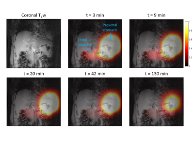 |
74 | Measurement of gastric emptying with dynamic 3D DMI using a deuterium body array at 7 T
Ayhan Gursan1, Arjan D. Hendriks1, Dimitri Welting1, Dennis W.J. Klomp1, and Jeanine J. Prompers1
1Department of Radiology, University Medical Center Utrecht, Utrecht, Netherlands
Gastric emptying abnormalities are frequently observed in diabetes patients. Here we explored whether dynamic 3D DMI could be a radiation-free alternative for scintigraphy to measure the gastric emptying rate. One volunteer was scanned after oral intake of deuterated glucose and proximal and distal gastric emptying was monitored. The distribution of the glucose load in the proximal and distal parts of the stomach and the rate of gastric emptying agreed well with scintigraphy results. DMI has potential to investigate gastric emptying abnormalities in patients with diabetes, while at the same time providing information on glucose uptake and metabolism in the liver.
|
||
2543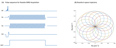 |
75 | Frequency and Phase Drift Correction of Rosette MRSI Data using Spectral Registration
Sneha Vaishali Senthil1,2, Brenden Toshihide Kadota 2, and Jamie Near2,3
1Integrated Program in Neuroscience, McGill University, Montreal, QC, Canada, 2Hurvitz Brain Sciences Program, Sunnybrook Research Institute, Toronto, ON, Canada, 3Medical Biophysics Department, Sunnybrook Research Institute, Toronto, ON, Canada
MRSI is a non-invasive in-vivo technique for mapping tissue concentrations in clinical and neuro-scientific research. Although, in vivo MRSI has made progress with respect to spatial resolution, acquisition time and the number of detectable metabolites; frequency and phase drifts in the acquired data are still a persisting problem, resulting in SNR losses, broadening of spectral peaks and deformities in line spectra of metabolites. In this abstract, we show how rosette MRSI sampling trajectories offer the possibility to perform frequency and phase drift correction which is otherwise not possible to do in most other commonly used cartesian and non-cartesian sampling trajectories.
|
||
2544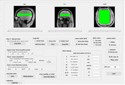 |
76 | Versatile Open source B0 shimtool
Mahrshi Jani1,2, Shengyue Su1, Manoj Kumar Sarma1, Ariane Fillmer3, and Anke Henning1,4
1Advance Imaging Research Center, University of Texas Southwestern Medical Center, Dallas, TX, United States, 2Department of Bioengineering, University of Texas at Arlington, Arlington, TX, United States, 3Medical Metrology, Physikalisch Technische Bundesanstalt, Berlin, Germany, 4Max Planck Institute for Biological Cybernetics, Tuebingen, Germany
In this work, we compare the spectroscopy results and metabolite maps for different shimming routine. We use vendor default shimming technique and our own shimming technique. We tried to do shimming in different regions of brain (i.e. prefrontal cortex, occipital, and insula), also we tried to do multivoxel shimming. We compared the frequency shift maps between vendor’s implemented shim routine and our own shim algorithm. Also, we compared metabolite maps we got from different shimmed region after shimming using vendors and our own shimming routine.
|
||
 Back to Meeting Home
 Back to the Program-at-a-Glance
The International Society for Magnetic Resonance in Medicine is accredited by the Accreditation Council for Continuing Medical Education to provide continuing medical education for physicians.
 Back to Meeting Home
 Back to the Program-at-a-Glance
 View the Presentation