Digital Poster
MRS, EPR & Hyperpolarization II
Joint Annual Meeting ISMRM-ESMRMB & ISMRT 31st Annual Meeting • 07-12 May 2022 • London, UK

| Computer # | ||||
|---|---|---|---|---|
1179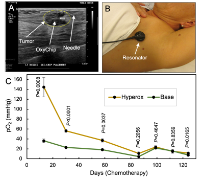 |
84 | Tumor oxygen levels and their response to hyperoxygenation in cancer patients measured using EPR oximetry with the OxyChip
Periannan Kuppusamy1, Benjamin B Williams2, Eunice Y Chen3, Maciej M Kmiec4, and Philip E Schaner2
1Radiology, Geisel School of Medicine at Dartmouth College, Lebanon, NH, United States, 2Medicine, Dartmouth-Hitchcock Medical Center, Lebanon, NH, United States, 3Dartmouth-Hitchcock Medical Center, Lebanon, NH, United States, 4Geisel School of Medicine at Dartmouth College, Lebanon, NH, United States
We report a first-in-humans clinical study of EPR oximetry using the OxyChip to establish its feasibility and utility for clinically useful tumor oxygen measurements in cancer patients. Repeated measurements from a cohort of 11 cancer patients in 33 sessions over a long period of time revealed variable levels of clinically significant hypoxia as well as variable responses to a hypoxia-mitigation intervention. Overall, in light of this variability, this study further underscores the need to provide individualized repeatable assessment of tumor oxygenation in the context of planned hyperoxygenation interventions to optimize clinical outcomes.
|
||
1180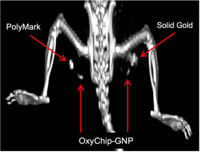 |
85 | OxyChip embedded with gold nanoparticles as a sensor for EPR oximetry and self-fiducial for anatomic registration
Maciej M Kmiec1, Kendra A Hebert1, Dan Tse1, Sassan Hodge1, Benjamin B Williams2, Philip E Schaner2, and Periannan Kuppusamy3
1Geisel School of Medicine at Dartmouth College, Lebanon, NH, United States, 2Medicine, Dartmouth-Hitchcock Medical Center, Lebanon, NH, United States, 3Radiology, Geisel School of Medicine at Dartmouth College, Lebanon, NH, United States
EPR oximetry using the OxyChip can provide direct and repeated measurement of tissue oxygen; however, a significant limitation is its inability to identify the location of the probe in the tissue. We report the development of an OxyChip embedded with gold nanoparticles to visualize the implant using clinical ultrasound or CT. In vitro characterization, imaging, and histopathology of the probe using tissue phantoms, excised tissues, and in vivo animal models demonstrated a substantial enhancement of CT contrast with OxyChip-GNP without compromising its oxygen-sensing properties or biocompatibility. The OxyChip-GNP can facilitate precisely localized in vivo oxygen measurements in the clinical setting.
|
||
1181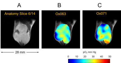 |
86 | Deuterated trityl spin probe for in vivo pO2 imaging with Time-Domain EPR
Nallathamby Devasahayam1, Shun Kishimoto1, Gadisetti Chandramouli2, Yasunori C Otowa1, Kota Yamashita1, Kazutoshi Yamamoto1, Jeffrey R Brender1, and Murali C Krishna1
1NCI, Bethesda, MD, United States, 2GenEpria Consulting, Inc, Columbia, MD, United States
In this work, we present our first 3D in vivo EPR oximetry study using Ox071 spin probe in comparison with Ox063 oximetry. The R2* change with [Ox071] and pO2 was calibrated using standard solutions. In vivo EPR imaging of a mouse tumor was performed on successive days by using either Ox071 or Ox063. The zero gradient signal level of Ox071 was about twice stronger and lasted almost twice longer compared to Ox063. The spin density, and pO2 maps and pO2 histograms of tumor regions marked by co-registration with MRI were similar between Ox063 and Ox071.
|
||
1182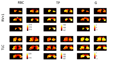 |
87 | 129Xe chemical shift mapping in the lungs with 3D density-weighted MRSI
Graham Norquay1, Guilhem J. Collier1, Rolf F. Schulte2, and Jim M. Wild1
1Infection, Immunity and Cardiovascular Disease, University of Sheffield, Sheffield, United Kingdom, 2GE Healthcare, Munich, Germany
3D density-weighted MRSI was used to regionally measure the 129Xe chemical shift from xenon in the lung airspaces (G), lung tissue/plasma (TP) and pulmonary red blood cells (RBC) at three lung inflation states. The 129Xe-RBC and 129Xe-G chemical shifts were both found to increase with increasing lung inflation (increase in alveolar pO2) while the 129Xe-TP shift was observed to be lung-inflation independent. The RBC chemical shift maps presented here may be used in patient populations to detect areas of low blood oxygenation in diseases presenting regional hypoxia in the lungs and other well-perfused organs such as the brain and kidneys.
|
||
1183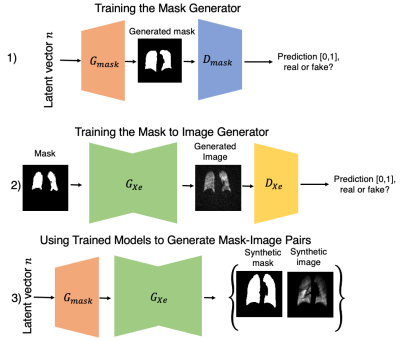 |
88 | A general framework of synthesizing 129Xe MRI data for improved segmentation model training
Junlan Lu1, Jesse Zhang2, Suphachart Leewiwatwong3, Isabelle Dummer4, David Mummy5, and Bastiaan Driehuys3
1Medical Physics, Duke University, Durham, NC, United States, 2Mathematics, Duke University, Durham, NC, United States, 3Biomedical Engineering, Duke University, Durham, NC, United States, 4Biomedical Engineering, McGill University, Montreal, QC, Canada, 5Radiology, Duke University, Durham, NC, United States
Quantitative analysis of hyperpolarized 129Xe MRI, segmentation of the thoracic cavity, a crucial step that is often the bottleneck in an otherwise fully automated pipeline. This problem is attractive to solve using deep learning methods, but they are limited by their large appetite for manually segmented training data. To this end, we propose a method to automatically synthesize both 129Xe ventilation MR images and their corresponding thoracic cavity masks using general adversarial networks. This data augmentation technique can accelerate the training of deep learning segmentation models.
|
||
1184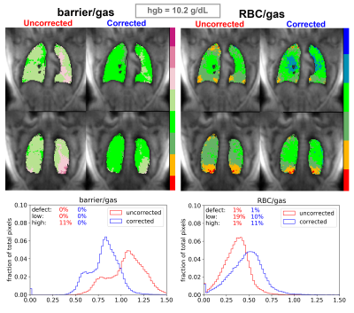 |
89 | Establishing a hemoglobin correction for 129Xe gas exchange MRI
Aryil Liam Bechtel1, Elianna Bier2, Junlan Lu3, Alexander Church4, Jennifer Korzekwinski4, David Mummy5, and Bastiaan Driehuys5
1Physics, Duke University, Durham, NC, United States, 2Biomedical Engineering, Duke University, Durham, NC, United States, 3Radiation Oncology, Clinical Science, Duke University, Durham, NC, United States, 4Duke University, Durham, NC, United States, 5Radiology, Clinical Science, Duke University, Durham, NC, United States Hyperpolarized 129Xe gas MRI provides a spatially resolved method of monitoring gas exchange function in the lungs via 3D reconstruction of RBC/gas and barrier/gas signals. However, the strength of these signals is affected by patient hemoglobin concentration (Hgb). Thus, correcting for Hgb is important for establishing normative healthy reference distributions and accurately assessing the degree of gas exchange impairment. Here, we use a 1D physical diffusion model to establish Hgb-dependent correction factors to standardize gas exchange MRI relative to a fixed Hgb value. This correction can result in substantial changes in the visualization and quantification of gas exchange MRI. |
||
1185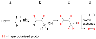 |
90 | Parahydrogen-Induced Polarization Relayed via Proton Exchange: Application to biological molecules.
Kolja Them1 and Jan Hövener1
1Section Biomedical Imaging, Molecular Imaging North Competence Center (MOIN CC), Department of Radiology and Neuroradiology, University Medical Center Schleswig-Holstein and Kiel University, Kiel, Germany Parahydrogen-induced polarization relayed via proton exchange (PHIP-X) is a new method to hyperpolarize a variety of molecules that participate in suitable fast proton exchange with allyl alcohol. The combination of the wide applicability of polarization-transfer via proton exchange and the strong polarization obtained using hydrogenative of PHIP opens up new avenues to hyperpolarize biological molecules such as glucose, lactate and pyruvate. Hence, PHIP-X provides a promisingnew platform for potential applications in metabolic MRI. |
||
1186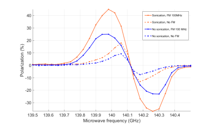 |
91 | Hyperpolarization of 129Xe gas via dissolution DNP: beneficial tips and tricks
Emma Linnea Wiström1, Andrea Capozzi1, Thanh Phong Lê1, Rolf Gruetter1, and Jean-Noël Hyacinthe2
1LIFMET, École polytechnique fédérale de Lausanne, Lausanne, Switzerland, 2Geneva School of Health Sciences (HES-SO), University of Applied Sciences and Arts Western Switzerland, Geneva, Switzerland Dissolution DNP (dDNP) is an alternative for hyperpolarizing 129Xe to the well acknowledged method spin exchange optical pumping (SEOP). As opposed to SEOP, DNP takes place in the solid state at very low temperatures. Therefore, it can potentially produce a large volume of hyperpolarized gas after dissolution. In this study, by implementing a new way of preparing the sample, we ease the overall process and establish a superior incorporation of the gas atoms into the solvent. Additionally, due to a surprisingly short radical electron T1, we show that a higher polarization was achieved by applying microwave frequency modulation. |
||
1187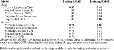 |
92 | Respiratory System Resistance Explained using Hyperpolarized 129Xe MRI Texture Features and Machine Learning
Marrissa J McIntosh1,2, Maksym Sharma1,2, Alexander M Matheson1,2, Harkiran K Kooner1,2, Rachel L Eddy3, Christopher Licskai4, David G McCormack4, Michael Nicholson4, Cory Yamashita4, and Grace Parraga1,2,4,5
1Department of Medical Biophysics, Western University, London, ON, Canada, 2Robarts Research Institute, London, ON, Canada, 3Centre for Heart Lung Innovation, University of British Columbia, Vancouver, BC, Canada, 4Division of Respirology, Department of Medicine, Western University, London, ON, Canada, 5School of Biomedical Engineering, Western University, London, ON, Canada
129Xe MRI ventilation images consist of embedded texture features that may help explain ventilation heterogeneity. We previously showed that 129Xe MRI ventilation features predicted response to biologic therapy in asthma and thus, we postulated that texture features may help explain central and peripheral airways resistance. We employed machine-learning techniques to identify specific 129Xe MRI features that were related to airway resistance. Ventilation texture analysis yielded four unique and two common features that independently explained central and peripheral airways resistance, respectively. These promising results suggest that 129Xe ventilation texture analysis may reveal hidden anatomic-physiologic measurements that lead to ventilation heterogeneity.
|
||
1188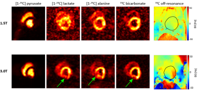 |
93 | Hyperpolarized 13C imaging at moderate field strengths: SNR considerations and in-vivo feasibility at 1.5T
Maximilian Fuetterer1, Julia Traechtler1, Andreas Dounas1, Tobias Hoh1, Mohammed Albannay1, and Sebastian Kozerke1
1Institute for Biomedical Engineering, University and ETH Zurich, Zürich ETH-Zentrum, Switzerland
Motivated by the fact that the hyperpolarized MRI signal does not quadratically scale with field strength as for conventional MRI at thermal equilibrium, the achievable SNR at reduced field strengths is explored. It is demonstrated that for larger coil diameters, sample noise dominates electrical noise at 1.5T and achievable SNR becomes comparable to 3T. By adaptively reducing acquisition bandwidth to the reduced spectral span and given longer T2* decay, effective SNR at 1.5T exceeds 3T. Feasibility is demonstrated with the first in-vivo data acquired on a clinical 1.5T system using hyperpolarized 13C pyruvate.
|
||
1189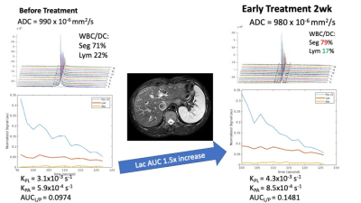 |
94 | Hyperpolarized [1-13C]Pyruvate MRI of Spleen: Monitoring Immune Activation for Cervical Cancer Patient following Radiotherapy Video Permission Withheld
Gigin Lin1, Ying-Chieh Lai1, Kuan-Ying Lu1, Albert P Chen2, and Ching-Yi Hsieh3
1Medical Imaging and Intervention, Chang Gung Memorial Hospital, Linkou, Taiwan, 2GE Healthcare, Toronto, ON, Canada, 3Medical Imaging Research Center, Institute for Radiological Research, Chang Gung University, Taoyuan, Taiwan
Successful cancer therapy depends on an optimal immune response, but noninvasive tools are lacking to monitor this dynamic process. In this proof-of-concept human study, we demonstrated hyperpolarized [1-13C]pyruvate MRI of the spleen monitoring the immune activation for cancer patients at the early time point following radiotherapy, by measuring the pyruvate-to-lactate conversion rate. The ADC-based cellularity of the spleen, however, did not detect remarkable changes.
|
||
1190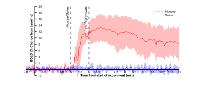 |
95 | Simultaneous fMRI and metabolic MRS of hyperpolarized [1-13C]pyruvate during nicotine stimulus in rat
Viivi Hyppönen1, Jessica Rosa1, and Mikko Kettunen1
1A. I. Virtanen Institute, University of Eastern Finland, Kuopio, Finland
fMRI is widely used to study cerebral metabolism to stimulus through combination of BOLD and hemodynamic changes. Hyperpolarized carbon can also be used to study cerebral metabolism, but it is currently poorly understood. Here we performed simultaneous 1H fMRI and 13C MRS experiments to compare their response to nicotine stimulus. Cortical BOLD signal increase was accompanied by increased bicarbonate labelling. Combined 1H fMRI and 13C MRS may therefore offer complementary information on brain response to stimulus.
|
||
1191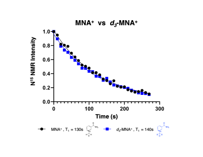 |
96 | 15N-enriched NAD+/ NADH Analogs as Redox-Responsive MRI Sensors
Emily Buchanan1,2,3, James Ratnakar1, Eul Hyun Suh1, Jun Chen1, Ivan Dimitrov1,4, Jae Mo Park1, A. Dean Sherry1,2, and Zoltan Kovacs1
1Advanced Imaging Research Center, UT Southwestern Medical Center, Dallas, TX, United States, 2Chemistry and Biochemistry, The University of Texas at Dallas, Richardson, TX, United States, 3Physical Measurement Laboratory, National Institute of Standards and Technology, Boulder, CO, United States, 4Phillips Medical Systems, Cleveland, OH, United States In this study, we prepared a 15N-enriched analog of NAD+ (N-methyl nicotinamide = MNA+) and demonstrated that it undergoes reduction by sodium dithionate to form MNAH. The 15N chemical shifts of the oxidized and reduced forms differ by 124.2ppm. DNP of MNA+ followed by dissolution and 15N NMR showed a favorable T1 relaxation time of 130s at 1T and 50s at 3T. Deuteration of the methyl protons only increased the T1 of 15N by ~10s. The long T1 of 15N in these NAD+/NADH mimetics and the large chemical shift difference offer the exciting potential for their use as redox sensors. |
||
1192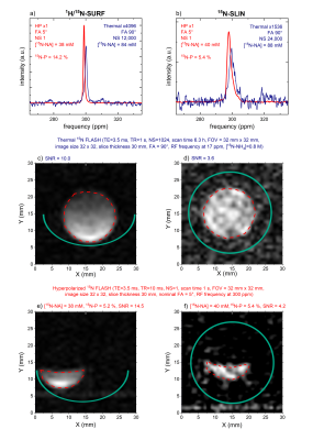 |
97 | Towards 15N molecular MRI with 15N-nicotinamide hyperpolarized by dissolution dynamic nuclear polarization (dDNP)
Josh Peters1, Arne Brahms2, Vivian Janicaud1,3, Mariia Anikeeva1, Eva Peschke1, Frowin Ellermann1, Arianna Ferrari1, Konrad Aden4, Stefan Schreiber4, Rainer Herges2, Jan-Bernd Hövener1, and Andrey N. Pravdivtsev1
1SBMI, MOIN CC, UKSH, Kiel University, Kiel, Germany, 2Otto Diels Institute for Organic Chemistry, Kiel University, Kiel, Germany, 3Universität zu Lübeck, Lübeck, Germany, 4Institute of Clinical Molecular Biology, UKSH, Kiel University, Kiel, Germany
We synthesized 1-15N nicotinamide (NA) and built a saddle-shaped mouse body linear coil for 15N imaging. We found a way to polarize 1-15N-NA by dissolution dynamic nuclear polarization to 9.1±4.4% and measured the effect of the radical load. The lifetime of hyperpolarized NA was field-dependent: 100 s at 1 T, 60 s at 7 T, and 30 s at 9.4 T. An effect of the pH on liquid-state polarization was found. N FLASH MRI of hyperpolarized NA was acquired in less than 1 s.
|
||
 Back to Meeting Home
 Back to the Program-at-a-Glance
The International Society for Magnetic Resonance in Medicine is accredited by the Accreditation Council for Continuing Medical Education to provide continuing medical education for physicians.
 Back to Meeting Home
 Back to the Program-at-a-Glance
 View the Presentation