Digital Poster
MRS & Hyperpolarization II
Joint Annual Meeting ISMRM-ESMRMB & ISMRT 31st Annual Meeting • 07-12 May 2022 • London, UK

| Computer # | ||||
|---|---|---|---|---|
1883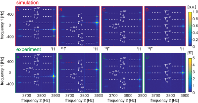 |
63 | Hyperpolarized multiple quantum coherences at ultra-low magnetic fields increase 15N parahydrogen-induced polarization
Andrey N. Pravdivtsev1, Nicolas Kempf2, Markus Plaumann3, Johannes Bernarding3, Klaus Scheffler2, Jan-Bernd Hövener1, and Kai Buckenmaier2
1SBMI, MOIN CC, UKSH, Kiel University, Kiel, Germany, 2High-Field Magnetic Resonance Center, Max Planck Institute for Biological Cybernetics, Tübingen, Germany, 3Institute for Biometrics and Medical Informatics, Otto-von-Guericke University, Magdeburg, Germany
We used signal amplification by reversible exchange of parahydrogen (SABRE) at low (~1 mT) and ultra-low (~1 μT, ULF) magnetic fields. We proposed and used ULF correlation spectroscopy (COSY) method to analyze PHIP spin order in real-time. Coherences up to the third-order were observed experimentally. Furthermore, we analyzed SABRE in alternating magnetic fields (alt-SABRE). We measured the evolution of 1H-15N zero-quantum coherences and have shown that they persist during field alternation and depend on the magnetic field strength. The resulting 15N-polarization in the alt-SABRE experiment was with magnetic was appoximately 30% higher.
|
||
1884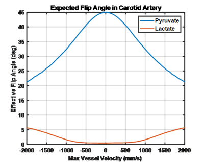 |
64 | Flow Effects on Spatial Spectral Excitation of Hyperpolarized Agents
Christopher Michael Walker1, Keith Michel1, Collin J Harlan1, Zhan Xu1, Gary Martinez1, Dawid Schellingerhout2, Stephen Y. Lai3, and James A. Bankson1
1Imaging Physics, MD Anderson Cancer Center, Houston, TX, United States, 2Neuroradiology Department, MD Anderson Cancer Center, Houston, TX, United States, 3Head & Neck Surgery, MD Anderson Cancer Center, Houston, TX, United States
Simulations were used to evaluate the effect of rapid vascular flow on spatial spectral pulses used for hyperpolarized MRI. Simulation results show that flow effects reduced the on-resonance excitation angle while increasing the off-resonance excitation in a velocity dependent manner. These excitation impacts will be particularly important when trying to estimate an arterial input function. Flow may also produce measurable lactate signal in flowing pyruvate, an effect that was observed in a hyperpolarized study of the thyroid.
|
||
1885 |
65 | Preliminary Assessment of Gas Exchange Efficiency in Lung Transplant Patients with Multi-breath Xenon Polarization Transfer Contrast Imaging
Faraz Amzajerdian1, Hooman Hamedani1, Ryan Baron1, Yi Xin1, Tahmina Achekzai1, Luis Loza1, Mostafa Ismail1, Ian Duncan1, Stephen Kadlecek1, Kai Ruppert1, and Rahim Rizi1
1University of Pennsylvania, Philadelphia, PA, United States
Quantifying the exchange of hyperpolarized xenon gas between the airways, lung parenchyma, and red blood cells may provide valuable insights into the progression of lung graft failure, enabling earlier diagnosis of chronic lung allograft dysfunction (CLAD). By combining Xenon polarization Transfer Contrast (XTC) imaging with a multi-breath model of fractional ventilation, gas exchange efficiency was assessed in four lung transplant recipients 3-12 months post-surgery in order to identify baseline metrics and trends.
|
||
1886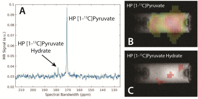 |
66 | Advancing SABRE Toward In Vivo Imaging: Phantom Chemical Conversion of Hyperpolarized [1-13C]-Pyruvate
Patrick Michael TomHon1, Keilian MacCulloch1, David Orestes Guarin2, Erin Hardy2, Austin Browning1, Carlos Dedesma3, Matthew S Rosen2, Yi-Fen Yen2, and Thomas Theis4
1Chemistry, North Carolina State University, Raleigh, NC, United States, 2Athinoula A. Martinos Center for Biomedical Imaging, Massachusetts General Hospital, Charlestown, MA, United States, 3Vizma Life Sciences, Chapel Hill, NC, United States, 4Chemistry, Physics, Biomedical Engineering, North Carolina State University, Raleigh, NC, United States
This work illustrates current advancements in SABRE hyperpolarized pyruvate toward in vivo imaging, demonstrating that significant levels of polarization achieved on pyruvate enable dynamic spectroscopy and imaging of downstream chemical products. Specifically in this study we show the monitoring of chemical conversion of pyruvate to pyruvate hydrate using a pH change and pyruvate to CO2 by reaction with H2O2. While the current work only focuses on the study of the chemical conversion of hyperpolarized substrate in a phantom, these studies lay the groundwork for future in vivo injections that mirror these chemical conversion processes.
|
||
1887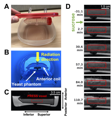 |
67 | Dynamic 1H MR spectroscopy of yeast metabolism during gamma irradiation using a 1.5T MR-Linac
Liam S. P. Lawrence1,2, Viktor Iakovenko3, Wendy Oakden1, Rachel W. Chan1, Greg J. Stanisz1,2,4, and Angus Z. Lau1,2
1Physical Sciences Platform, Sunnybrook Research Institute, Toronto, ON, Canada, 2Department of Medical Biophysics, University of Toronto, Toronto, ON, Canada, 3Department of Medical Physics, Sunnybrook Health Sciences Centre, Toronto, ON, Canada, 4Department of Neurosurgery and Paediatric Neurosurgery, Medical University, Lublin, Poland
1H MR spectroscopy (MRS) can detect metabolic changes in treated tumours that could indicate therapeutic response. MR response assessment might inform radiation dose plan adaptation on MRI linear accelerators (MR-Linacs). We report the first MRS experiments on a 1.5T MR-Linac. Dynamic spectra of yeast during fermentation were acquired with and without irradiation, to measure real-time changes in metabolism. Ethanol production was detectable. The Larmor frequency drifted by 33 Hz over 11 minutes during irradiation, degrading spectral quality. While no differences in ethanol production were detected between irradiation/control experiments, the phantom setup could be optimized to increase dose and improve repeatability.
|
||
1888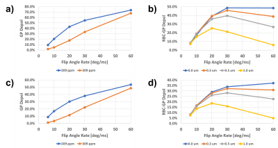 |
68 | Investigating the feasibility to assess the alveolar-volume-to-blood membrane thickness with hyperpolarized xenon-129 MRI using simulations
Kai Ruppert1, Tahmina S. Achekzai1, Luis Loza1, Faraz Amzajerdian1, Yi Xin1, Hooman Hamedani1, Ryan J. Baron1, Mostafa Ismail1, Ian F. Duncan1, Stephen Kadlecek1, and Rahim R. Rizi1
1University of Pennsylvania, Philadelphia, PA, United States
The thickness of the membrane separating the alveolar volume from the capillary blood is difficult measure with the chemical shift saturation recovery (CSSR) hyperpolarized xenon-129 MRI technique. In this work, we used a 1D gas exchange simulation to investigate the concept of measuring the gas-phase depolarization induced by the application of long RF pulses of variable power to extract information about the thickness of this membrane. Our simulations predict that measuring the red blood cell-induced gas-phase depolarization enables an estimation of membrane thickness even when the temporal lag between the rise in dissolved-phase signals cannot be accurately determined.
|
||
1889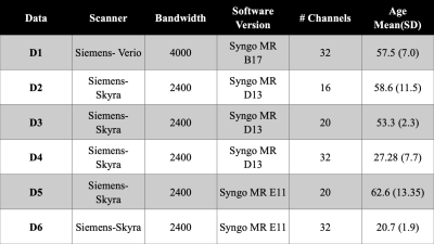 |
69 | The Effect of Differences in MRS Parameters on Data Harmonization of Normative Data
Marcia Sahaya Louis1,2, Huijun Vicky Liao2, Ajay Joshi1, and Alexander Lin2
1ECE, Boston University, Boston, MA, United States, 2Radiology, Brigham and Women's Hospital, Boston, MA, United States
Building normative databases for MR spectroscopy studies is much needed to further the use of MRS in large clinical studies. The effect of differences in MRS parameters is not well documented and there are no established data harmonization methods for MRS data. Therefore, the goal of this study is to identify the effects of variations such as dwell time, coil type, and software version and describe a method for harmonizing data in healthy controls that will ameliorate those effects.
|
||
1890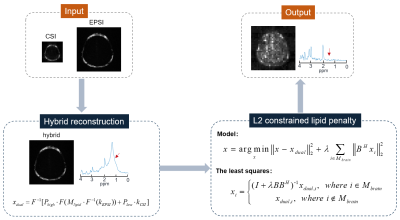 |
70 | Integrating hybrid CSI/EPSI acquisitions with L2 regularization for lipid removal of MRSI
Yiling Liu1, Jianfeng Bao2, Hao Chen1, Guiqin Liu3, Jingliang Chen2, and Zhiyong Zhang1
1School of Biomedical Engineering, Shanghai Jiao Tong University, Shanghai, China, 2The First Affiliated Hospital of Zhengzhou University, Zhengzhou University, Zhengzhou, China, 3Renji Hospital Affiliated to Shanghai Jiaotong University School of Medicine, Shanghai Jiao Tong University, Shanghai, China
Placements of lipid saturation bands not only demand a skillful MRSI scanning operation but also restrict the full brain MRSI coverage. Therefore, MRSI acquisition without lipid suppression while doing post-removal of lipid becomes clinically attractive and valuable. In this work, we propose to integrate hybrid CSI/EPSI acquisitions with L2 regularization for fast lipid removal of MRSI. The hybrid CSI/EPSI acquisitions rely on the same excitation and refocusing evolution with the modified CSI and EPSI sequences. Phantom and in-vivo brain studies demonstrate the effectiveness and low computational cost of the proposed method.
|
||
1891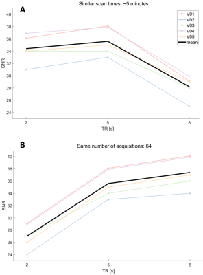 |
71 | semi-LASER MRS at 3T with TR=5s: All the Signal, Similar Scan Time, and Minimal T1 Weighting
Alex Ensworth1,2, Laura R. Barlow3,4, Piotr Kozlowski1,2,3,4, Erin L. MacMillan3,4,5, and Cornelia Laule1,2,3,6
1Physics and Astronomy, University of British Columbia, Vancouver, BC, Canada, 2International Collaboration on Repair Discoveries (ICORD), University of British Columbia, Vancouver, BC, Canada, 3Radiology, University of British Columbia, Vancouver, BC, Canada, 4UBC MRI Research Centre, University of British Columbia, Vancouver, BC, Canada, 5Philips Canada, Mississauga, ON, Canada, 6Pathology & Laboratory Medicine, University of British Columbia, Vancouver, BC, Canada The choice of repetition time (TR) in MR spectroscopy studies is widely debated. A shorter TR results in more clinically acceptable scan times, however due to T1 effects, the metabolite concentrations are reduced. We used semi-LASER MRS to compare the SNR and absolute metabolite concentrations for TR=2,5 and 8s. We demonstrate it is possible to achieve similar SNR and scan time using a TR of 5s with half the acquisitions compared to a TR of 2s, where the metabolite concentrations are 20% lower. Using a TR of 5s for any MRS study is feasible and recommended to minimize T1 effects. |
||
1892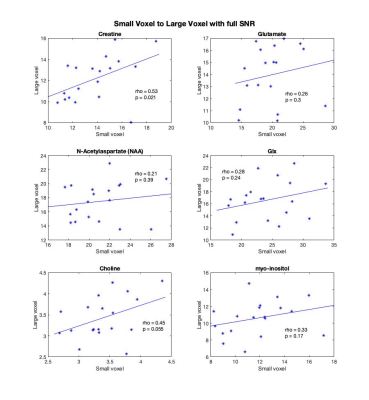 |
72 | Investigating metabolite regional dependencies in the frontal lobe: overlapping small and large voxels
Marilena M DeMayo1,2,3,4,5, Alexander McGirr2,3,4, Ben Selby3,6, Frank MacMaster7, Chantel T Debert3,6, and Ashley D Harris1,3,5
1Alberta Children's Hospital Research Institute, University of Calgary, Calgary, AB, Canada, 2Department of Psychiatry, University of Calgary, Calgary, AB, Canada, 3Hotchkiss Brain Institute, University of Calgary, Calgary, AB, Canada, 4Mathison Centre for Mental Health Research and Education, University of Calgary, Calgary, AB, Canada, 5Department of Radiology, University of Calgary, Calgary, AB, Canada, 6Department of Clinical Neurosciences, University of Calgary, Calgary, AB, Canada, 7Provincial Addiction and Mental Health (PAMH) Portfolio, University of Calgary, Calgary, AB, Canada
Small anatomically specific voxels in cerebral cortex require a large number of averages to quantify metabolites by magnetic resonance spectroscopy. It is unclear whether cortical regional variability in metabolite concentration requires small voxels, or whether a larger voxel which incorporates the region of interest can provide adequately representative measures with fewer averages. Concentrations of glutamate, glx, creatine, choline, myo-inositol and N-acetyl-aspartate (NAA) were correlated within subjects between an overlapping small (15x15x15mm) and large (30x30x30mm) voxel. There was a significant correlation between creatine measures, while choline showed a positive trend. There was no correlation between glutamate, glx, NAA, and myo-inositol.
|
||
1893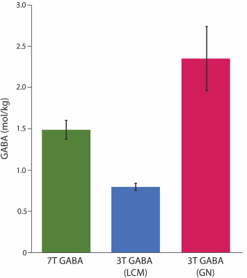 |
73 | A comparison of human brain GABA levels measured at 3T and 7T
Tiffany Bell1,2,3, Dana Goerzen4, Masoumeh Dehghani4, Jamie Near4,5,6, and Ashley D Harris1,2,3
1Department of Radiology, University of Calgary, Calgary, AB, Canada, 2Hotchkiss Brain Institute, University of Calgary, Calgary, AB, Canada, 3Alberta Children's Hospital Research Institute, University of Calgary, Calgary, AB, Canada, 4Centre d’Imagerie Cerebrale, Douglas Mental Health Institute, Montreal, QC, Canada, 5Department of Medical Biophysics, University of Toronto, Toronto, ON, Canada, 6Sunnybrook Research Institute, University of Toronto, Toronto, ON, Canada
Measuring GABA in the human brain in vivo using magnetic resonance spectroscopy (MRS) is not trivial due to its low abundance in the brain and the overlap of signal from higher concentration metabolites. At 3T, an editing sequence is typically used to isolate the GABA signal. Data acquired at 7T has higher SNR and better spectral resolution, therefore editing is often not used to measure GABA at this field strength. Here we compare GABA levels acquired at 7T using STEAM to GABA levels acquired at 3T using GABA-edited MEGA-PRESS and find little agreement between the two measures.
|
||
1894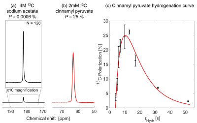 |
74 | Parahydrogen-Based Hyperpolarization of a Pyruvate Ester to 25% Within an MRI System Using SAMBADENA
Henri de Maissin1,2, Vladislav Ivantaev1, Christoph A. Müller1,2, Obaid Mohiuddin1, Jürgen Hennig3, Dominik von Elverfeldt3, Jan-Bernd Hövener4, Strefan Glöggler5,6, and Andreas Benjamin Schmidt1,2
1Radiology - Medical Physics, University Medical Center Freiburg, Freiburg, Germany, 2German Cancer Consortium (DKTK) partner site Freiburg, German Cancer Center (DKFZ), Heidelberg, Germany, 3University Medical Center Freiburg, Freiburg, Germany, 43Section Biomedical Imaging, Molecular Imaging North Competence Center (MOINCC), Department of Radiology and Neuroradiology, University Medical Center Schleswig-Holstein and Kiel University, Kiel, Germany, 5Max Planck Institute for Biophysical Chemistry, Göttingen, Germany, 6Center for Biostructural Imaging of Neurodegeneration, University Medical Center Göttingen, Göttingen, Germany
Parahydrogen (pH2) and Synthesis Amid the Magnet Bore Allow Dramatically Enhanced Nuclear Alignment (SAMBADENA) and have recently enabled hyperpolarization (HP) of 13C and subsequent in vivo administration of the produced contrast agent inside an MRI system. pH2 Induced Polarization (PHIP) by Side-Arm Hydrogenation (PHIP-SAH) has extended the portfolio of PHIP agents to metabolically-active molecules such as acetate, lactate, and pyruvate, but typically requires hydrogenation in organic solvents like chloroform. Here we present our new, solvent-compatible SAMBADENA setup and preliminary results of the 13C HP in chloroform of the PHIP-SAH molecule cinnamyl-pyruvate – opening the pathway towards metabolic studies with SAMBADENA.
|
||
 Back to Meeting Home
 Back to the Program-at-a-Glance
The International Society for Magnetic Resonance in Medicine is accredited by the Accreditation Council for Continuing Medical Education to provide continuing medical education for physicians.
 Back to Meeting Home
 Back to the Program-at-a-Glance
 View the Presentation