Digital Poster
Diffusion Artifacts, Acquisition & Applications I
Joint Annual Meeting ISMRM-ESMRMB & ISMRT 31st Annual Meeting • 07-12 May 2022 • London, UK

Digital Poster
Diffusion Artifacts, Acquisition & Applications I
| Computer # | ||||
|---|---|---|---|---|
1622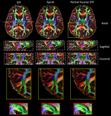 |
77 | Effects of T2*-blurring on effective resolution of diffusion MRI with spiral and EPI readout trajectories at 7T
Sajjad Feizollah1,2 and Christine L. Tardif1,2,3
1Department of Neurology and Neurosurgery, McGill University, Montreal, QC, Canada, 2McConnell Brain Imaging Center, Montreal Neurological Institute, McGill University, Montreal, QC, Canada, 3Department of Biomedical Engineering, McGill University, Montreal, QC, Canada
Several strategies have been used to increase the SNR of diffusion-weighted imaging including the use of partial Fourier EPI and spiral trajectories. Although these methods increase the SNR and sequence efficiency significantly, they affect the image quality and resulting diffusion measures causing loss of microstructural information of fine structures. Blurring caused by the T2* decay is one of the important effects resulting in low effective resolutions. Shorter T2* at ultra-high magnetic fields reduces the effective resolution in spite of providing higher SNR. This study shows about 60% lower effective resolution than the nominal resolution using PF-EPI and spiral at 7T.
|
||
1623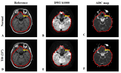 |
78 | Head-tilting in diffusion weighted imaging to reduce susceptibility-induced signal dropout in the frontal cortex: feasibility study.
Jae-Kyun Ryu1, Chuluunbaatar Otgonbaatar2, Seok-Jin Yeo3, Jeonghak Song4, Eunseok Jang4, Jaebin Lee4, and Hackjoon Shim1,4
1Medical Imaging AI Research Center, Canon Medical Systems Korea, Seoul, Korea, Republic of, 2Department of Radiology, Seoul National University, Seoul, Korea, Republic of, 3Department of Biomedical Engineering, Sungkyunkwan University, Suwon, Korea, Republic of, 4Magnetic Resonance Business Unit, Canon Medical Systems Korea, Seoul, Korea, Republic of
Diffusion-weighted MRI is a highly sensitive to alteration in the movement of water molecules and allows the assessment of various pathologies. Interpretation of the changes can be obscured by signal-dropout due to tissue-air susceptibility difference at the boundaries of nasal cavities, especially in the prefrontal region. The head tilting (chin-up) method during the brain scan could be considered as an accessible way without additional hardware- or sequence to prevent those susceptibility-induced signal-dropout. We demonstrated DWI with head-tilting (up to 20°) resulted in had less severe signal dropout and improved image quality than normal scan in the prefrontal cortex.
|
||
1624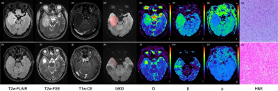 |
79 | Characterizing Heterogeneity of Gliomas with a Fractional-order Calculus Diffusion Model Video Not Available
Wenbo Sun1, Dan Xu1, Yunfei Zhang2, Yongming Dai2, and Haibo Xu1
1Department of Radiology, Zhongnan Hospital of Wuhan University, Wuhan, China, 2Central Research Institute, United Imaging Healthcare, Shanghai, China
This study aimed to investigate whether the FROC diffusion model could help predict molecular biomarkers in gliomas. It was found that FROC has the potential in comprehensively characterizing heterogeneity of gliomas, not only in glioma grading, but also in predicting tumor cell proliferation rate and isocitrate dehydrogenase-1 (IDH-1) gene mutation status.
|
||
1625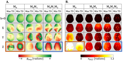 |
80 | Phase Stabilization with Motion Compensated Gradient Waveforms for Brain Diffusion Weighted Imaging (DWI)
Ariel J Hannum1,2,3, Tyler E Cork1,2,3, Merlin J Fair1,2, Kawin Setsompop1,4, and Daniel B Ennis1,2
1Department of Radiology, Stanford University, Stanford, CA, United States, 2Division of Radiology, Veterans Administration Health Care System, Palo Alto, CA, United States, 3Department of Bioengineering, Stanford University, Stanford, CA, United States, 4Department of Electrical Engineering, Stanford University, Stanford, CA, United States
Conventional brain DWI acquisitions are sensitive to physiological motion, which causes shot-to-shot phase variations between images. This makes accelerated, multi-shot imaging harder to achieve. We propose that motion compensated gradient waveforms will improve shot-to-shot phase stability. We found that both velocity and acceleration gradient moment nulling, particularly when diffusion encoding along the z-axis, improves shot-to-shot phase consistency at different cardiac trigger-delay times and reduces phase variation between repetitions for a fixed trigger-delay. We conclude that motion-compensated diffusion encoding gradients improve the phase stability between image shots.
|
||
1626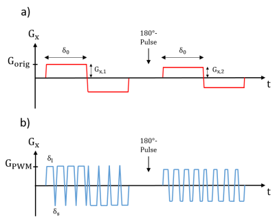 |
81 | Pulse-width modulation for simultaneous concomitant field compensation and diffusion weighting in double diffusion encoding
Julian Rauch1,2, Frederik B. Laun3, Dominik Ludwig1, Maxim Zaitsev4,5, Mark E. Ladd1,2,6, Peter Bachert1,2, and Tristan A. Kuder1
1Division of Medical Physics in Radiology, German Cancer Research Center (DKFZ), Heidelberg, Germany, 2Faculty of Physics and Astronomy, Heidelberg University, Heidelberg, Germany, 3Institute of Radiology, University Hospital Erlangen, Friedrich-Alexander-Universität Erlangen-Nürnberg (FAU), Erlangen, Germany, 4Medical Physics, Department of Radiology, Faculty of Medicine, Medical Center University of Freiburg, Freiburg, Germany, 5High Field Magnetic Resonance Center, Center for Medical Physics and Biomedical Engineering Medical University of Vienna, Vienna, Austria, 6Faculty of Medicine, Heidelberg University, Heidelberg, Germany
Intravoxel dephasing caused by Maxwell or concomitant fields can lead to image artifacts as signal voids or falsify the quantitation. In this study, a framework for compensation of concomitant field effects for double diffusion encoding sequences with single pairs of bipolar gradients on each axis is presented. Using pulse-width modulated oscillating gradients with an optimized timing of the oscillating lobes allows for simultaneous diffusion weighting and compensation of the self-squared terms of the concomitant fields as well as for the cross terms. Simulations with realistic parameters reveal the possibility of a signal gain with the proposed method without significant drawbacks.
|
||
1627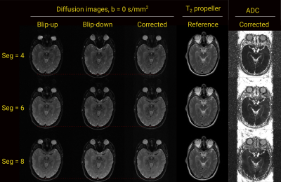 |
82 | Segmented accelerated multi-shot diffusion imaging combined with reverse polarity gradient (RPG) correction
Jens Johansson1, Kerstin Lagerstrand2,3, Pär-Arne Svensson4, Jr-yuan George Chiou5, Bruno Madore5, Hanna Hebelka1,4, and Stephan Maier1,5
1Department of Radiology, Institute of Clinical Sciences, Sahlgrenska Academy, University of Gothenburg, Gothenburg, Sweden, 2Medical radiation sciences, Institute of Clinical Sciences, Sahlgrenska Academy, University of Gothenburg, Gothenburg, Sweden, 3Department of Medical Physics and Biomedical Engineering, Sahlgrenska University Hospital, Gothenburg, Sweden, 4Department of Radiology, Sahlgrenska University Hospital, Gothenburg, Sweden, 5Department of Radiology, Brigham and Women's Hospital, Boston, MA, United States
The inherent low bandwidth along the phase encoding direction in diffusion-weighted imaging with echo-planar signal readout can cause severe image distortions. These artifacts arise near tissue boundaries and are caused by static magnetic field inhomogeneities. Correction of such artifacts can considerably improve clinical value. We combine a previously presented segmented accelerated multi-shot diffusion imaging method with reverse polarity gradient (RPG) for markedly improved reduction of geometric distortions.
|
||
1628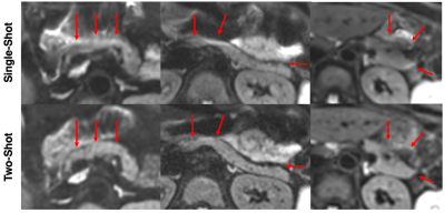 |
83 | Multishot EPI and a Deep Learning-Based Noise Reduction Strategy for High Resolution Pancreatic DWI
Matthew J. Middione1, Alimohammad S. Moalem1, Cheng William Hong1, Arnaud Guidon2, Daniel B. Ennis1, and Ryan L. Brunsing1
1Department of Radiology, Stanford University, Stanford, CA, United States, 2GE Healthcare, Boston, MA, United States DWI of the pancreas is challenging due to artifacts from physiologic motion, image distortion, and blurring, but has promising applications in pancreatic cancer detection. We conducted a pilot study of pancreatic DWI comparing single-shot and multi-shot EPI protocols as well as multi-shot EPI protocols with and without a new commercially available deep learning (DL) based denoising reconstruction method. Image quality was subjectively scored with key metrics. Multi-shot EPI reduced perceived distortion within the pancreatic bed, while the combination of multi-shot EPI and DL reconstruction subjectively reduced noise. |
||
1629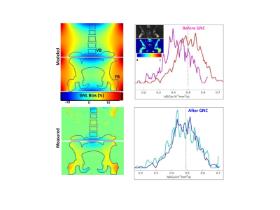 |
84 | Deployment of prospective ADC correction of gradient nonlinearity bias in myelofibrosis clinical trial
Dariya Malyarenko1, Yuxi Pang1, Ajit Devaraj2, Johannes Peeters3, Harry Friel2, Kristen M Pettit4, Moshe Talpaz4, Brian D Ross1, Gary D Luker1, and Thomas L Chenevert1
1Radiology, University of Michigan, Ann Arbor, MI, United States, 2Philips Healthcare, Highland Heights, OH, United States, 3Philips MR Sclinical Science, Best, Netherlands, 4Internal Medicine, University of Michigan, Ann Arbor, MI, United States
Apparent diffusion coefficient (ADC) metric is evaluated as a potential alternative to biopsy for disease grading and therapy response assessment in bone marrow of myelofibrosis (MF) patients. Spatially dependent bias in diffusion weighting due to systematic gradient nonlinearity (GNL) results in false heterogeneity of ADC maps over the imaged bone space. Here we illustrate deployment of prospective GNL bias correction based on technology developed in an academic industrial partnership to reduce technical variability of ADC in a MF clinical imaging trial.
|
||
1630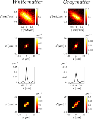 |
85 | MR determination of the diffusion propagator: measurements on mouse spinal cord using a benchtop scanner
Alfredo Ordinola1, Walker Jackson2, and Evren Özarslan1
1Department of Biomedical Engineering, Linköping University, Linköping, Sweden, 2Department of Biomedical and Clinical Sciences, Linköping University, Linköping, Sweden
We demonstrate the experimental determination of the diffusion propagator, indicating a conditional probability density function with two spatial arguments. To this end, a recently introduced method was implemented on a benchtop MR scanner and incorporated into imaging sequences. The data involving two independent wavenumbers were transformed from the measurement domain to the spatial domain, yielding an apparent diffusion propagator. Experiments on freely diffusing water provides accurate determination of the diffusion propagator while apparent propagators measured in mouse spinal cord reveal significant differences between white and gray matter regions.
|
||
1631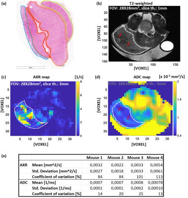 |
86 | Filter-exchange spectroscopy and imaging sensitively detect necrotic cell death in vitro and in vivo
Athanasia Kaika1, Geoffrey J. Topping1, Simone Ballke2, Thomas Theodor Metzler2, Irina Heid3, Mathias Schillmaier1, Rickmer Braren3, and Franz Schilling1
1Department of Nuclear Medicine, TUM School of Medicine, Klinikum Rechts der Isar, Technical University of Munich, Munich, Germany, 2Institute of Pathology, TUM School of Medicine, Technical University of Munich, Munich, Germany, 3Institute of Radiology, TUM School of Medicine, Klinikum Rechts der Isar, Technical University of Munich, Munich, Germany Filter-exchange spectroscopy (FEXSY) and imaging (FEXI) are sensitive to transmembrane water exchange, which is commonly altered during cell death1. Yeast cells in different permeabilization stages were studied with FEXI, DWI and CFDA-AM/ propidium iodide (PI) staining using flow cytometry. Cell populations with 3.8% PI positive staining had 44% increased AXR, but unchanged ADC. At 33% nonvital cells, detection of AXR became unreliable and ADC increased. In vivo FEXI and DWI in EL4 lymphoma, with H&E-confirmed necrotic tumor cells and edema, showed AXR and ADC variations across the tumor, possibly due to differing vitality cell stages. |
||
 Back to Meeting Home
 Back to the Program-at-a-Glance
The International Society for Magnetic Resonance in Medicine is accredited by the Accreditation Council for Continuing Medical Education to provide continuing medical education for physicians.
 Back to Meeting Home
 Back to the Program-at-a-Glance
 View the Presentation