Digital Poster
Diffusion Artifacts, Acquisition & Applications II
Joint Annual Meeting ISMRM-ESMRMB & ISMRT 31st Annual Meeting • 07-12 May 2022 • London, UK

Digital Poster
Diffusion Artifacts, Acquisition & Applications II
| Computer # | ||||
|---|---|---|---|---|
1717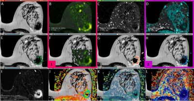 |
86 | Evaluation of breast cancer using IVIM-Kurtosis and compartmental tracer kinetic models through semi-automated segmentation
Natalia Noemí Massaccesi Bove1,2, Trinidad González Padin1,2, Daniel Fino1,2,3, Nicolás Moyano Brandi1,2, Clara Lisazo1,2, Leandro Enrique Salcedo1,2,4, Federico Julián González Nicolini2,3,5, Pedro Pablo Ariza1,2, Roberto Andres Isoardi2,3,5, María Cecilia Lorca1,2, Cristian Allard6, and Verónica R. Saucedo1,2
1MRI Department, Fundación Argentina para el Desarrollo en Salud, Mendoza, Argentina, 2MRI Department, Fundación Escuela de Medicina Nuclear, Mendoza, Argentina, 3Instituto Balseiro, Universidad Nacional de Cuyo, San Carlos de Bariloche, Argentina, 4Universidad de Mendoza, Mendoza, Argentina, 5GQNYCS, Comisión Nacional de Energía Atómica, CABA, Argentina, 6GE Healthcare, Santiago de Chile, Chile
This study aims to evaluate the correlation between the IVIM-DKI models (which describe pure and pseudo diffusion) and the quantitative and semiquantitative parameters from dynamic contrast-enhanced (DCE) MRI (that analyze vascular permeability and pharmacokinetics). A Python algorithm was implemented to adjust the intensities of 12 b-values DWI to a biexponential function modeling the IVIM and DKI. Then, the statistical association between these parameters and the DCE MRI was evaluated with a Pearson’s correlation coefficient. The f and D* factors are both good biomarkers for the evaluation of perfusion properties, according to the examined correlation between all the parameters.
|
||
1718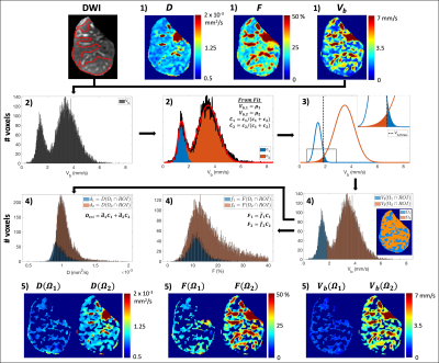 |
87 | 2D (b-M1) Data Sampling and Blood Velocity Standard Deviation Distribution Fitting for Repeatable Tri-exponential IVIM Quantification
Gregory Simchick1,2, Ruiqi Geng1,2, and Diego Hernando1,2
1Radiology, University of Wisconsin-Madison, Madison, WI, United States, 2Medical Physics, University of Wisconsin-Madison, Madison, WI, United States
There is growing evidence of tri-exponential IVIM-DWI signal behavior in the liver. However, tri-exponential estimates often suffer from the instability associated with separating three decaying signal components sampled along a single dimension (i.e., b-value). In this work, standard monopolar and noise-optimized 2D (b-M1) data sampling schemes were acquired. From each acquisition, tri-exponential ROI-based fitting and a blood velocity standard deviation distribution (BVD) fitting method were performed. The estimated tri-exponential parameters were compared across acquisitions and fitting methods. Repeatable tri-exponential IVIM estimates were obtained using the 2D data sampling combined with BVD fitting. A new, physical tri-exponential model is also proposed.
|
||
1719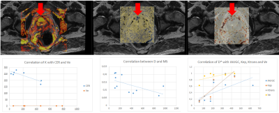 |
88 | Comparison between IVIM-DKI, MRE and DCE-Tofts quantitative techniques at 3.0 T multiparametric prostate protocol
Trinidad González Padin1,2, Nicolas Moyano Brandi1,2, María Paula Del Pópolo1,2, Natalia Noemi Massaccesi Bove1,2, Clara Chacon1,2, Federico Julian Gonzalez1,3,4, Roberto Andrés Isoardi1,3,4,5, Pedro Pablo Ariza1,2, Andrés Dominguez1, Eduardo H,M.S.G de Figuereido6, and Daniel Fino Villamil1,2,4,5
1MRI department, Fundación Escuela de Medicina Nuclear, Mendoza, Argentina, 2MRI department, Fundación Argentina para el Desarrollo en Salud, Mendoza, Argentina, 3GQNYCS, Comisión Nacional de Energía Atómica, Buenos Aires, Argentina, 4Instituto Balseiro, Universidad Nacional de Cuyo, San Carlos de Bariloche, Argentina, 5FCEN, Universidad Nacional De Cuyo, Mendoza, Argentina, 6General Electric- Healthcare, Sao Paulo-SP, Brazil, Brazil
Since there is no clear relationship between the coefficients of the DWI biexponential model and permeability and pharmacokinetics, we aim to correlate the coefficients associated to the IVIM-DKI model, MRE, and quantitative and semiquantitative maps of the multiparametric prostate protocol. Through a Python algorithm, the DWI data acquisition was fitted to an IVIM-DKI function after a semi-automatic segmentation. Then, PR, PCa and healthy tissue were evaluated, and the IVIM-DKI parametric maps were contrasted with the DCE and MRE through a Pearson’s correlation test. Empirical relationships were established between the pseudo-perfusion models, semiquantitative maps and the variables of the compartmental models.
|
||
1720 |
89 | Multi-parameter Magnetic Resonance Quantitative Evaluation of Pancreatic Cancer with Vascular Invasion Video Not Available
Mi Zhou1, Long-Lin Yin1, and Yunzhu Wu2
1Department of Radiology, Sichuan Provincial People's Hospital, University of Electronic Science and Technology of China, Chengdu, China, 2MR Scientific Marketing, SIEMENS Healthineers, Shanghai, China
In this study, the sensitivity, specificity and accuracy of quantitative parameters derived from T2WI+DKI+DCE combined model for pancreatic cancer vascular invasion were evaluated based on the pathological results as the gold standard. Combined T2WI+DKI+DCE quantitative analysis can improve the specificity and accuracy of diagnostic efficiency of pancreatic cancer vascular invasion. There were significant differences in MD, MK, Ktrans, Kep and Ve between vascular invasion group and non-vascular invasion group. The diffusion parameters and perfusion information are highly consistent, which can provide reliable evidence for preoperative non-invasive diagnosis and treatment of pancreatic cancer.
|
||
1721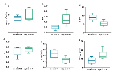 |
90 | Value of integrated 18F-FDG PET with fractional order calculus model-based MRI and DKI to evaluate the proliferation of lung squamous cell carcinoma Video Not Available
Zhun Huang1, Nan Meng2, Fangfang Fu3, Yan Bai3, Wei Wei3, Ting Fang4, Pengyang Feng5, Ziqiang Li6, Yu Luo2, Jianmin Yuan7, Yang Yang8, Zhe Wang7, and Meiyun Wang*9
1Department of Radiology, Henan University People’s Hospital & Henan Provincial People’s Hospital, School of Basic Medicine, Zhengzhou, China, 2Department of Radiology, Zhengzhou University People’s Hospital & Henan Provincial People’s Hospital, Academy of Medical Sciences, Zhengzhou, China, 3Henan Provincial People’s Hospital, Zhengzhou, China, 4Department of Radiology, Zhengzhou University People’s Hospital & Henan Provincial People’s Hospital, Zhengzhou, China, 5Department of Radiology, Henan University People’s Hospital & Henan Provincial People’s Hospital, Zhengzhou, China, 6XinxiangMedical University & Henan Provincial People’s Hospital, Xinxiang&Zhengzhou, Zhengzhou, China, 7Central Research Institute, UIH Group, Shanghai, China., Shang Hai, China, 8Beijing United Imaging Research Institute of Intelligent Imaging, Bei Jing, China, 9Henan Provincial People's Hospital, Zhengzhou, China
18F-FDG-PET/MRI combines the functional and metabolic characteristics of PET with the good soft-tissue contrast of MRI. The results showed that high Ki-67-expressing tumors showed lower D and μ values and higher MK and SUVmax values. In addition, the diagnostic efficacy of all parameters is similar.
|
||
1722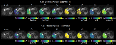 |
91 | Inter-scanner reproducibility and variability assessment of advanced liver diffusion MRI metrics
Francesco Grussu1, Kinga Bernatowicz1, Ignasi Barba2, Marco Palombo3, Juan Francisco Corral4,5, Marta Vidorreta6, Xavier Merino4,5, Richard Mast4,5, Núria Roson4,5, Nahúm Calvo‐Malvar4,7, Manuel Escobar Amores4,5, Josep R. Garcia-Bennett7, and Raquel Perez-Lopez1,5
1Radiomics Group, Vall d'Hebron Institute of Oncology, Vall d'Hebron Barcelona Hospital Campus, Barcelona, Spain, 2NMR Lab, Vall d'Hebron Institute of Oncology, Vall d'Hebron Barcelona Hospital Campus, Barcelona, Spain, 3Cardiff University Brain Research Imaging Centre, Cardiff University, Cardiff, United Kingdom, 4Institut de Diagnòstic per la Imatge (IDI), Catalonia, Spain, 5Department of Radiology, Hospital Universitari Vall d'Hebron, Barcelona, Spain, 6Siemens Healthineers, Madrid, Spain, 7Hospital Universitari de Bellvitge, L'Hospitalet de Llobregat, Spain
Innovative liver Diffusion-Weighted (DW) MRI aims to increase sensitivity and biological specificity of routine DW imaging, but may feature lower reproducibility due to longer scan times and acquisition of highly DW images. We assess inter-scanner reproducibility and variability of metrics from two novel approaches, T2-Intra-Voxel Incoherent Motion-Kurtosis (T2-IVIM-Kurtosis) and Diffusion-Relaxation Hepatic Imaging via Generalised Assessment of DiffusiOn Simulations (DR-HIGADOS), in two 1.5T scanners (Siemens Avanto; Philips Ingenia). Both methods are reproducible across scanners. Cellularity, intra-cellular diffusivity and vascular fraction show the highest measurement variability, implying that larger cohorts may be required in studies that focus on these indices.
|
||
1723 |
92 | Repeatability of Diffusion Kurtosis Tensor Parameters in Muscles of the Lower Legs
Ethan Mathew1,2, Chad Quarles1, Sudarshan Ragunathan1, and Richard Dortch1
1Barrow Neurological Institute, Tempe, AZ, United States, 2Arizona State University, Tempe, AZ, United States
This study examines the repeatability of quantitative measures obtained from the lower legs using a diffusion kurtosis tensor imaging sequence with a scan time of just 13 minutes. Test-retest data was acquired, and kurtosis parameters were calculated for both sets of data. Muscle groups were segmented, and repeatability was assessed by muscle group. In both the medial gastrocnemius and tibialis anterior, kurtosis fractional anisotropy and fractional anisotropy showed highest repeatability, as well as mean diffusivity in the medial gastrocnemius.
|
||
1724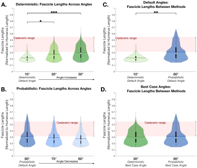 |
93 | Sensitivity comparison of probabilistic and deterministic DTI tractography for robust characterization of arm skeletal muscle geometry
Divya Joshi1,2, Julius PA Dewald1,2,3, and Carson Ingo2,4
1Department of Biomedical Engineering, Northwestern University, Chicago, IL, United States, 2Department of Physical Therapy and Human Movement Sciences, Northwestern University Feinberg School of Medicine, Chicago, IL, United States, 3Department of Physical Medicine and Rehabilitation, Northwestern University Feinberg School of Medicine, Chicago, IL, United States, 4Department of Neurology, Northwestern University Feinberg School of Medicine, Chicago, IL, United States
The robustness of probabilistic versus deterministic DTI tractography in characterization of skeletal muscle structure is not yet established. We investigated the sensitivity to turning angle when probabilistic and deterministic methods were used to estimate fascicle lengths and fractional anisotropy in the arm’s biceps muscle (n=8). Results produced with deterministic methods were highly sensitive to the turning angle, but those produced with probabilistic methods were not. Furthermore, the recommended angle in probabilistic methods yielded findings consistent with ground truth measurements, while the turning angle in deterministic methods needed to be altered from the recommended value to achieve similar results.
|
||
1725 |
94 | Model-based reconstruction of IVIM-DTI in skeletal muscle: Improving IVIM-DTI parameter map quality in low-perfused tissues
Susanne Rauh1, Oliver Maier2, Oliver Gurney-Champion3, Melissa Hooijmans3, Rudolf Stollberger2, Aart Nederveen3, and Gustav Strijkers1
1Department of Biomedical Engineering and Physics, Amsterdam UMC, Amsterdam, Netherlands, 2Institute of Medical Engineering, Graz University of Technology, Graz, Austria, 3Department of Radiology and Nuclear Medicine, Amsterdam UMC, Amsterdam, Netherlands IVIM and DTI techniques are both sensitive to a variety of muscle disorders. Their combination can give a more complete description of ordered diffusion processes and perfusion. Model-based reconstruction can overcome many short-comings of image-based fitting such as Rician noise bias or low SNR. In this study we investigate the feasibility of model-based IVIM-DTI reconstruction in skeletal muscle, a low-perfused tissue. Results were compared to image-based least-square fitting. The parameter maps show more anatomical detail with model-based reconstruction. The mean parameter values are comparable to image-based fitting. Model-based reconstruction is sensitive to subtle changes after exercise in the underlying parameters. |
||
1726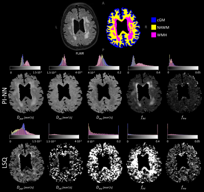 |
95 | Three-component IVIM fitting in cerebrovascular disease using physics-informed neural networks: repeatability and accuracy
Paulien H.M. Voorter1,2, Walter H. Backes1,2,3, Oliver J. Gurney-Champion4, Sau-May Wong1, Julie Staals3,5, Robert-Jan van Oostenbrugge2,3,5, Merel M. van der Thiel1,2, Jacobus F.A. Jansen1,2,6, and Gerhard S. Drenthen1,2
1Department of Radiology & Nuclear Medicine, Maastricht University Medical Center, Maastricht, Netherlands, 2School for Mental Health & Neuroscience, Maastricht University, Maastricht, Netherlands, 3School for Cardiovascular Disease, Maastricht University, Maastricht, Netherlands, 4Department of Radiology and Nuclear medicine, Amsterdam University Medical Center, Cancer Center Amsterdam, University of Amsterdam, Amsterdam, Netherlands, 5Department of Neurology, Maastricht University Medical Center, Maastricht, Netherlands, 6Department of Electrical Engineering, Eindhoven University of Technology, Eindhoven, Netherlands
Next to parenchymal diffusion and microvascular pseudo-diffusion, a third diffusion component is present in cerebral intravoxel incoherent motion (IVIM) imaging, representing interstitial fluid. Fitting the three-component IVIM model using conventional fitting methods strongly suffers from image noise. Therefore, we explored the applicability of a physics-informed neural network (PI-NN) fitting approach, previously shown to be more robust to noise. Using test-retest data from sixteen patients with cerebrovascular disease, we found higher repeatability of all IVIM parameters using PI-NN. Furthermore, simulations showed that PI-NN provided more accurate IVIM parameters. Hence, using PI-NN is promising to obtain tissue markers of cerebrovascular disease.
|
||
1727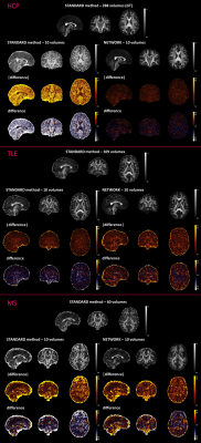 |
96 | A generalized deep learning network for fractional anisotropy reconstruction: application to epilepsy and multiple sclerosis
Marta Gaviraghi1, Antonio Ricciardi2, Fulvia Palesi3, Wallace Brownlee2, Paolo Vitali4, Ferran Prados2, Baris Kanber2, and Claudia AM Gandini Wheeler-Kingshott2,3,5
1Department of Electrical, Computer and Biomedical Engineering, University of Pavia, Pavia, Italy, 2NMR Research Unit, Queen Square Multiple Sclerosis Centre, Department of Neuroinflammation, UCL Queen Square Institute of Neurology, UCL, London, United Kingdom, 3Department of Brain and Behavioural Sciences, University of Pavia, Pavia, Italy, 4IRCCS Policlinico San Donato, Milano, Italy, 5Brain Connectivity Centre, IRCCS Mondino Foundation, Pavia, Italy
Quantitative maps obtained from Diffusion Tensor are very useful for investigating microstructural changes that occur in brain diseases. However, the long acquisition times required for a fully sampled diffusion-weighted space makes their clinical use unfeasible. Here we have adapted a U-net that obtains reliable Fractional Anisotropy (FA) maps from a reduced set of 10 Diffusion Weighted volumes (that can be acquired in less than 1 min). Our network was applied to two independent, clinical datasets, without retraining, and produced FA that retained clinical sensitivity and characteristic FA value distributions in the brain white matter.
|
||
1728 |
97 | Brain Extracellular Free Water Correlates with MRSI Biomarkers of Neuro-inflammation in the Brain of HIV Clade-C Infected Individuals
Teddy Salan1, Sameer Vyas2, Deepika Aggarwal2, Paramjeet Singh2, and Varan Govind1
1Radiology, University of Miami, Miami, FL, United States, 2Post Graduate Institute of Medical Education & Research, Chandigarh, India Free water (FW) imaging is a diffusion-weighted MRI technique that differentiates extracellular from intracellular water compartments. Increased FW fraction is generally associated with neuroinflammation. However, establishment of a direct link between FW and biomarkers of neuroinflammation in human studies is not feasible. In this work, we use MRSI to find correlations between FW and metabolic biomarkers of inflammation in HIV infected individuals. Our results show that FW had the most significant positive correlations with myo-inositol, a strong biomarker of gliosis and inflammation. This corroborates previous findings that elevated FW can be interpreted as a sign of inflammation in the brain. |
||
1729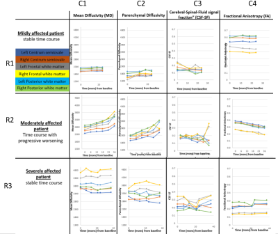 |
98 | Developing a robust quantitative diffusion MRI protocol for longitudinal assessment of neurodegenerative disorders
Amritha Nayak1,2, Diana Bharucha-Goebel3,4, M.Okan Irfanoglu2, Gilberto Averion3, Dimah Saade3, Carsten Bönnemann3, and Carlo Pierpaoli2
1Henry Jackson Foundation for advancement in Military Medicine Inc, Rockville, MD, United States, 2QMI, NIBIB, National Institutes of Health, Bethesda, MD, United States, 3NNDCS, NINDS, National Institutes of Health, Bethesda, MD, United States, 4Children's National Hospital, Division of Neurology, Washington DC, DC, United States
In this work, we have developed a quantitative diffusion MRI (dMRI) pipeline, to include acquisition, processing, and analysis, robust enough to evaluate the evolution of a neurodegenerative disease in each individual patient.
|
||
1730 |
99 | A diffusion MRI tractometry approach in cerebral small vessel disease
Maxime Chamberland1, Anil M Tuladhar2, Anna Dewenter3, Mengfei Cai2, Mina A Jacob2, Annemieke ter Telgte2,4, Kim Wiegertjes2, Marcel Zwiers1, José P. Marques1, Chantal M.W. Tax5,6, Marco Duering3,7, Frank-Erik de Leeuw2, and David G. Norris1
1Radboud University, Donders Institute for Brain, Cognition, and Behavior, Nijmegen, Netherlands, 2Department of Neurology, Radboud University, Donders Institute for Brain, Cognition, and Behavior, Nijmegen, Netherlands, 3University Hospital, LMU Munich, Institute for Stroke and Dementia Research (ISD), Munich, Germany, 4VASCage, Research Centre on Vascular Ageing and Stroke, Innsbruck, Austria, 5University Medical Center Utrecht, Image Science Institute, Utrecht, Netherlands, 6School of Physics, Cardiff University Brain Research Imaging Centre, Cardiff, United Kingdom, 7Department of Biomedical Engineering, University of Basel, Medical Image Analysis Center (MIAC) and qbig, Basel, Switzerland
In a cohort of 54 patients with sporadic cerebral small vessel disease, associations between diffusion MRI measures and processing speed were investigated both at low and high b-values, revealing complementary microstructural information. Associations with processing speed found by skeletonization were confirmed with an independent tractometry approach, which was further utilized to identify a set of association tracts associated with cognition.
|
||
 Back to Meeting Home
 Back to the Program-at-a-Glance
The International Society for Magnetic Resonance in Medicine is accredited by the Accreditation Council for Continuing Medical Education to provide continuing medical education for physicians.
 Back to Meeting Home
 Back to the Program-at-a-Glance
 View the Presentation