Digital Poster
Diffusion Analysis, Tractography & Microstructure II
Joint Annual Meeting ISMRM-ESMRMB & ISMRT 31st Annual Meeting • 07-12 May 2022 • London, UK

Digital Poster
Diffusion Analysis, Tractography & Microstructure II
| Computer # | ||||
|---|---|---|---|---|
0975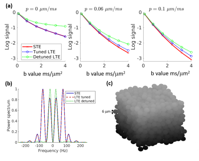 |
95 | Effects of exchange on spherical vs. linear diffusion tensor encodings: A simulation study
Jonathan Scharff Nielsen1 and Manisha Aggarwal1
1Department of Radiology, Johns Hopkins University School of Medicine, Baltimore, MD, United States
To investigate the influence of exchange on microscopic anisotropy estimates derived from kurtosis differences between linear-tensor-encoding (LTE) and spherical-tensor-encoding (STE) diffusion weighting waveforms, random walk simulations were performed in a substrate of packed spheres of varying permeability. A pronounced μFA bias was observed with spectrally detuned LTE due to restricted diffusion and kurtosis time dependence, decreasing with rate of exchange, but also with tuned LTE due to inherent differences in exchange sensitivity, increasing with exchange. Our results suggest a need for caution in interpreting MDE-derived microscopic anisotropy estimates in the presence of exchange.
|
||
0976 |
96 | Characterisation of restricted diffusion and exchange using the velocity autocorrelation function
Arthur Chakwizira1, Samo Lasič2, Alexis Reymbaut3, Carl-Fredrik Westin4, Filip Szczepankiewicz5, and Markus Nilsson5
1Medical Radiation Physics, Lund, Lund University, Lund, Sweden, 2Danish Research Centre for Magnetic Resonance, Centre for Functional and Diagnostic Imaging and Research, Copenhagen University Hospital, Amager and Hvidovre, Copenhagen, Denmark, 3Random Walk Imaging, AB, Lund, Sweden, 4Department of Radiology, Brigham and Women's hospital, Harvard Medical School, Boston, MA, United States, 5Clinical Sciences Lund, Lund University, Lund, Sweden
Temporal velocity correlations of diffusing particles carry information about the structure of the diffusion environment. Studying high-order velocity autocorrelation functions can aid in understanding how signal is influenced by restricted diffusion and exchange, and in turn how to design experiments that are sensitive/independent to these phenomena. In this work, we employ numerical simulations on a variety of substrates to demonstrate this notion. We find that the fourth order velocity autocorrelation bears a distinctive signature of exchanging systems. In addition, we highlight that the effect of exchange on the second order velocity autocorrelation is negligible when exchange is barrier-limited.
|
||
0977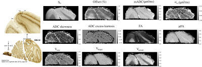 |
97 | High Resolution Ex Vivo Diffusion Tensor Distribution MRI of Neural Tissue
Kulam Najmudeen Magdoom1,2,3, Michal E. Komlosh1,2,3, Saleem Kadharbatcha1,3, Dario Gasbarra4, and Peter J. Basser2,3
1The Henry M. Jackson Foundation for the Advancement of Military Medicine (HJF) Inc.,, Bethesda, MD, United States, 2Eunice Kennedy Shriver National Institute of Child Health and Human Development, National Institutes of Healt, Bethesda, MD, United States, 3Center for Neuroscience and Regenerative Medicine, Uniformed Services University of Health Sciences, Bethesda, MD, United States, 4University of Helsinki, Helsinki, Finland
Neural tissue microstructure plays a key role in developmental, physiological, and pathophysiological processes. Diffusion tensor distribution (DTD) MRI is a promising approach to resolve sub-voxel microstructural features using multiple diffusion encodings. In this study, we have applied a novel DTD framework and pulse sequence to investigate its capabilities in revealing neural tissue microstructure. We present high resolution data acquired using an excised visual cortex and cervical spinal cord of a macaque monkey. The results show DTD MRI untangles size, shape and orientation heterogeneity within a voxel and parcellates tissue consistent with histological findings.
|
||
0978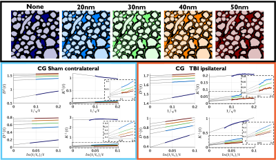 |
98 | Time-dependent diffusion and kurtosis in the extra-axonal space from 3D electron microscopy substrates of injured rat brain white matter
Ricardo Coronado-Leija1, Hong-Hsi Lee2, Ali Abdollahzadeh3, Jussi Tohka3, Alejandra Sierra3, Els Fieremans1, and Dmitry S Novikov1
1Radiology, New York University School of Medicine, New York, NY, United States, 2Radiology, Massachusetts General Hospital, Harvard Medical School, Boston, MA, United States, 3University of Eastern Finland, Kuopio, Finland In this work, we perform Monte Carlo simulations of diffusion in the extra-axonal space segmented from realistic 3D electron microscopy substrates. Simulations in sham and TBI rat brains confirm the universality of the power-law functional form of the axial and radial time-dependent diffusion $$$D^{\parallel,\perp}(t)$$$ and kurtosis $$$K^{\parallel,\perp}(t)$$$. We characterize the changes caused by TBI, finding that the dependence of long-time asysmptote $$$D^{\perp}_\infty$$$ on the extra-axonal volume fraction follows Archie's law. We also validate the theoretically predicted relationship between the power-law tails of $$$D(t)$$$ and $$$K(t)$$$. |
||
0979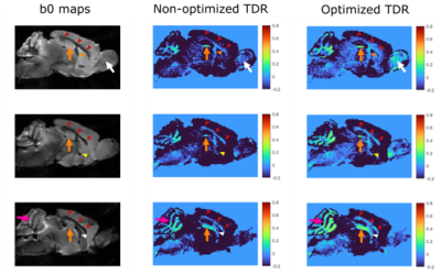 |
99 | Optimised Temporal Diffusion Ratio for Imaging Restricted Diffusion
William Richard Warner1, Andrada Ianus2, Noam Shemesh2, Malwina Molendowska3, Derek Jones3, Flavio Dell'Acqua4, Marco Palombo3,5, and Ivana Drobnjak1
1Computer Science Department, University College London, London, United Kingdom, 2Champalimaud Research, Champalimaud Centre for the Unknown, Lisbon, Portugal, 3Cardiff University Brain Research Imaging Centre, Cardiff, United Kingdom, 4NatBrainLab, King's College London, London, United Kingdom, 5School of Computer Science and Informatics, Cardiff University, Cardiff, United Kingdom
Temporal Diffusion Ratio (TDR) is a recently proposed dMRI technique, with potential for mapping areas of restricted diffusion. We optimise the TDR diffusion sequences in simulation to provide maximum contrast for a range of restricted structures, finding sequence shapes and the optimal HARDI subset size for calculating TDR. We then demonstrate the results experimentally in-vivo on the human brain, and ex-vivo using a pre-clinical scanner on the mouse brain.
|
||
0980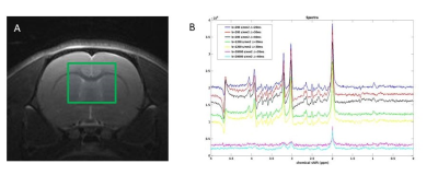 |
100 | Probing complex morphologies at decreasing diffusion times using Diffusion-weighted MR Spectroscopy
Nathalie Just1, Samo Lasič2,3, Ditte Bentsen Christensen4, Julien Valette5, Tim Dyrby2,6, Hartwig Siebner2, and Henrik Lundell2
1Danish Research Center for Magnetic Resonance, Copenhagen University Hospital - Amager and Hvidovre, Hvidovre, Denmark, 2Danish Research Center for Magnetic Resonance, Copenhagen University Hospital Amager and Hvidovre, Hvidovre, Denmark, 3Random Walk Imaging, Lund, Sweden, 4HYPERMAG, DTU, Copenhagen, Denmark, 5MIRCen, Commissariat à l' energie atomique et aux Energies Alternatives, Fontenay-aux-Roses, France, 6Department of Applied Mathematics and Computer Science, Technical University of Denmark, Kongens Lyngby, Denmark
Metabolite diffusion provide the unique ability to study the intracellular environment of specific cell types of the brain. Multiple studies suggest that the diffusivity along dendrites and axons is time dependent due to variations in diameter. We explore the signal due to such effects in Monte Carlo simulations in settings feasible for preclinical PGSE measurements and find that time dependent kurtosis, but not diffusivity provide the most potent source of contrast to this effect. We observe similar effects of intraneuronal NAA diffusion in rat.
|
||
0981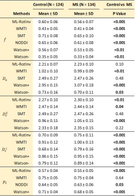 |
101 | How to understand diffusion MRI changes in the white matter of Multiple Sclerosis patients?
Ying Liao1, Santiago Coelho1, Wafaa Sweidan1, Jenny Chen1, Dmitry S. Novikov1, and Els Fieremans1
1Radiology, NYU School of Medicine, New York, NY, United States
Conventional diffusion MRI (dMRI) techniques, such as DTI and DKI, are sensitive to pathology but lack specificity. In brain white matter, the “Standard Model” framework of dMRI may provide specificity to microstructural changes. Generally, clinical dMRI is noisy and limited, making SM estimation challenging. Thus, different constraints and techniques have been introduced to robustly extract SM parametric maps. Here, we employ a large clinical dataset of Multiple Sclerosis patient data (N = 134) and noise propagation experiments to study the sensitivity and specificity of these techniques.
|
||
0982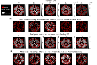 |
102 | Violation of axial-symmetric assumption in DKI and consequences for biophysical parameter estimates across white matter
Jan Malte Oeschger1, Karsten Tabelow2, and Siawoosh Mohammadi1,3
1Institute of Systems Neurosciences, University Medical Center Hamburg-Eppendorf, Hamburg, Germany, 2Weierstrass Institute for Applied Analysis and Stochastics, Berlin, Germany, 3Department of Neurophysics, Max Planck Institute for Human Cognitive and Brain Sciences, Leipzig, Germany
The recently introduced axial-symmetric DKI framework is less noise sensitive but unable to capture more complex fiber configurations causing an AxDKI inherent bias in those voxels. We found that the parallel and perpendicular kurtosis are most often affected by this inherent bias and that it translates to the AxDKI based biophysical parameters. Bias-free estimation of the biophysical parameters with the AxDKI framework, therefore, is difficult if not impossible at this point. However, the parallel and perpendicular diffusivity and the mean kurtosis were largely bias-free, encouraging use of the AxDKI framework in studies where these parameters are focused on.
|
||
0983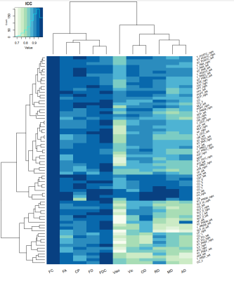 |
103 | Test-retest reliability of whole-brain and bundle-level microstructural properties
Timo Roine1,2 and Sila Genc3
1Department of Neuroscience and Biomedical Engineering, Aalto University, Espoo, Finland, 2Turku Brain and Mind Center, University of Turku, Turku, Finland, 3Cardiff University Brain Research Imaging Centre (CUBRIC), School of Psychology, Cardiff University, Cardiff, United Kingdom
We investigated the test-retest reliability of bundle- and voxel-/fixel-level microstructural metrics with fixel-based analysis, neurite orientation dispersion and density imaging, and diffusion tensor imaging. For the bundle-level analyses, TractSeg was used to automatically segment 72 fiber bundles of the brain. Our results indicate that most of the metrics, especially those measured with fixel-based analysis, are highly robust and show excellent test-retest reliability both in local and bundle-level analyses. In general, the reproducibility was higher in the white matter in contrast to gray matter and for acquisitions with multi-shell compared to single-shell data.
|
||
0984 |
104 | Diffusion dispersion and microscopic fractional anisotropy reveal acute sensitivity to mild traumatic brain injury in a mouse model
Naila Rahman1,2, Kathy Xu2, Arthur Brown2,3, and Corey Baron1,2
1Medical Biophysics, Western University, London, ON, Canada, 2Robarts Research Institute, London, ON, Canada, 3Anatomy and Cell Biology, Western University, London, ON, Canada
Imaging markers of mild to moderate concussion are notoriously difficult to detect in vivo. Advanced diffusion MRI (dMRI) techniques have shown increased sensitivity and specificity to microstructural changes in various disease and injury models. Oscillating gradient spin-echo (OGSE) dMRI is sensitive to structural disorder and microscopic anisotropy (µA) dMRI is sensitive to water diffusion anisotropy independent of neuron fiber orientation. In this work, we demonstrate that both microscopic fractional anisotropy and diffusion dispersion show acute sensitivity to concussion, while traditional diffusion MRI markers do not.
|
||
0985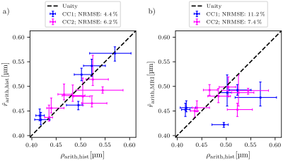 |
105 | Predicting the arithmetic mean radius from the MRI-visible axon radius
Laurin Mordhorst1, Mohammad Ashtarayeh1, Maria Morozova2,3, Sebastian Papazoglou1, Björn Fricke1, Tobias Streubel1,2, Carsten Jäger2, Henriette Rusch3, Ludger Starke4, Thomas Gladytz4, Ehsan Tasbihi4, Thoralf Niendorf4,5, Nikolaus Weiskopf2,6, Markus Morawski2,3, and Siawoosh Mohammadi1,2
1Institute of Systems Neuroscience, University Medical Center Hamburg-Eppendorf, Hamburg, Germany, 2Department of Neurophysics, Max Planck Institute for Human Cognitive and Brain Sciences, Leipzig, Germany, 3Paul Flechsig Institute of Brain Research, University of Leipzig, Leipzig, Germany, 4Berlin Ultrahigh Field Facility, Max Delbrück Center for Molecular Medicine in the Helmholtz Association, Berlin, Germany, 5Experimental and Clinical Research Center, a joint cooperation between the Charité Medical Faculty and the Max Delbrück Center for Molecular Medicine in the Helmholtz Association, Berlin, Germany, 6Felix Bloch Institute for Solid State Physics, Faculty of Physics and Earth Sciences, Leipzig, Germany
The axon radius is a main determinant of the conduction velocity of action potentials. Robust, MRI-based axon radius estimation is sensitive to a tail-weighted estimate of the ensemble-average axon radius, i.e., the effective axon radius ($$$r_{\text{eff}}$$$). It is unclear how $$$r_{\text{eff}}$$$ translates into the arithmetic mean axon radius ($$$r_{\text{arith}}$$$), which may be more indicative of the conduction velocity than $$$r_{\text{eff}}$$$. We investigate the feasibility to predict $$$r_{\text{arith}}$$$ from $$$r_{\text{eff}}$$$ using linear regression on high-resolution, large-scale light-microscopy images of a human corpus callosum sample and validate this linear relationship with microscopy and diffusion-weighted MRI images of another human corpus callosum sample.
|
||
0986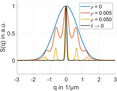 |
106 | On Measuring the Pore Distribution Function and the Occurrence of Signal Peaks When Utilizing Long Diffusion Gradients
Christoph Martin Stuprich1, Tristan Anselm Kuder2, Bernhard Hensel3, Michael Uder1, and Frederik Bernd Laun1
1Radiology, University Hospital Erlangen, Friedrich Alexander University Erlangen, Erlangen, Germany, 2German Cancer Research Center(DKFZ) Heidelberg, Heidelberg, Germany, 3Friedrich Alexander University Erlangen, Erlangen, Germany
In Diffusion MR, short gradient pulses are often ascribed to generate oscillating signal curves. These characteristic oscillations or “peaks” appear if a certain characteristic length-scale in the underlying tissue is present. Mitra and Halperin mention the possibility of “Bragg Peaks” when using long gradient pulses. In this work we explicitly show and interpret the occurrence of such signal peaks under application of long gradient pulses in theory and simulation.
|
||
0987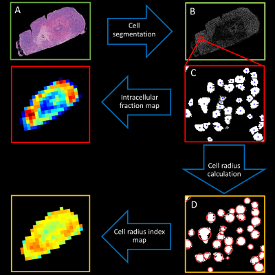 |
107 | VERDICT MRI for estimation of intracellular volume fraction and cell radius: comparison with histology in a mouse model of neuroendocrine tumor
Lukas Lundholm1, Mikael Montelius1, Oscar Jalnefjord1,2, Eva Forssell-Aronsson1,2, and Maria Ljungberg1,2
1Medical Radiation Sciences, University of Gothenburg, Gothenburg, Sweden, 2Medical Physics and Biomedical Engineering, Sahlgrenska University Hospital, Gothenburg, Sweden
VERDICT MRI provides estimates of intracellular volume fraction and cell radius non-invasively which may facilitate e.g., tumor grade classification and longitudinal studies without the need for biopsy. Tumors of a human SI-NET animal model were irradiated and measured with diffusion MRI. Colormaps of cell radius index and intracellular fraction were derived from both VERDICT analysis of the MR data and histological analysis of stained tumor slices. VERDICT maps of intracellular fraction corresponded well with histology in necrotic tissue, however the cell radius index was poorly estimated in these regions. Further work is needed to optimize VERDICT for different tissue types.
|
||
 Back to Meeting Home
 Back to the Program-at-a-Glance
The International Society for Magnetic Resonance in Medicine is accredited by the Accreditation Council for Continuing Medical Education to provide continuing medical education for physicians.
 Back to Meeting Home
 Back to the Program-at-a-Glance
 View the Presentation